Cell Cycle and Checkpoint Controls
The role of cell cycle checkpoint proteins is to integrate internal and external factors to determine whether the cell is prepared for progression of the cell cycle.
What is the cell cycle?
The cell cycle, or cell-division cycle, is the series of events that take place in a cell leading to its division and duplication (replication) to produce two daughter cells. The passage of a cell through the cell cycle is controlled by proteins in the cytoplasm.
Cyclin-dependent kinases and tumor suppressor proteins are stimulators and modulators of cell division. Recent studies have examined the consequences of epigenetic marks and cell cycle control, which has led to more research regarding cell division cancer, emphasizing the fact that the cell division process requires accurate checkpoints to avoid genetic damage.
What are the main cell cycle checkpoints?
The cell cycle is based on three main checkpoints:
- Phase G1 – DNA integrity and cell size
- Phase G2 – DNA damage and chromosome duplication
- Phase M – Attachment of kinetochore and a spindle fiber
The key role of checkpoint proteins is to detect DNA damage and send a signal to delay cell cycle advance until the damaged chromosomes are repaired (Figure 1).
What controls the cell cycle at key checkpoints?
There are two key classes of regulatory molecules:
- Cyclins – a group of proteins that control the progression of cells through the cell cycle by activating cyclin-dependent kinase (CDK) enzymes
- CDKs – a family of protein kinases that are involved in regulating transcription, mRNA processing, and the differentiation of nerve cells
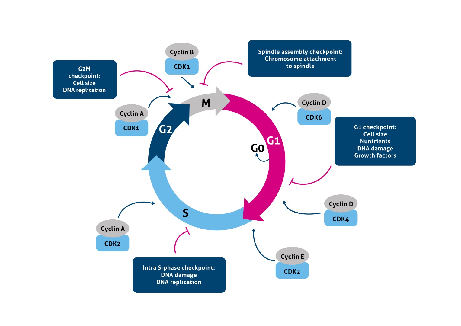
Figure 1. The cell cycle and its checkpoints.
What are the best markers to study a certain cell cycle stage?
A few suggestions below:
1. CDC25A/B/C as DNA markers are good markers to study the G1/S phase (Figure 2)
Figure 2. Immunofluorescent analysis of (4% PFA) fixed human breast cancer tissue using 55031-1-AP (KD/KO validated CDC25A antibody) at a dilution of 1:50 and Alexa Fluor 488-conjugated AffiniPure Goat Anti-Rabbit IgG(H+L).
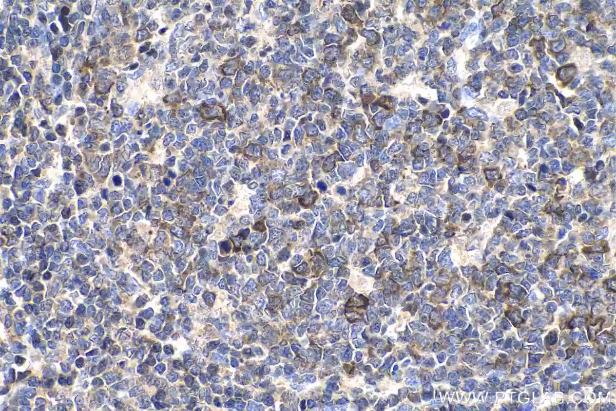
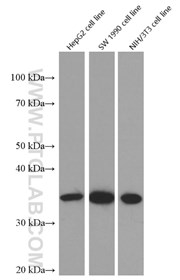
2. High accumulation of Cyclin B1 in the nuclei can be used as a marker for studying the G2/M phase (Figure 3). Also, the expression of Cyclin D should be decreased at the G2/M checkpoint (Figure 4).
 |
|
Figure 3. Immunohistochemical analysis of paraffin-embedded human tonsillitis tissue slide using 28603-1-AP (Cyclin B1 antibody) at dilution of 1:500 (under 40x lens). Heat mediated antigen retrieval with Tris-EDTA buffer (pH 9.0).. |
 |
|
Figure 4. Western blot of Knockout validated Cyclin D1 antibody in HepG2, SW 1990, and NIH/3T3 cell lines with 60186-1-Ig at a dilution of 1:10000 incubated at room temperature for 1.5 hours. |

3) Cyclin D1 is required for G1/S cell cycle transition and can also be used as a G2/M checkpoint marker
CCND1 (Cyclin D1), also known as PRAD1 or BCL1, belongs to the highly conserved cyclin family, whose members are characterized by an ebb and flow in protein abundance throughout the cell cycle. CCND1 forms a complex with and functions as a regulatory subunit of CDK4 or CDK6, whose activity is required for cell cycle G1/S transition. The CCND1 gene, located on 11q13, has been reported to be overexpressed in mantle cell lymphoma (MCL) due to the chromosomal translocation. CCND1 has been shown to interact with tumor suppressor protein Rb and the expression of this gene is positively regulated by Rb. Over-expression of CCND1 correlates with the early onset of cancer and risk of tumor progression and metastasis.
4) To mark mitotic structures, use an antibody against phospho histone 3 (pHH3).
5) In immunohistochemistry, antibodies specific for the Ki-67 antigen will label all cycling cells from G1 to M (Figure 5).
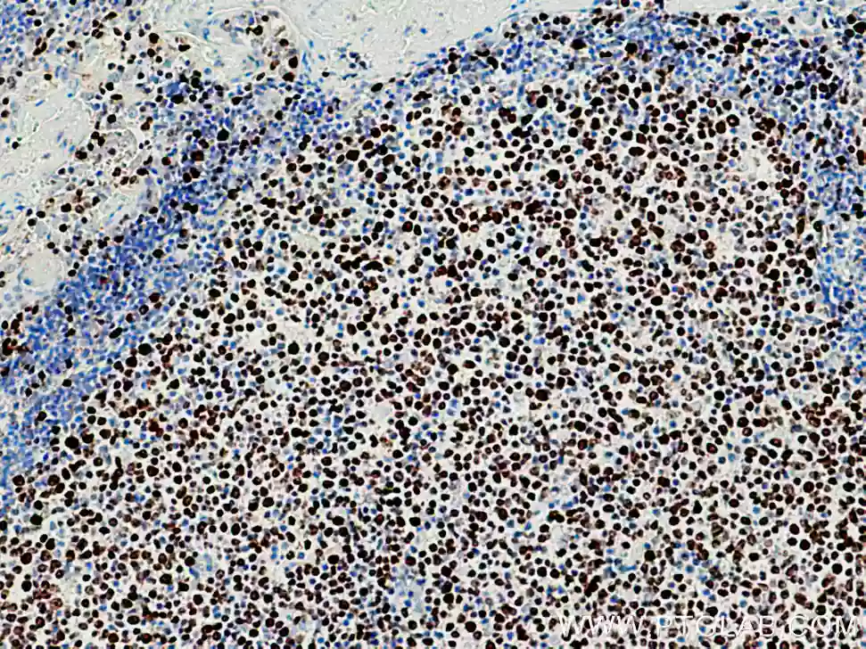
Figure 5. Immunohistochemical analysis of paraffin-embedded human tonsillitis tissue slide using 27309-1-AP (KI67 antibody) at dilution of 1:16000 (under 10x lens). Heat mediated antigen retrieval with Tris-EDTA buffer (pH 9.0).
6) Cyclin A/CDK2 (Figure 6, CDK2 antibody 0122-1-AP) have the highest abundance at the G2 phase of the cell cycle whereas Cyclin B/CDK1 are highest at the M phase of the cell cycle.
CDK2 (Cyclin-dependent kinase 2) is also named CDKN2 and belongs to the protein kinase superfamily, CMGC Ser/Thr protein kinase family, CDC2/CDKX subfamily. It is involved in the control of the cell cycle. It is essential for meiosis but is not required for mitosis. It has 2 isoforms produced by alternative splicing.
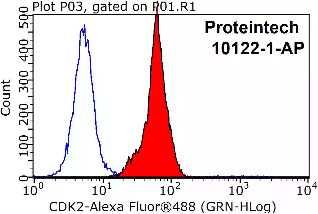
Figure 6. 1X10^6 HepG2 cells were stained with a 0.2ug CDK2 antibody (10122-1-AP, red) and control antibody (blue). Fixed with 90% MeOH blocked with 3% BSA (30 min). Alexa Fluor 488-conjugated AffiniPure Goat Anti-Rabbit IgG(H+L) with dilution 1:1000.
Cyclin related antibodies:
|
Product ID |
Antigen Name |
Cellular Function |
|
CDK1-Specific |
Cell cycle (G2–M) |
|
|
CDK2 |
Cell cycle (G1–S) |
|
|
CDK2 |
Cell cycle (G1–S) |
|
|
CDK3 |
Cell cycle (G0–G1–S) |
|
|
CDK4 |
Cell cycle (G1–S) |
|
|
CDK6 |
Cell cycle (G1–S) |
|
|
CDK6 |
Cell cycle (G1–S) |
|
|
CDK8 |
Transcription |
|
|
CDK9 |
Transcription |
|
|
CDK10 |
Transcription, cell cycle (G2–M) |
Related Content
Cancer stem cells as a key to cure cancer
Molecular markers for liver cancer

Support
Newsletter Signup
Stay up-to-date with our latest news and events. New to Proteintech? Get 10% off your first order when you sign up.