Immunohistochemistry Overview
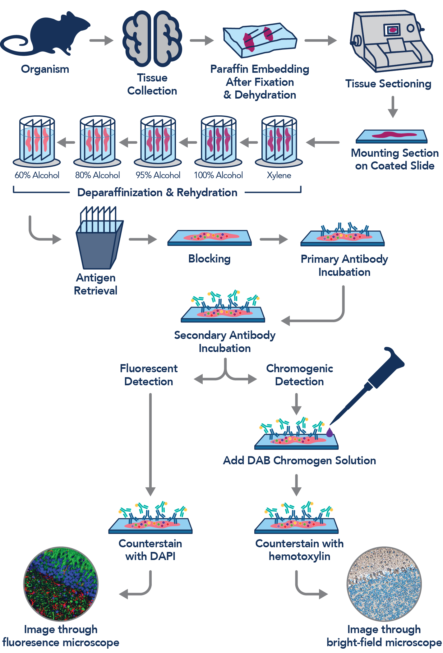
An overview of the immunohistochemistry workflow.
What is Immunohistochemistry (IHC)?
Immunohistochemistry (IHC) is an antibody application which allows you to visualize proteins in tissue while retaining its microstructure. It helps to demonstrate the exact position and distribution of the protein of interest in the analyzed tissue section. The advantage of this visualization is that it allows for comparison between, for example, healthy and diseased tissues. Briefly, in an IHC experiment, the antigen of interest is localized by the binding of an antibody. The antibody-antigen interaction is then further visualized via chromogenic or fluorescent detection.

Immunohistochemistry: Factors to consider before you start
The IHC protocol contains many steps that may require optimization to ensure specific antibody binding and optimal visualization of the target protein. As IHC protocol contains many different variable factors, it is challenging to find the best working conditions to obtain strong and specific staining.
The Main Steps Of Immunohistochemistry
Overview of the main steps of IHC using a chromogenic labeled secondary antibody on paraffin-embedded tissue slides.