at dilution of 1:1000 incubated at room temperature for 1.5 hours.")
 at dilution of 1:10000 incubated at room temperature for 1.5 hours.")
 at dilution of 1:600 incubated at room temperature for 1.5 hours.")
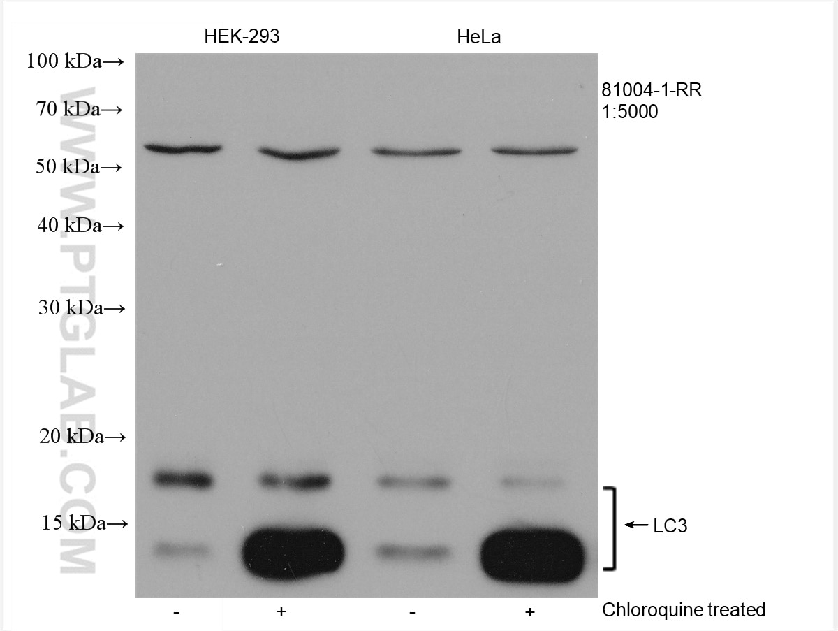
 at dilution of 1:5000 incubated at room temperature for 1.5 hours.")
 at dilution of 1:5000 incubated at room temperature for 1.5 hours.")
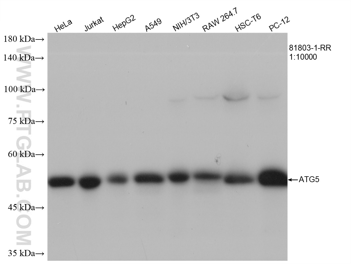
 at dilution of 1:10000 incubated at room temperature for 1.5 hours.")
 at dilution of 1:3000 incubated at room temperature for 1.5 hours.")
 at dilution of 1:3000 incubated at room temperature for 1.5 hours.")
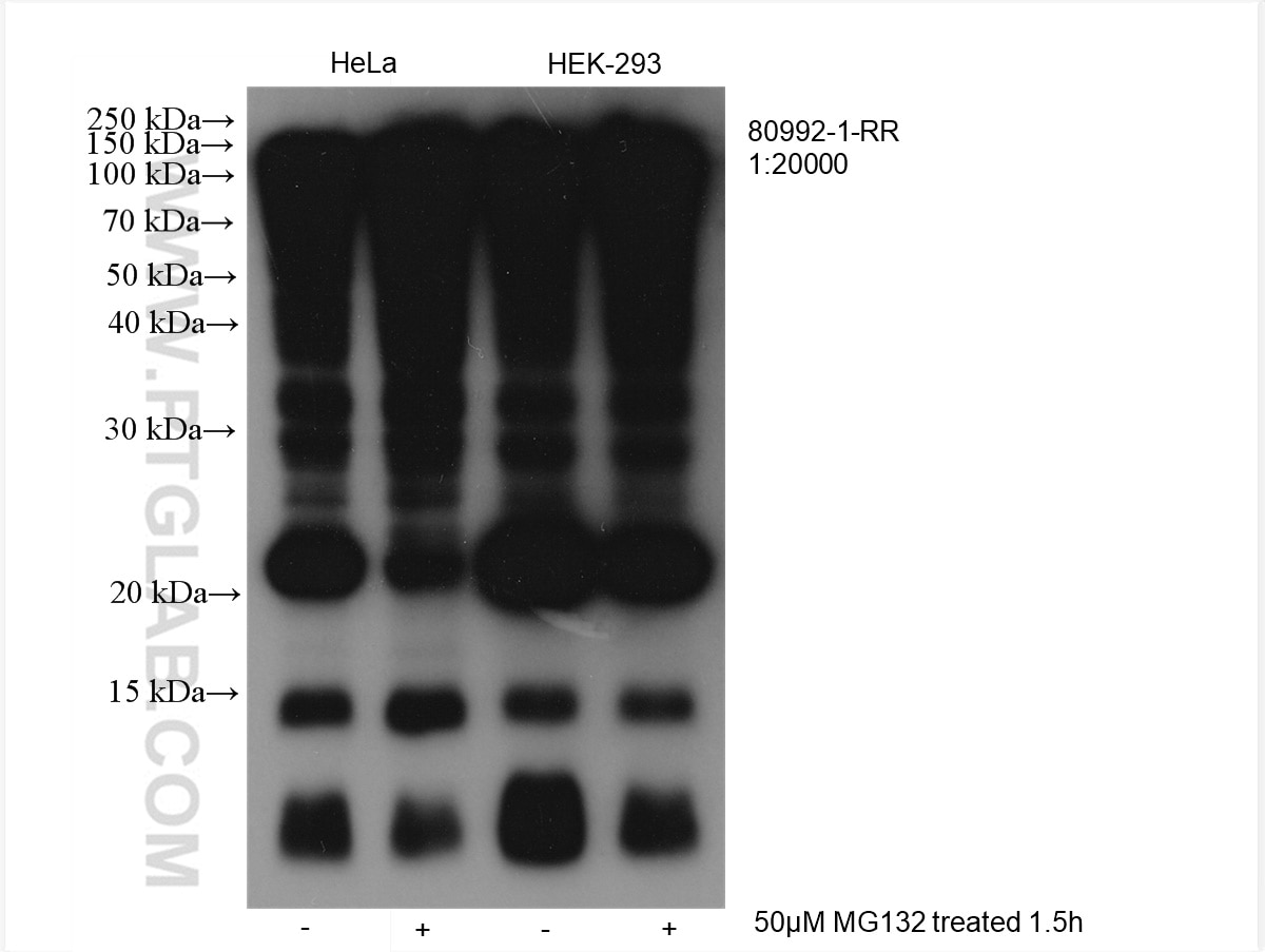
 at dilution of 1:20000 incubated at room temperature for 1.5 hours.")

 with NIH/3T3 cells lysate 1920 ug.")
 with HeLa cells lysate 1800 ug.")
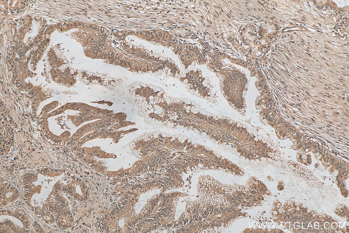
 at dilution of 1:500 (under 10x lens).")
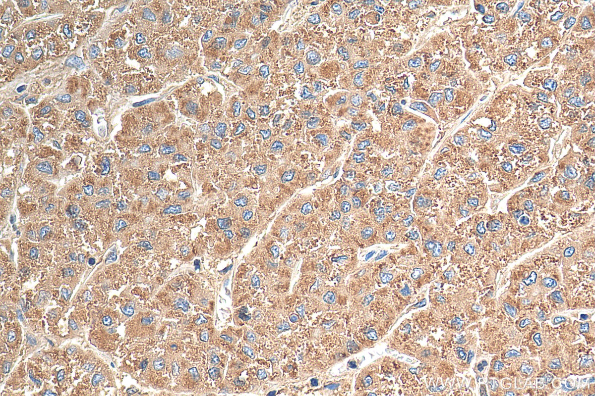
 at dilution of 1:100 (under 10x lens).")
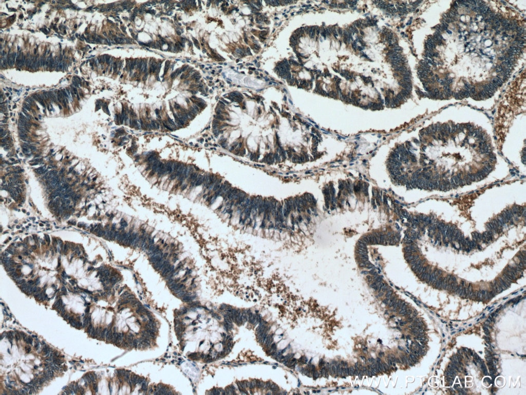
 at dilution of 1:500 (under 10x lens). Heat mediated antigen retrieval with Tris-EDTA buffer (pH 9.0).")
 at dilution of 1:400 (under 10x lens). Heat mediated antigen retrieval with Tris-EDTA buffer (pH 9.0).")
 at dilution of 1:1000 (under 10x lens). Heat mediated antigen retrieval with Tris-EDTA buffer (pH 9.0).")
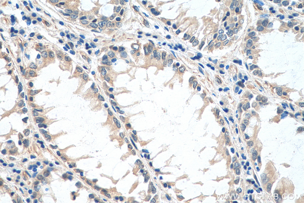
 at dilution of 1:400 (under 40x lens). Heat mediated antigen retrieval with Tris-EDTA buffer (pH 9.0).")
 at dilution of 1:200 (under 10x lens). Heat mediated antigen retrieval with Tris-EDTA buffer (pH 9.0).")
 at dilution of 1:200 (under 10x lens). Heat mediated antigen retrieval with Tris-EDTA buffer (pH 9.0).")
 at dilution of 1:200 (under 40x lens). Heat mediated antigen retrieval with Tris-EDTA buffer (pH 9.0).")
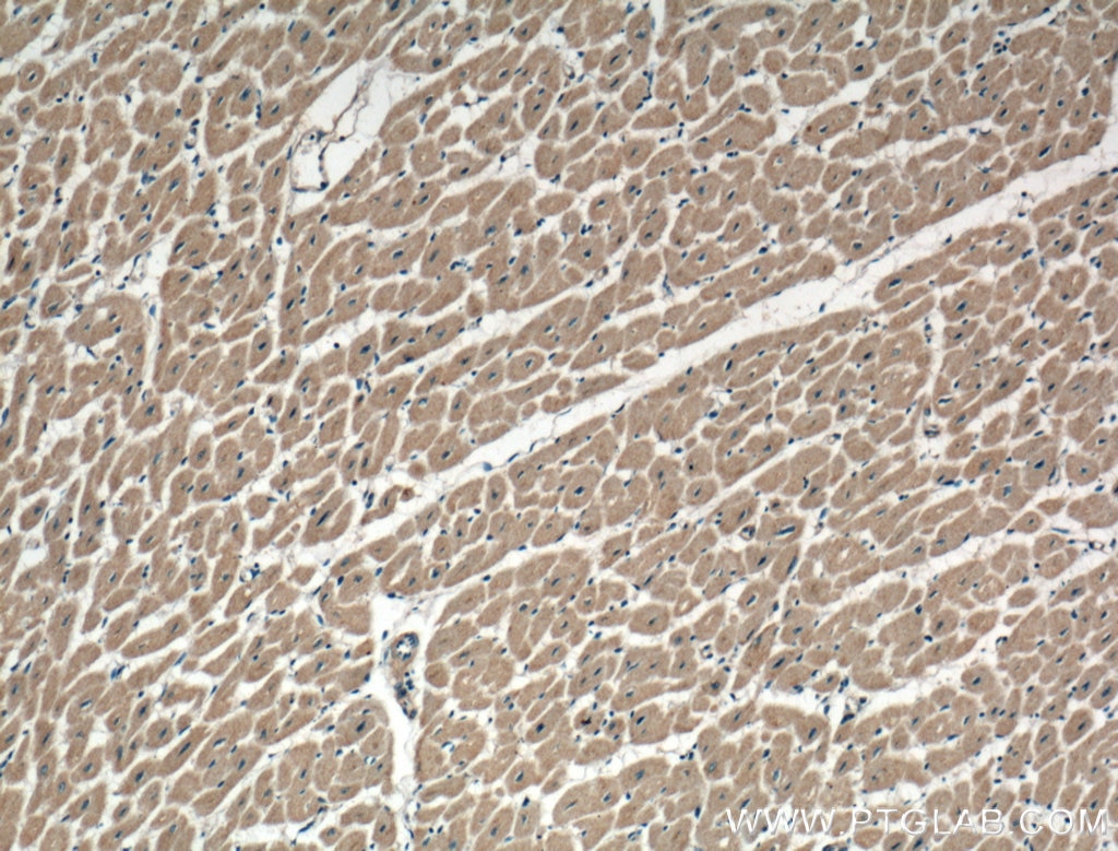
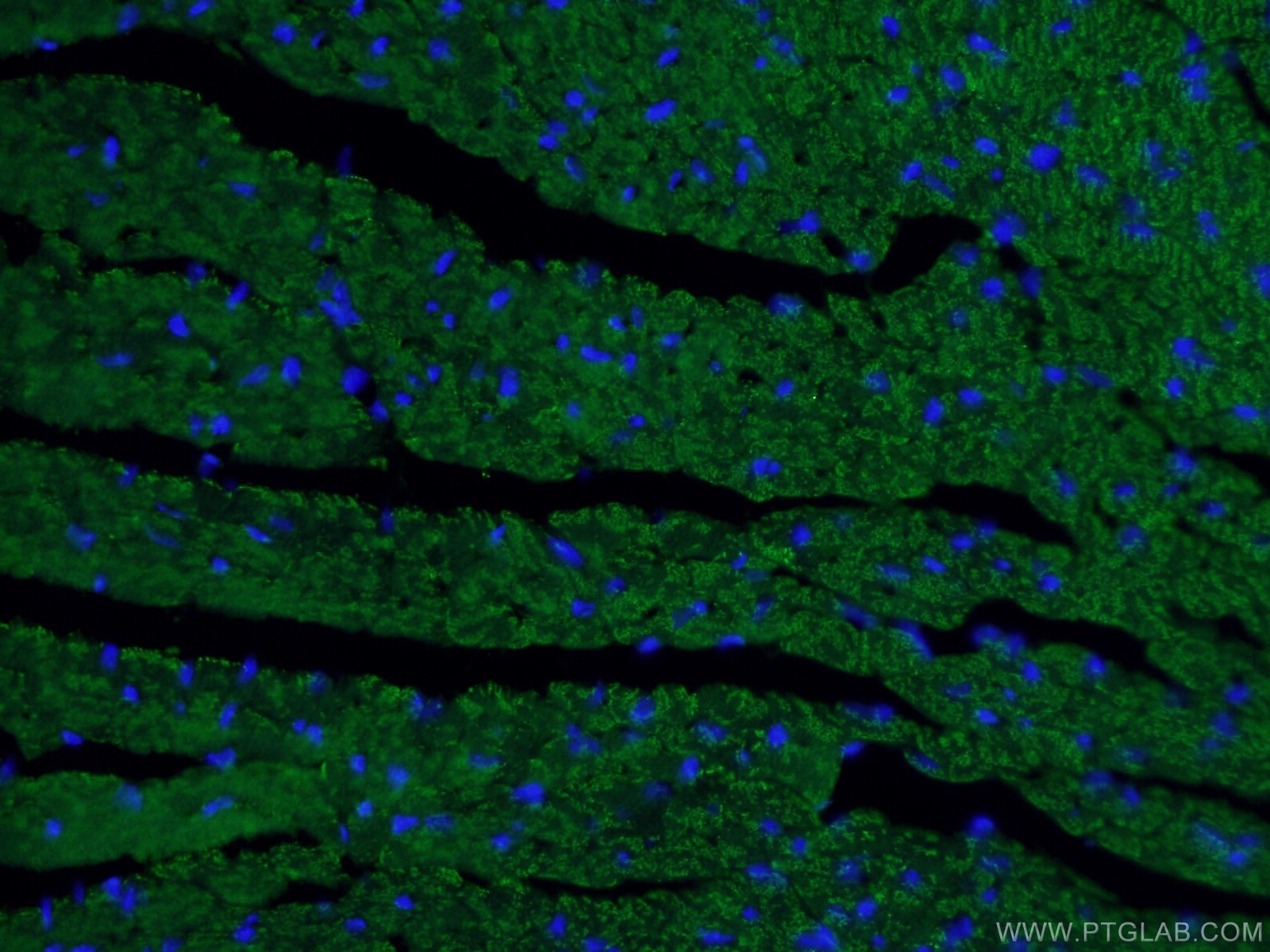
 fixed mouse heart tissue using 66665-1-Ig (Beclin 1 antibody) at dilution of 1:100 and Alexa Fluor 488-Conjugated AffiniPure Goat Anti-Mouse IgG(H+L).")
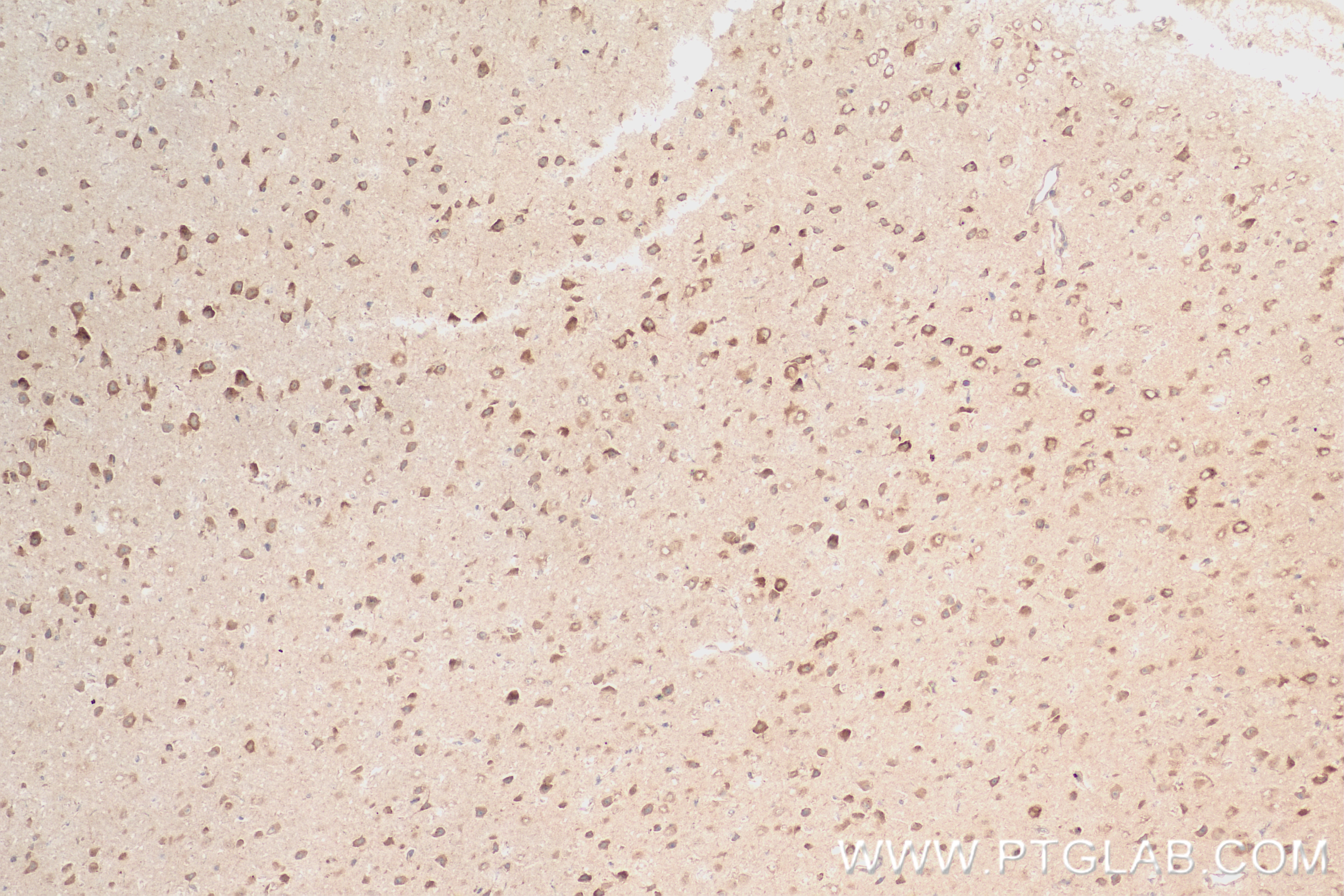
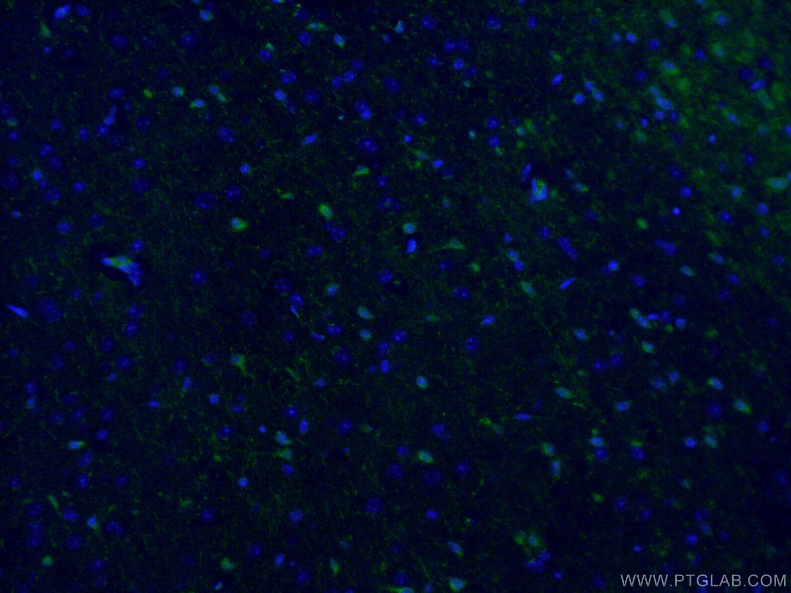
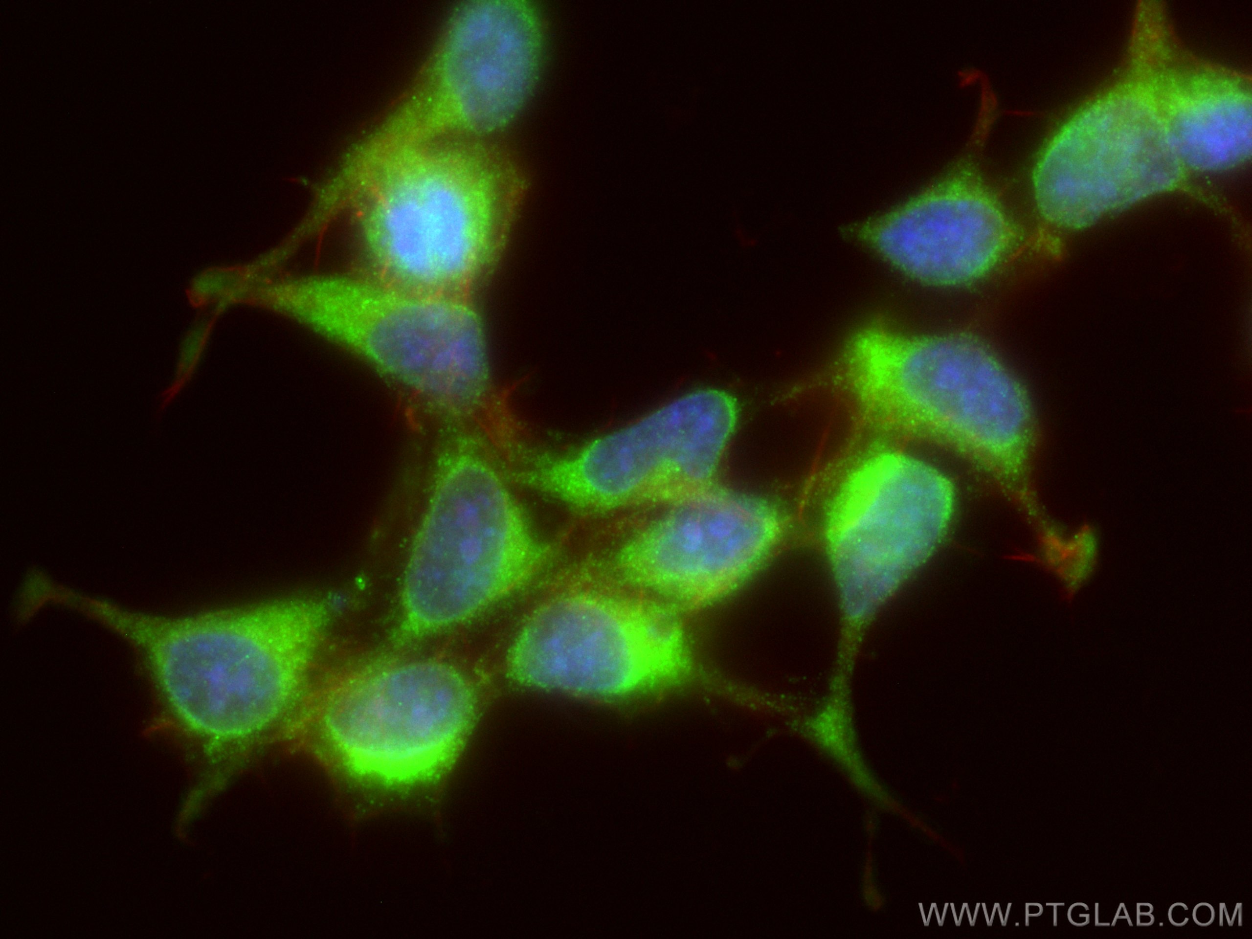
 fixed mouse brain tissue using 26276-1-AP (ATG9A antibody) at dilution of 1:50 and Alexa Fluor 488-Conjugated AffiniPure Goat Anti-Rabbit IgG(H+L).")
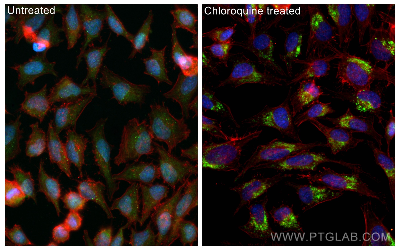
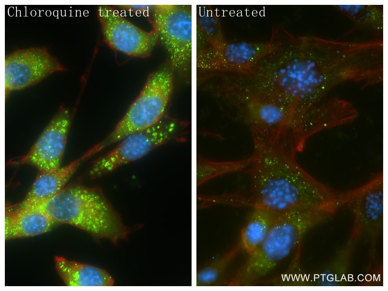
 fixed Chloroquine treated HeLa cells using LC3 antibody (81004-1-RR, Clone: 5P12 ) at dilution of 1:1000 and CoraLite®488-Conjugated AffiniPure Goat Anti-Rabbit IgG(H+L), CoraLite®594 Beta Actin antibody (CL594-66009, Clone: 2D4H5, red).")
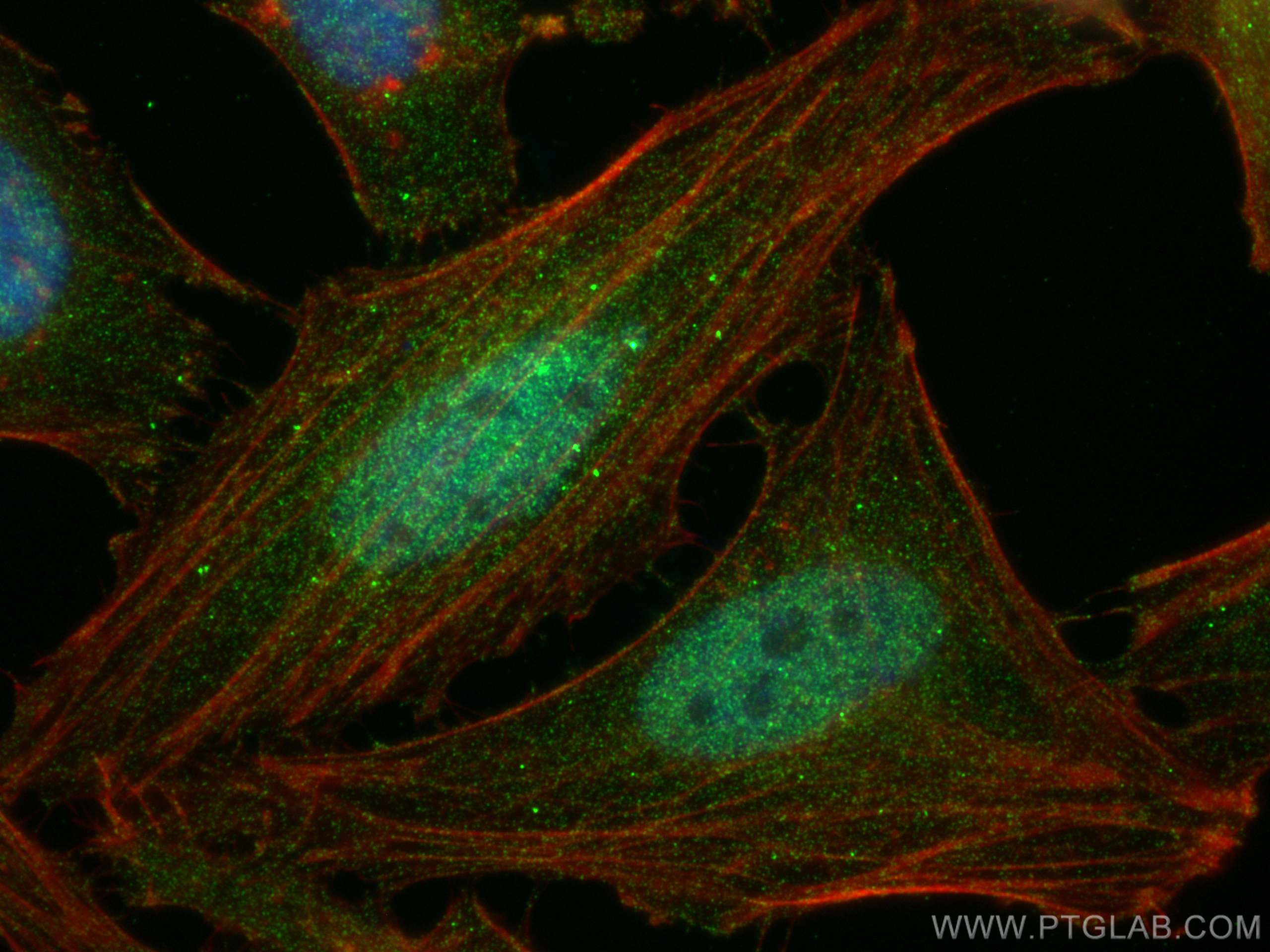
 fixed NIH/3T3 cells using P62,SQSTM1 antibody (84826-1-RR, Clone: 241992C4 ) at dilution of 1:500 and CoraLite®488-Conjugated Goat Anti-Rabbit IgG(H+L) (SA00013-2), CL594-Phalloidin (red).")
 fixed HEK-293 cells using ATG16L1 antibody (29445-1-AP) at dilution of 1:200 and CoraLite®488-Conjugated AffiniPure Goat Anti-Rabbit IgG(H+L), CL594-Phalloidin (red).")
 fixed HeLa cells using ubiquitin antibody (80992-1-RR, Clone: 6H6 ) at dilution of 1:1000 and CoraLite®488-Conjugated AffiniPure Goat Anti-Rabbit IgG(H+L), CL594-phalloidin (red).")
Product Information
The Autophagy Expanded Antibody Kit provides a cost-effective tool for studying key proteins involved in the autophagy pathway. Perfect for researchers starting a new project, screening multiple prospective targets or those who simply require less volume.
Kit Components
The Autophagy Expanded Antibody Kit contains antibodies against 10 key protein targets playing critical roles in the autophagy pathway.
| Antigen | Catalog No. | Host, clonality | Tested Reactivity | Applications | Volume |
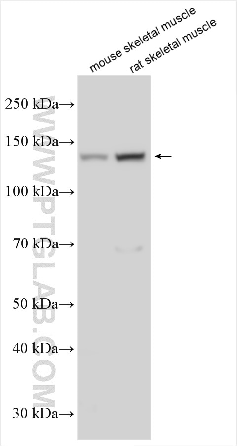
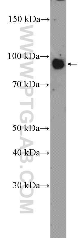
| ULK1 | 20986-1-AP | Rabbit polyclonal | H, M, R | WB, IHC, ELISA | 20 uL |
| Beclin 1 | 66665-1-Ig | Mouse monoclonal | H, M, R | WB, IP, IHC, IF, ELISA | 20 uL |
| ATG9A | 67096-1-Ig | Mouse monoclonal | H | WB, IHC, IF/ICC, ELISA | 20 uL |
| LC3B | 81004-1-RR | Rabbit monoclonal | H, M, R, Pg | WB, IHC, IF/ICC, ELISA | 20 uL |
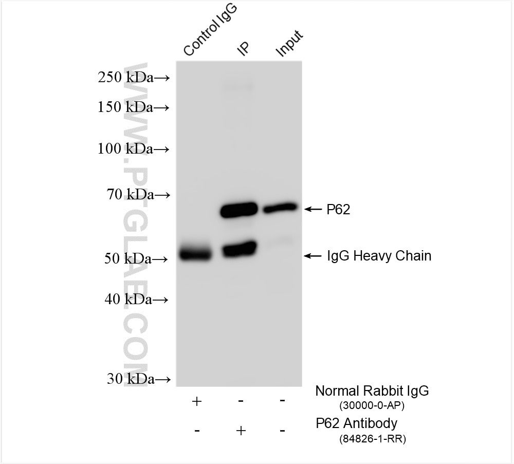
| p62 | 84826-1-RR | Rabbit monoclonal | H, M, R | WB, IHC, IF/ICC, IP, ELISA | 20 uL |
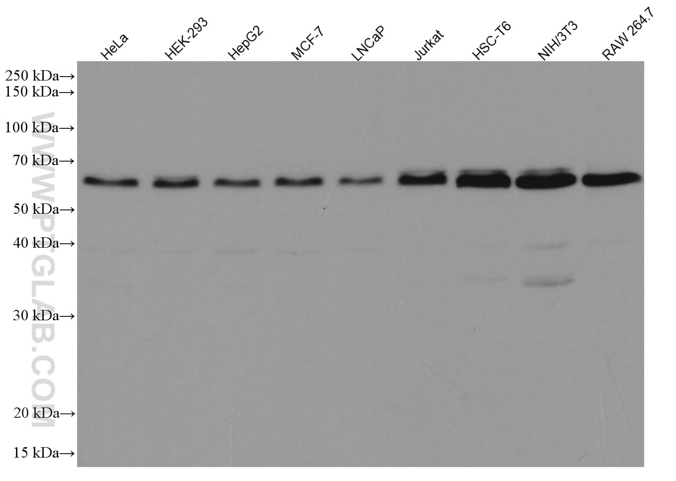
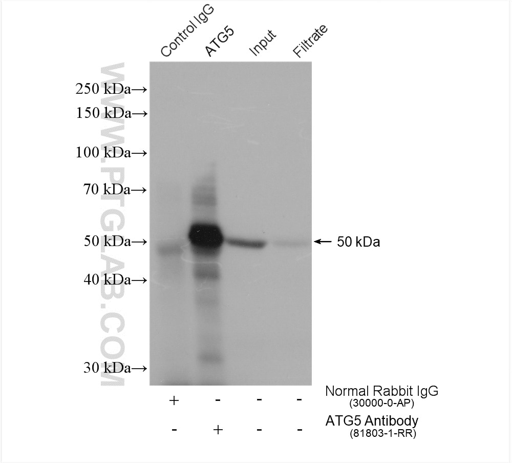
| ATG5 | 81803-1-RR | Rabbit monoclonal | H, M, R | WB, IP, IHC, ELISA | 20 uL |
| ATG16 L1 | 29445-1-AP | Rabbit polyclonal | H, M, R | WB, IHC, IF, ELISA | 20 uL |
| ATG12 | 11264-1-AP | Rabbit polyclonal | H, M | WB, IHC, IF/ICC, ELISA | 20 uL |
| Ubiquitin | 80992-1-RR | Rabbit monoclonal | H, M, | WB, IHC, IF/ICC, ELISA | 20 uL |
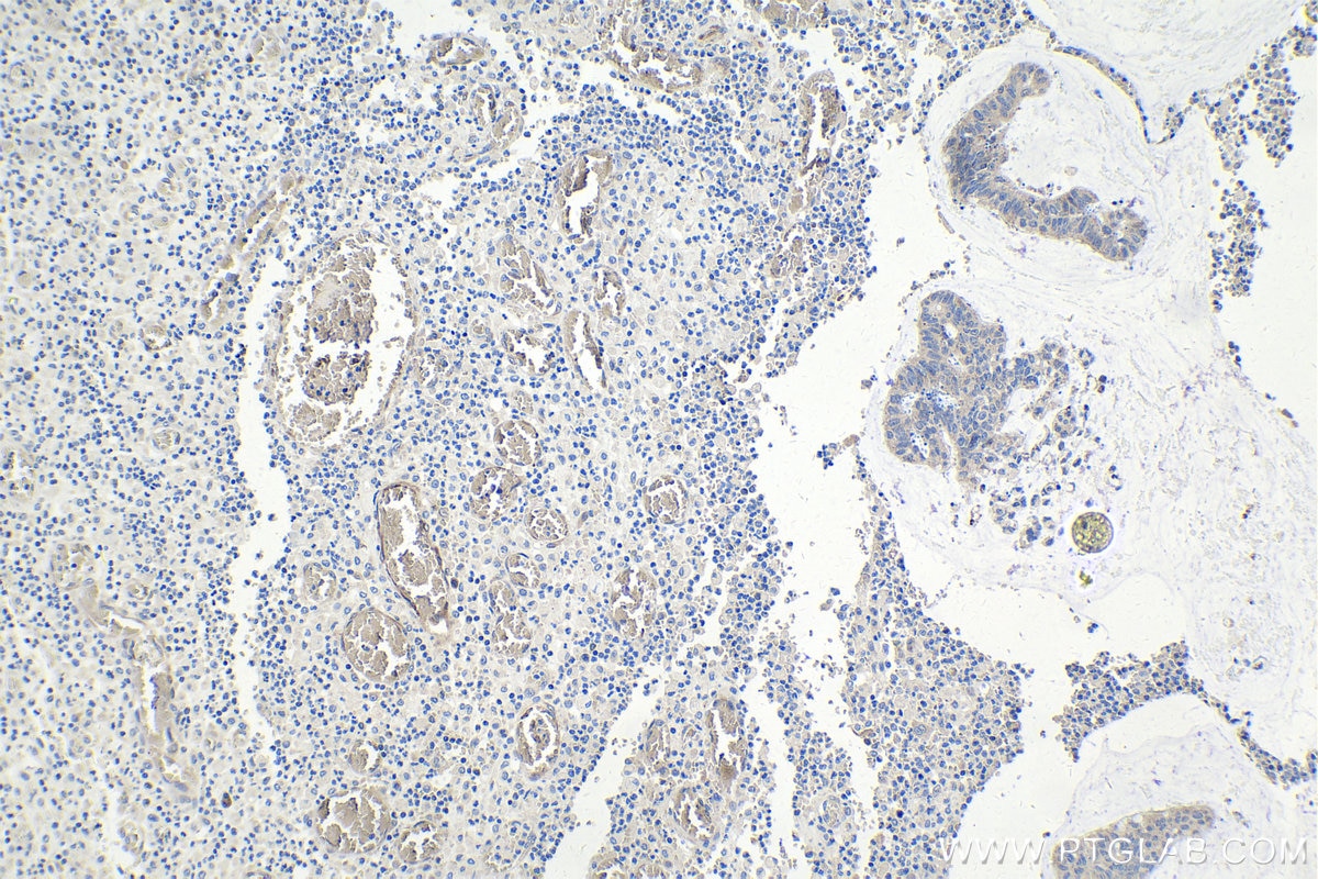
| LAMP1 | 21997-1-AP | Rabbit polyclonal | H | WB, IHC, ELISA | 20 uL |
Also see our 'Autophagy Essentials Antibody Kit' on the following page https://www.ptglab.com/products/Autophagy-Essentials-Antibody-Kit-PK30004.htm
Storage
Store at -20°C. Stable for one year from the date of receipt.
Background Information
Autophagy is a highly dynamic process consisting of the following three steps: (1) autophagosome formation, (2) autophagosome-lysosome fusion, and (3) degradation. It can be induced by multiple signaling pathways related to various triggers including nutrient deprivation, growth factor signaling, and cellular stress. ULK1 and Beclin 1 are critical for the initiation of autophagy. The process of autophagosome formation proceeds through the steps of initiation, nucleation, elongation, closure, and ultimately fusion, each of which is regulated by various ATG proteins. Ubiquitination of various autophagy-related proteins and regulatory proteins are critical for the precise regulation of the autophagy pathway.
The ideal approach for measuring autophagy is to assess autophagic flux, which represents the rate of degradation of the autophagic pathway. The most widely used method for measuring autophagic flux is to detect the processing of the autophagosomal membrane protein, LC3. Analyzing autophagy substrates such as p62/SQSTM1 is often recommended in addition to measuring LC3-II turnover for accurate assessment of autophagic flux. The fusion of autophagosomes with lysosomes can be monitored by analyzing the autophagosomal marker LC3 and the lysosomal marker, LAMP simultaneously.
Standard Protocols
Click here to view our standard protocols for various applications including WB, IP, IHC, IF, FC, and ELISA.
Cited in Article as
PK30005, Autophagy Expanded Antibody Kit, Proteintech, IL, USA
Documentation
| Datasheet |
|---|
| Autophagy Expanded Antibody Kit Datasheet |