Intracellular Flow Cytometry
Introducing our NEW range of antibodies for intracellular flow cytometry
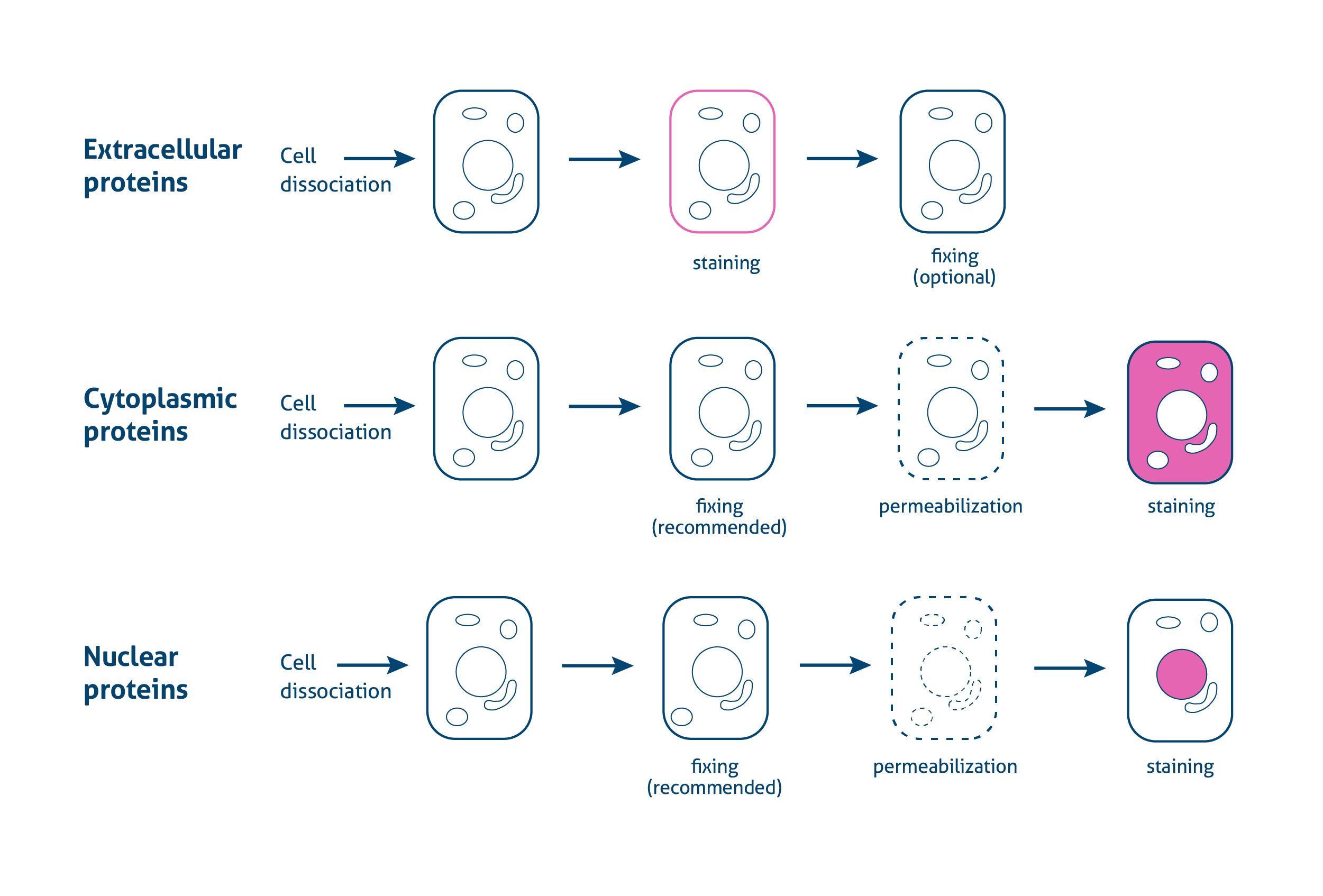
Intracellular flow cytometry
Unlike traditional flow cytometry for cell surface markers staining, intracellular flow cytometry brings us a powerful tool to stain intracellular target proteins, such as phosphorylated signal transducers, transcription factors and cytokines.
Through fixation and permeabilization, antibodies are able to penetrate the cell and nuclear membranes to stain target proteins within the cell. Proteintech offers a range of antibodies for intracellular flow cytometry single cell analysis with optimized fixation and permeabilization reagents for reproducible results.
Cytoplasmic Target Detection
Intracellular flow cytometry is a powerful tool to simultaneously detect multiple targets within an individual cell. Intracellular flow cytometry is a powerful tool to analyze T-, B-, NK- cell differentiation, activation and plasticity during immune or inflammatory response.
An important group of cytoplasmic targets are cytokines, small secreted molecules which are mediators of immune and inflammatory responses. Unlike ELISA or WB, intracellular flow techniques can pinpoint the cellular source of a particular cytokine within a larger, heterogenous population when combined with traditional surface labeling. This is achieved by using protein transport inhibitors, like Brefeldin A or monensin, to trap cytokines in within the secretory cells. Besides cytokines, other cytoplasmic proteins can also be detected through intracellular flow. Proteintech provides various fluorochrome labeled antibodies which cover a large number of targets and different colors, as well as relative reagents for cytoplasmic targets intracellular flow analysis.
Besides cytokines, other cytoplasmic proteins can also be detected through intracellular flow. Proteintech provides various fluorochrome labeled antibodies which cover a large number of targets and different colors, as well as relative reagents for cytoplasmic targets intracellular flow analysis.
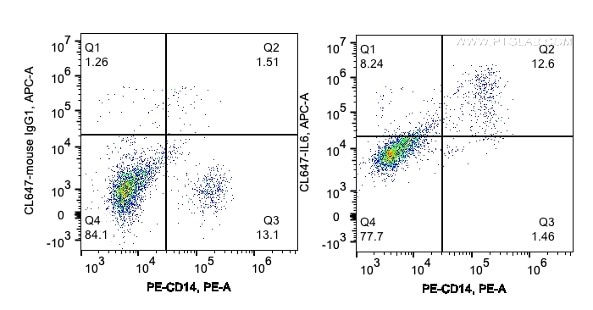
Human PBMCs are treated by LPS and protein transporter Brefeldin A. 1*10^6 stimulated human PBMCs were surface stained with PE Anti-human CD14 (PE-65056, Clone: UCHM-1), fixed by 4% PFA and then permeabilized by 1X Flow Cytometry permeabilization buffer (PF00011-C). Cells were then intracellularly stained with 1ul CoraLite®647-conjugated Anti-human IL6 (CL647-69001, Clone: 4G3E6), or CoraLite®647-conjugated mouse IgG1 isotype control.
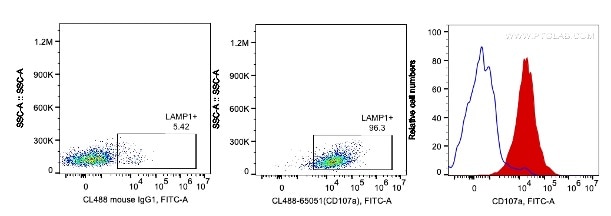
Human PBMCs are stimulated by PHA for 3 days. Cells were intracellularly stained with 5ul CoraLite488 Anti-human CD107a (H4A3) (red), or 0.2ug CoraLite488 mouse IgG1 isotype control (blue). Cells were fixed by 4% PFA and permeabilized with 1X intracellular flow cytometry perm buffer (PF00011-C). Lymphocytes are gated.
Example Protocol for Cytoplasmic Staining
Required reagents:
- 4% PFA Fix buffer (4% paraformaldehyde dissolve in 1x PBS, adjust pH to 7.4)
- 10x Flow Cytometry Permeabilization buffer (PF00011-C) link all catalog numbers to landing page dilute to 1X concentrate in distilled water prior to use
- Flow cytometry staining buffer (PF00012)
- 1x PBS
- Flow cytometry antibodies
Experiment procedures:
- Collect cells from cell culture incubator. For suspension cells, spin down cells by centrifuge 400g for 5 min, and wash the cells by 1xPBS twice. For adherent cells, digest cells by Trypsin for 5 min, add cell culture medium to stop digestion and wash the cells by 1xPBS twice. Aliquot cells in each 5mL round bottom polystyrene FACS tubes at a cell concentration suitable for staining.
- Stain surface marker antibodies with the recommended optimal concentration of fluorochrome labeled antibodies.
- Wash cell with 1mL staining buffer, centrifuge at 400g for 5 minutes and discard supernatant.
- Resuspend cells with 200 µl 4% PFA Fix buffer and vortex briefly, incubate in dark at 4°C for 30 min to 1 hour.
- Spin down cells by centrifuge 400g, 5 min, resuspend cells with 2mL 1x Flow Cytometry Permeabilization buffer and incubate at room temperature for 5 minutes in dark.
- Spin down cells by centrifuge 400g, 5 min, resuspend cells with 100 µl 1x Flow Cytometry Permeabilization buffer. Add the recommended amount of fluorochrome-labeled antibody for detection of intracellular antigen(s) to cells and incubate in the dark at room temperature for 20-60 minutes
Note: Antibodies for intracellular staining should always be prepared in 1x Flow Cytometry Permeabilization buffer.
- After incubation, wash cell in 1mL staining buffer and spin down cells by centrifuge 400g, 5 min, resuspend cells with 500 µl 1x Flow Cytometry staining buffer for Flow Cytometer analysis.
Optional: If the primary antibody is unconjugated, spin down cells by centrifuge 400g, 5 min, resuspend cells with 100 µl 1x Flow Cytometry Permeabilization buffer and add 0.1 µg secondary antibody to each tube and incubate in dark for 15 minutes at 4°C. After incubation, spin down cells by centrifuge 400g, 5 min, resuspend cells with 500 µl 1x Flow Cytometry Staining buffer for flow cytometer analysis.
Related
- Flow cytometry antibodies
- Supporting reagents and kits
- Introduction to flow cytometry
- Flow cytometry applications
- Flow cytometry sample preparation
- Flow cytometry multicolor panel design
- Protocol for studying extracellular and intracellular proteins
- Experimental protocol to study cell viability and apoptosis
- Spectra viewer
Nuclear Targets Detection
Similar to intracellular protocols for cytoplasmic detection, nuclear staining for transcription factor and other nuclear proteins requires cellular fixation and permeabilization after surface marker staining. Depending on the nature of the transcription factor and its location on the nucleus, DNA, or other proteins, different fixation and permeabilization steps may be required.
The fixation and permeabilization of cells can compromise cell surface marker staining, which makes the choice of compatible buffers even more critical. To further enable easy transcription factor detection, Proteintech recommends using Foxp3 / Transcription Factor Staining Buffer Kit (Catalog #PF00011) which is specially formulated for optimal resolution and low background in your analysis of nuclear targets by flow cytometry. Through fixation and permeabilization reagents included in this kit, antibodies are able to penetrate the cell and nuclear membranes to stain target proteins within the cell. Combining with Proteintech’s featured Intracellular flow cytometry antibodies, this kit will bring better intracellular flow cytometry results.
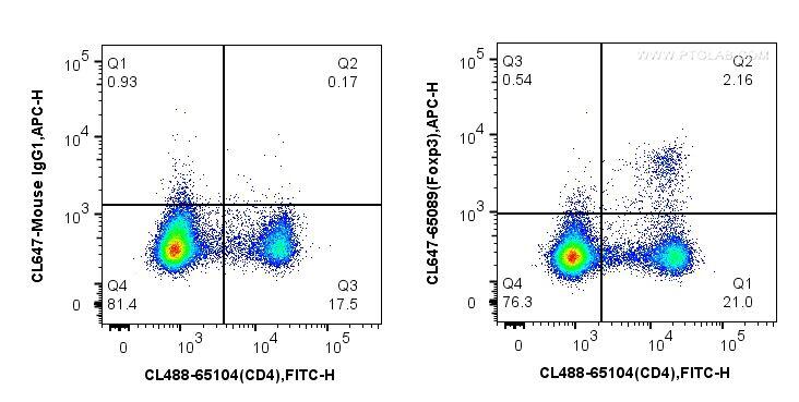
1X10^6 mouse splenocytes were surface stained with PE Anti-Mouse CD4 (GK1.5) (PE-65104, Clone: GK1.5) and then fixed by Transcription Factor fixation buffer and permeabilization buffer (PF00011-C). Cells were then stained with 5 ul CoraLite®647-conjugated Anti-Mouse Foxp3 (CL647-65089, Clone: 3G3), or CoraLite®647-conjugated mouse IgG1 isotype control.
Example Protocol for Nuclear Staining
Required reagents:
Foxp3 / Transcription Factor Staining Buffer Kit (PF00011) includes PF00011-A, PF00011-B, PF00011-C
- 4x Transcription Factor Fix/Perm concentrate (PF00011-A), dilute to 1X concentrate in 1x Transcription Factor Fix/Perm dilute (PF00011-B) prior to use
- 10x Flow Cytometry Permeabilization buffer (PF00011-C), dilute to 1X concentrate in distilled water prior to use
- Flow cytometry staining buffer (PF00012)
- 1x PBS
- Flow cytometry antibodies
Experiment procedures:
- Collect cells from cell culture incubator. For suspension cells, spin down cells by centrifuge 400g for 5 min, and wash the cells by 1xPBS twice. For adherent cells, digest cells by Trypsin for 5 min, add cell culture medium to stop digestion and wash the cells by 1xPBS twice. Aliquot cells in each 5mL round bottom polystyrene FACS tubes at a cell concentration suitable for staining.
- Stain surface marker antibodies with the recommended optimal concentration of fluorochrome labeled antibodies.Wash cell in 1mL staining buffer, centrifuge at 400g for 5 minutes and discard supernatant.
- Resuspend cells with 200 µl 1x Transcription Factor Fix/Perm buffer and vortex briefly, incubate in dark at 4°C for 30 min to 1 hour.
- Spin down cells by centrifuge 400g, 5 min, resuspend cells with 2mL 1x Flow Cytometry Permeabilization buffer and incubate at room temperature for 5 minutes in dark.
- Spin down cells by centrifuge 400g, 5 min, resuspend cells with 100 µl 1x Flow Cytometry Permeabilization buffer. Add the recommended amount of fluorochrome-labeled antibody for detection of intracellular antigen(s) to cells and incubate in the dark at room temperature for 20-60 minutes
Note: Antibodies for intracellular staining should always be prepared in 1x Flow Cytometry Permeabilization buffer.
- After incubation, wash cell in 1mL staining buffer and spin down cells by centrifuge 400g, 5 min, resuspend cells with 500 µl 1x Flow Cytometry staining buffer for Flow Cytometer analysis.
Optional: If the primary antibody is unconjugated, spin down cells by centrifuge 400g, 5 min, resuspend cells with 100 µl 1x Flow Cytometry Permeabilization buffer and add 0.1 µg secondary antibody to each tube and incubate in dark for 15 minutes at 4°C. After incubation, wash cells with 1ml staining buffer and spin down cells by centrifuge 400g, 5 min, resuspend cells with 500 µl 1x Flow Cytometry Staining buffer for flow cytometer analysis.
Related
- Flow cytometry antibodies
- Supporting reagents and kits
- Introduction to flow cytometry
- Flow cytometry applications
- Flow cytometry sample preparation
- Flow cytometry multicolor panel design
- Protocol for studying extracellular and intracellular proteins
- Experimental protocol to study cell viability and apoptosis
- Spectra viewer
Fixation and Permeabilization Reagents
Foxp3 / Transcription Factor Staining Buffer Kit
The Foxp3 / Transcription Factor Staining Buffer Kit contains specially formulated buffers and solutions for optimal resolution and low background in your analysis of nuclear antigens by flow cytometry.
Foxp3 / Transcription Factor Fix/Perm Concentrate (4X)
Provided in a concentrated soltion which, when diluted with Foxp3 / Transcription Factor Fix/Perm Diluent (1X) provide best results during intracellular staining of transcription factors using fluorescently conjugated antibodies.
Foxp3 / Transcription Factor Fix/Perm Diluent (1X)
Intended for use as a diluent for Foxp3 / Transcription Factor Fix/Perm Concentrate (4X).
Flow Cytometry Perm Buffer (10X)
Provided as a concentrate which, when diluted with distilled water to a 1X solution, provide best results during intracellular staining of cytokines and other cytoplasmic antigens, by maintaining membrane permeabilization throughout staining and washing steps.
Flow Cytometry Staining Buffer (1X)
Ready-to-use Flow Staining Buffer (1X) is recommended immunofluorescent staining protocols using viable or fixed cells in suspension for flow cytometry.
RBC Lysis Buffer (10X) is a concentrated ammonium chloride-based lysing reagent. The diluted 1X working solution will lyse red blood cells in single cell suspensions with minimal effects on leukocytes.
Related
- Flow cytometry antibodies
- Supporting reagents and kits
- Introduction to flow cytometry
- Flow cytometry applications
- Flow cytometry sample preparation
- Flow cytometry multicolor panel design
- Protocol for studying extracellular and intracellular proteins
- Experimental protocol to study cell viability and apoptosis
- Spectra viewer
Isotype Controls
| Subclass | Species | Isotype Control |
|---|---|---|
| IgG | Rabbit | IgG control Polyclonal antibody |
| IgG1 | Mouse | Mouse IgG1 isotype control Monoclonal antibody |
| IgG2a | Mouse | Mouse IgG2a isotype control Monoclonal antibody |
| IgG2b | Mouse | Mouse IgG2b isotype control Monoclonal antibody |
| IgG1 | Rat | CoraLite®647 Rat IgG1 Isotype Control (HRPN) |
| IgG2a | Rat | CoraLite®647 Rat IgG2a Isotype Control (2A3) |
| IgG2b | Rat | CoraLite®647 Rat IgG2b Isotype Control (LTF-2) |
Related
- Flow cytometry antibodies
- Supporting reagents and kits
- Introduction to flow cytometry
- Flow cytometry applications
- Flow cytometry sample preparation
- Flow cytometry multicolor panel design
- Protocol for studying extracellular and intracellular proteins
- Experimental protocol to study cell viability and apoptosis
- Spectra viewer