• Phare
  • Validé par KD/KO

Anticorps Polyclonal de lapin anti-VDAC1/Porin

VDAC1/Porin Polyclonal Antibody for FC, IF, IHC, WB, ELISA

Hôte / Isotype

Lapin / IgG

Réactivité testée

Humain, rat, souris et plus (6)

Applications

WB, PLA, IHC, IF, FC, CoIP, ELISA

Conjugaison

Non conjugué

N° de cat : 55259-1-AP

Synonymes

hVDAC1, Plasmalemmal porin, Porin, PORIN 31 HL, Porin 31HL, Porin 31HM, VDAC, VDAC 1, VDAC1, VDAC1/Porin


Filter:
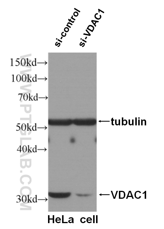
  • Western Blot (WB) analysis of HeLa cells using VDAC1/Porin Polyclonal antibody (55259-1-AP)

    WB analysis of HeLa cells using 55259-1-AP

    WB result of VDAC1 antibody (55259-1-AP, 1:2000) with si-Control and si-VDAC1 transfected HeLa cells.

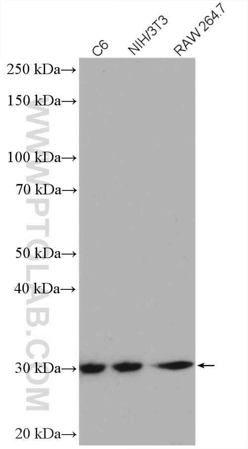
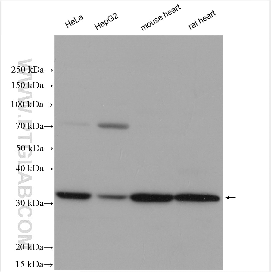
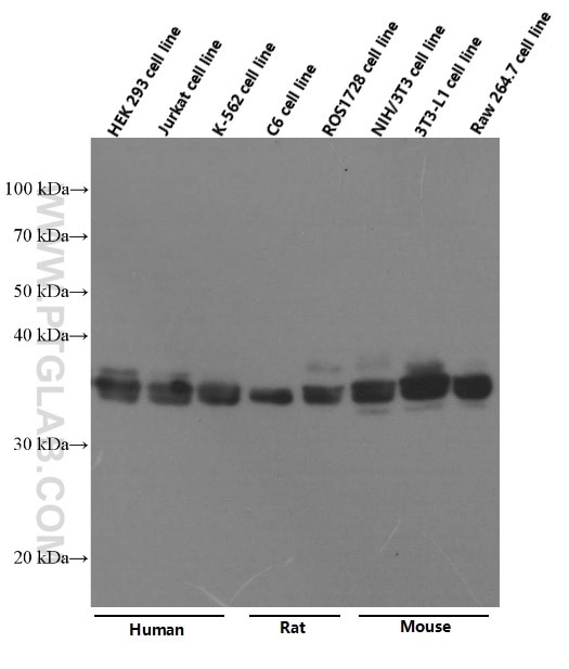
  • Western Blot (WB) analysis of various lysates using VDAC1/Porin Polyclonal antibody (55259-1-AP)

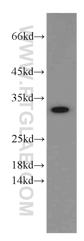
    WB analysis using 55259-1-AP

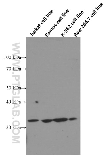
    Various lysates were subjected to SDS PAGE followed by western blot with 55259-1-AP (VDAC1/Porin antibody) at dilution of 1:3000 incubated at room temperature for 1.5 hours.

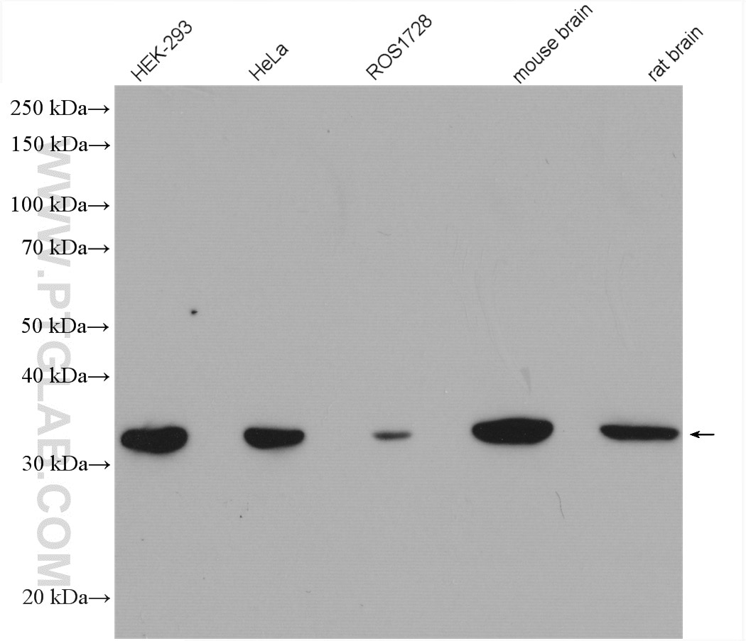
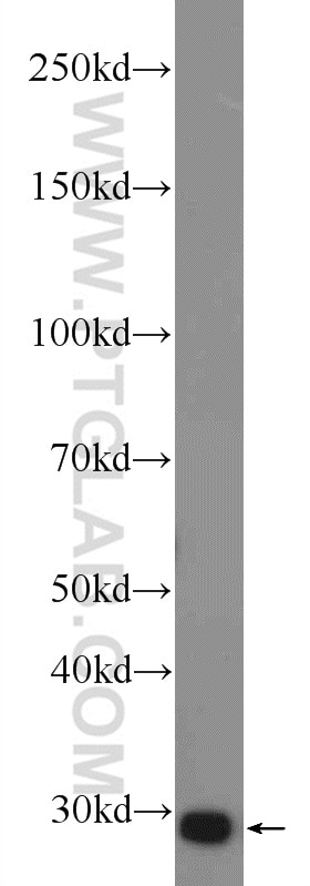
  • Western Blot (WB) analysis of various lysates using VDAC1/Porin Polyclonal antibody (55259-1-AP)

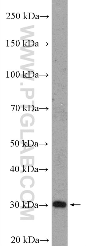
    WB analysis using 55259-1-AP

    Various lysates were subjected to SDS PAGE followed by western blot with 55259-1-AP (VDAC1/Porin antibody) at dilution of 1:5000 incubated at room temperature for 1.5 hours.

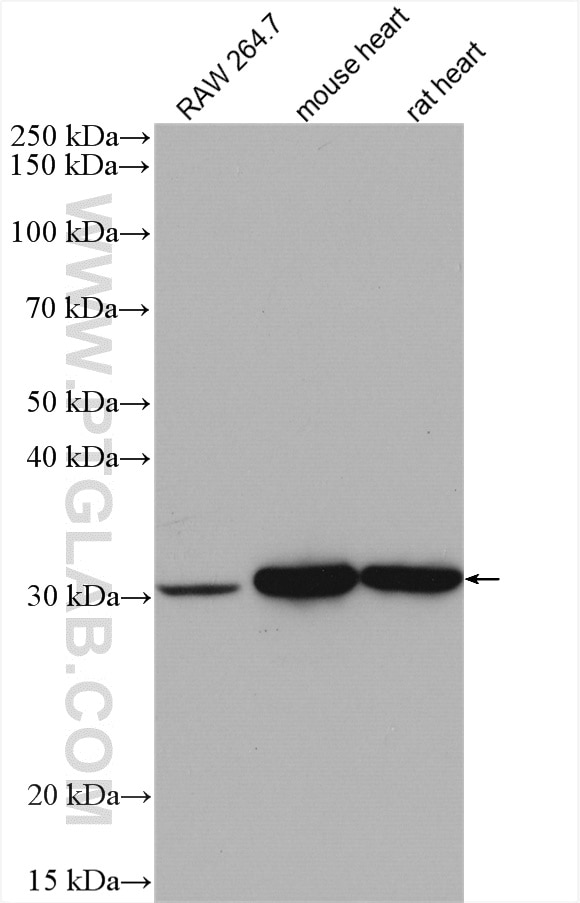
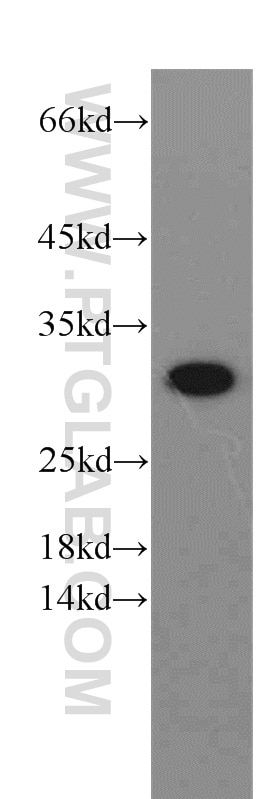
  • Western Blot (WB) analysis of various lysates using VDAC1/Porin Polyclonal antibody (55259-1-AP)

    WB analysis using 55259-1-AP

    Various lysates were subjected to SDS PAGE followed by western blot with 55259-1-AP (VDAC1/Porin antibody) at dilution of 1:3000 incubated at room temperature for 1.5 hours.

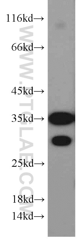
  • Western Blot (WB) analysis of various lysates using VDAC1/Porin Polyclonal antibody (55259-1-AP)

    WB analysis using 55259-1-AP

    Various lysates were subjected to SDS PAGE followed by western blot with 55259-1-AP (VDAC1/Porin antibody) at dilution of 1:1500 incubated at room temperature for 1.5 hours.

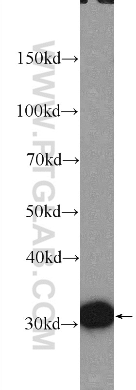
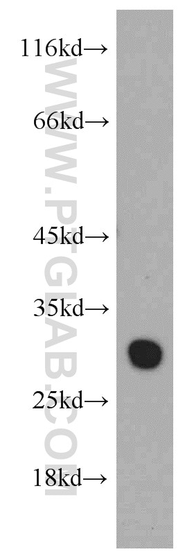
  • Western Blot (WB) analysis of rat liver tissue using VDAC1/Porin Polyclonal antibody (55259-1-AP)

    WB analysis of rat liver using 55259-1-AP

    rat liver tissue were subjected to SDS PAGE followed by western blot with 55259-1-AP (VDAC1/Porin antibody at dilution of 1:600 incubated at room temperature for 1.5 hours.

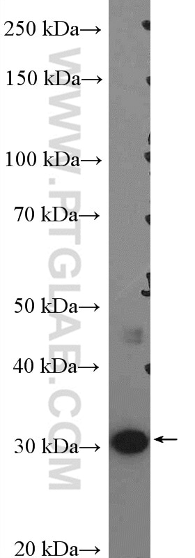
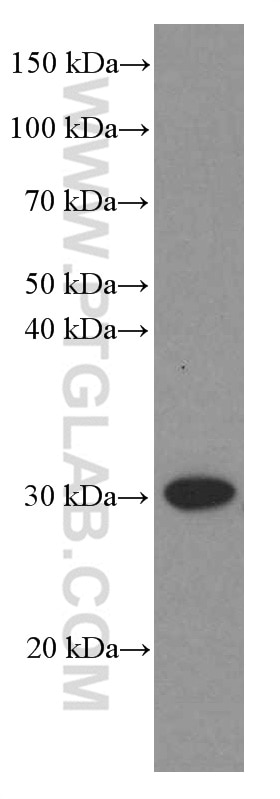
  • Western Blot (WB) analysis of HepG2 cells using VDAC1/Porin Polyclonal antibody (55259-1-AP)

    WB analysis of HepG2 using 55259-1-AP

    HepG2 cells were subjected to SDS PAGE followed by western blot with 55259-1-AP (VDAC1/Porin antibody) at dilution of 1:600 incubated at room temperature for 1.5 hours.

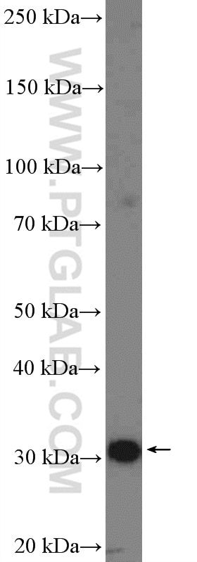
  • Western Blot (WB) analysis of human skeletal muscle tissue using VDAC1/Porin Polyclonal antibody (55259-1-AP)

    WB analysis of human skeletal muscle using 55259-1-AP

    human skeletal muscle tissue were subjected to SDS PAGE followed by western blot with 55259-1-AP (VDAC1/Porin antibody) at dilution of 1:600 incubated at room temperature for 1.5 hours.

  • Western Blot (WB) analysis of mouse liver tissue using VDAC1/Porin Polyclonal antibody (55259-1-AP)

    WB analysis of mouse liver using 55259-1-AP

    mouse liver tissue were subjected to SDS PAGE followed by western blot with 55259-1-AP (VDAC1/Porin antibody at dilution of 1:600 incubated at room temperature for 1.5 hours.

  • Western Blot (WB) analysis of A431 cells using VDAC1/Porin Polyclonal antibody (55259-1-AP)

    WB analysis of A431 using 55259-1-AP

    A431 cells were subjected to SDS PAGE followed by western blot with 55259-1-AP (VDAC1/Porin antibody at dilution of 1:600 incubated at room temperature for 1.5 hours.

  • Western Blot (WB) analysis of mouse heart tissue using VDAC1/Porin Polyclonal antibody (55259-1-AP)

    WB analysis of mouse heart using 55259-1-AP

    mouse heart tissue were subjected to SDS PAGE followed by western blot with 55259-1-AP (VDAC1/Porin antibody at dilution of 1:600 incubated at room temperature for 1.5 hours.

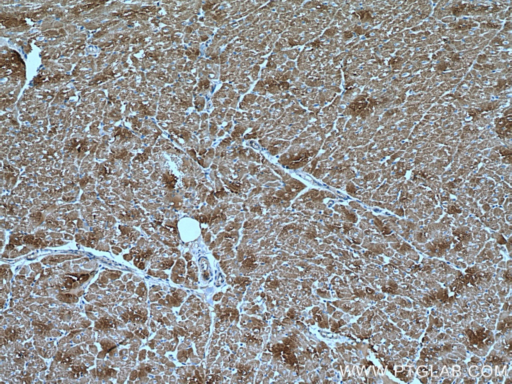
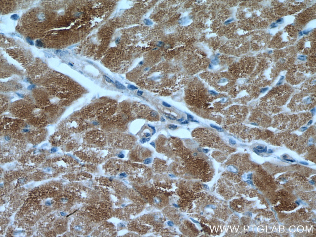
  • Immunohistochemistry (IHC) staining of human heart tissue using VDAC1/Porin Polyclonal antibody (55259-1-AP)

    IHC staining of human heart using 55259-1-AP

    Immunohistochemical analysis of paraffin-embedded human heart tissue slide using 55259-1-AP (VDAC1/Porin antibody) at dilution of 1:200 (under 10x lens). Heat mediated antigen retrieval with Tris-EDTA buffer (pH 9.0).

  • Immunohistochemistry (IHC) staining of human heart tissue using VDAC1/Porin Polyclonal antibody (55259-1-AP)

    IHC staining of human heart using 55259-1-AP

    Immunohistochemical analysis of paraffin-embedded human heart tissue slide using 55259-1-AP (VDAC1/Porin antibody) at dilution of 1:200 (under 40x lens). Heat mediated antigen retrieval with Tris-EDTA buffer (pH 9.0).

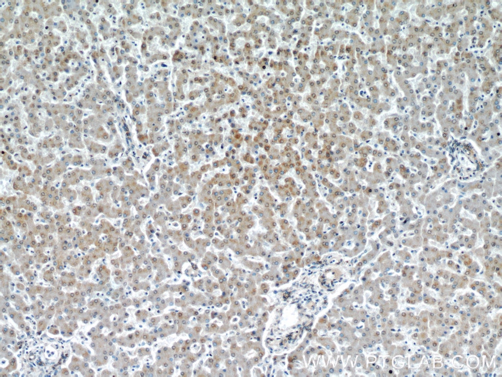
  • Immunohistochemistry (IHC) staining of human liver tissue using VDAC1/Porin Polyclonal antibody (55259-1-AP)

    IHC staining of human liver using 55259-1-AP

    Immunohistochemical analysis of paraffin-embedded human liver using 55259-1-AP (VDAC1/Porin antibody) at dilution of 1:50 (under 10x lens).

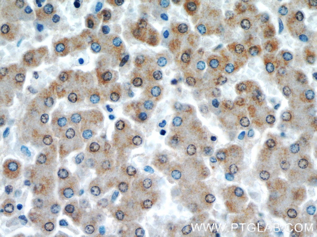
  • Immunohistochemistry (IHC) staining of human liver tissue using VDAC1/Porin Polyclonal antibody (55259-1-AP)

    IHC staining of human liver using 55259-1-AP

    Immunohistochemical analysis of paraffin-embedded human liver using 55259-1-AP (VDAC1/Porin antibody) at dilution of 1:50 (under 40x lens).

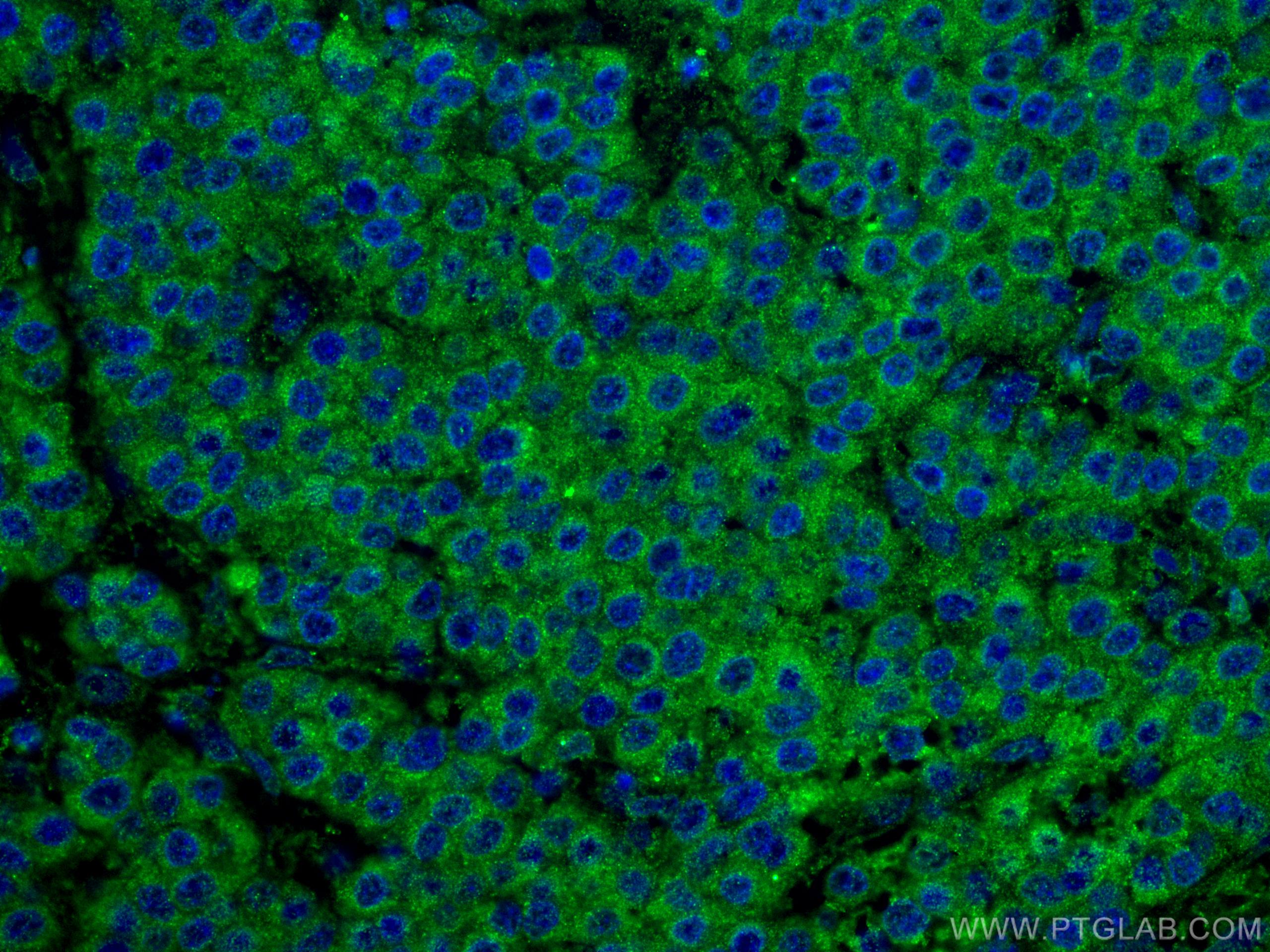
  • Immunofluorescence (IF) / fluorescent staining of human liver cancer tissue using VDAC1/Porin Polyclonal antibody (55259-1-AP)

    IF Staining of human liver cancer using 55259-1-AP

    Immunofluorescent analysis of (4% PFA) fixed human liver cancer tissue using VDAC1/Porin antibody (55259-1-AP) at dilution of 1:200 and CoraLite®488-Conjugated AffiniPure Goat Anti-Rabbit IgG(H+L).

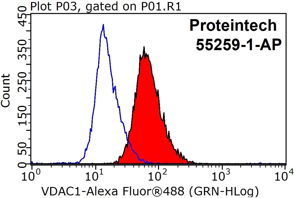
  • Flow cytometry (FC) experiment of HepG2 cells using VDAC1/Porin Polyclonal antibody (55259-1-AP)

    FC experiment of HepG2 using 55259-1-AP

    1X10^6 HepG2 cells were stained with 0.2ug VDAC1/Porin antibody (55259-1-AP, red) and control antibody (blue). Fixed with 90% MeOH blocked with 3% BSA (30 min). Alexa Fluor 488-conjugated AffiniPure Goat Anti-Rabbit IgG(H+L) with dilution 1:1000.

"VDAC1/Porin Antibodies" Comparison

View side-by-side comparison of VDAC1/Porin antibodies from other vendors to find the one that best suits your research needs.

Applications testées

Résultats positifs en WBcellules RAW 264.7, cellules A431, cellules HEK-293, cellules HeLa, cellules HepG2, cellules NIH/3T3, cellules ROS1728, tissu cardiaque de rat, tissu cardiaque de souris, tissu cérébral de rat, tissu cérébral de souris, tissu de muscle squelettique humain, tissu hépatique de rat, tissu hépatique de souris
Résultats positifs en IHCtissu cardiaque humain, tissu hépatique humain
il est suggéré de démasquer l'antigène avec un tampon de TE buffer pH 9.0; (*) À défaut, 'le démasquage de l'antigène peut être 'effectué avec un tampon citrate pH 6,0.
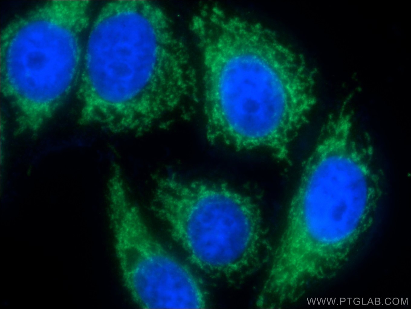
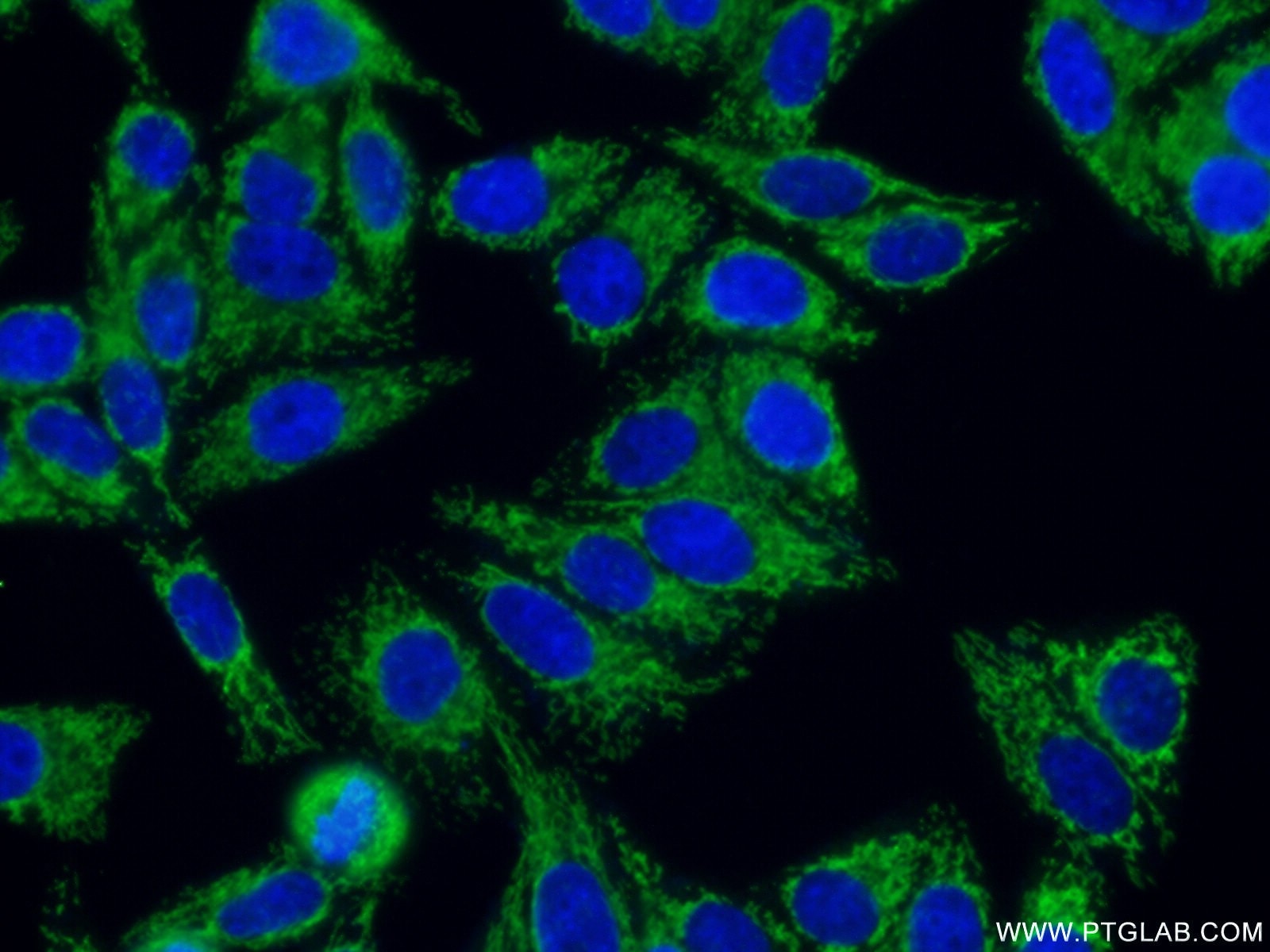
Résultats positifs en IFtissu de cancer du foie humain,
Résultats positifs en cytométriecellules HepG2

Dilution recommandée

ApplicationDilution
Western Blot (WB)WB : 1:1000-1:6000
Immunohistochimie (IHC)IHC : 1:50-1:500
Immunofluorescence (IF)IF : 1:50-1:500
Flow Cytometry (FC)FC : 0.20 ug per 10^6 cells in a 100 µl suspension
It is recommended that this reagent should be titrated in each testing system to obtain optimal results.
Sample-dependent, check data in validation data gallery

Informations sur le produit

55259-1-AP cible VDAC1/Porin dans les applications de WB, PLA, IHC, IF, FC, CoIP, ELISA et montre une réactivité avec des échantillons Humain, rat, souris

Réactivité Humain, rat, souris
Réactivité citéerat, bovin, canin, Humain, porc, poulet, singe, souris, Hamster
Hôte / Isotype Lapin / IgG
Clonalité Polyclonal
Type Anticorps
Immunogène Peptide
Nom complet voltage-dependent anion channel 1
Masse moléculaire calculée 31 kDa
Poids moléculaire observé 31 kDa
Numéro d’acquisition GenBankNM_003374
Symbole du gène VDAC1
Identification du gène (NCBI) 7416
Conjugaison Non conjugué
Forme Liquide
Méthode de purification Purification par affinité contre l'antigène
Tampon de stockage PBS avec azoture de sodium à 0,02 % et glycérol à 50 % pH 7,3
Conditions de stockageStocker à -20°C. Stable pendant un an après l'expédition. L'aliquotage n'est pas nécessaire pour le stockage à -20oC Les 20ul contiennent 0,1% de BSA.

Informations générales

VDAC1, also named as VDAC, porin 31HM, porin 31HL and plasmalemmal porin, belongs to the eukaryotic mitochondrial porin family. It adopts an open conformation at low or zero membrane potential and a closed conformation at potentials above 30-40 mV, to form a channel through the mitochondrial outer membrane and also the plasma membrane. Unlike other membrane transport proteins, porins are large enough to allow passive diffusion. Studies have shown that VDAC1 is subject to both phosphorylation and acetylation (PMID: 23233904). The apparent molecular weight of VDAC1 is 30-37 kDa (PMID: 14573604; 23754752; 25681439). Hypoxic conditions were found to trigger cleavage of the VDAC1 C-terminal to yield a 26-kDa truncated but active form (PMID: 22389449; 23233904). This antibody is specific to VDAC1.

Protocole

Product Specific Protocols
WB protocol for VDAC1/Porin antibody 55259-1-APDownload protocol
IHC protocol for VDAC1/Porin antibody 55259-1-APDownload protocol
IF protocol for VDAC1/Porin antibody 55259-1-APDownload protocol
Standard Protocols
Click here to view our Standard Protocols

Publications

SpeciesApplicationTitle
monkeyWB

Nat Commun

An ESCRT-dependent step in fatty acid transfer from lipid droplets to mitochondria through VPS13D-TSG101 interactions.

Authors - Jingru Wang
humanWB

Nucleic Acids Res

The human tRNA taurine modification enzyme GTPBP3 is an active GTPase linked to mitochondrial diseases.

Authors - Gui-Xin Peng
IHC

Mol Biol Evol

Functional Attenuation of UCP1 as the Potential Mechanism for Thickened Blubber Layer in Cetaceans

Authors - Ming Zhou
mouseWB

Redox Biol

Adiponectin/AdiopR1 signaling prevents mitochondrial dysfunction and oxidative injury after traumatic brain injury in a SIRT3 dependent manner.

Authors - Shenghao Zhang
humanWB

Autophagy

KAT7-mediated CANX (calnexin) crotonylation regulates leucine-stimulated MTORC1 activity.

Authors - Guokai Yan
mouseWB

Autophagy

Mycobacterium bovis induces mitophagy to suppress host xenophagy for its intracellular survival.

Authors - Yinjuan Song

Avis

The reviews below have been submitted by verified Proteintech customers who received an incentive forproviding their feedback.


FH

Ying (Verified Customer) (04-22-2021)

good antibody. works very well with mouse cell lines.

  • Applications: Western Blot
  • Primary Antibody Dilution: 1:2000
VDAC1/Porin Antibody Western Blot validation (1:2000 dilution) in (Cat no:55259-1-AP)