COXIV Monoclonal antibody

COXIV Monoclonal Antibody for WB, IHC, IF/ICC, ELISA

Host / Isotype

Mouse / IgG1

Reactivity

human and More (2)

Applications

WB, IHC, IF/ICC, ELISA

Conjugate

Unconjugated

CloneNo.

2A7B2

Cat no : 66110-1-Ig

Synonyms

Cell and organelle markers, COX IV 1, COX4, COX4I1, COXIV, Cytochrome c oxidase polypeptide IV, Cytochrome c oxidase subunit 4 isoform 1 mitochondrial, Mitochondria Marker, Mitochondrial Marker, Mitochondrion Marker


Filter:
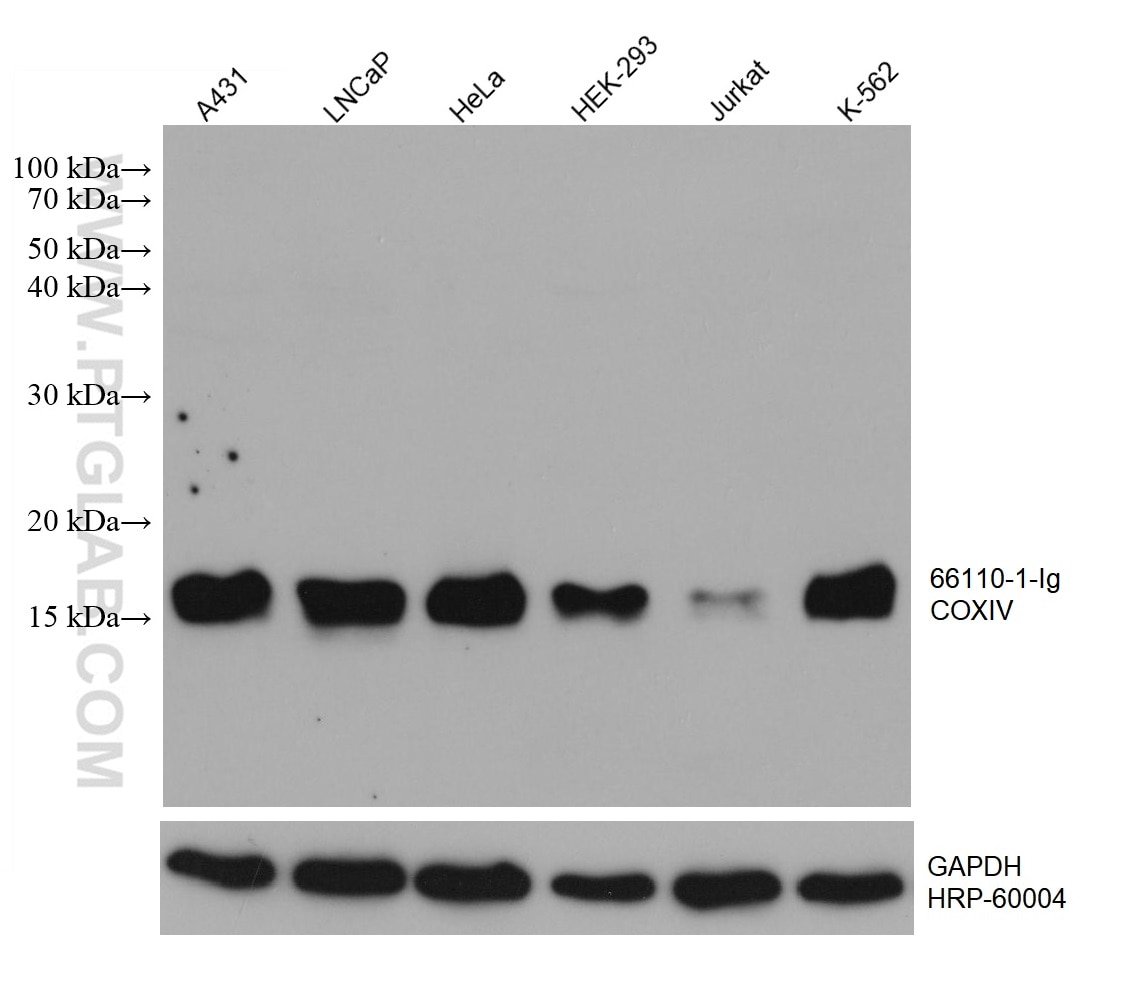
  • Western Blot (WB) analysis of various lysates using COXIV Monoclonal antibody (66110-1-Ig)

    WB analysis using 66110-1-Ig

    Various lysates were subjected to SDS PAGE followed by western blot with 66110-1-Ig (COXIV antibody) at dilution of 1:20000 incubated at room temperature for 1.5 hours. The membrane was stripped and reblotted with HRP-conjugated GAPDH Monoclonal antibody (HRP-60004) as loading control.

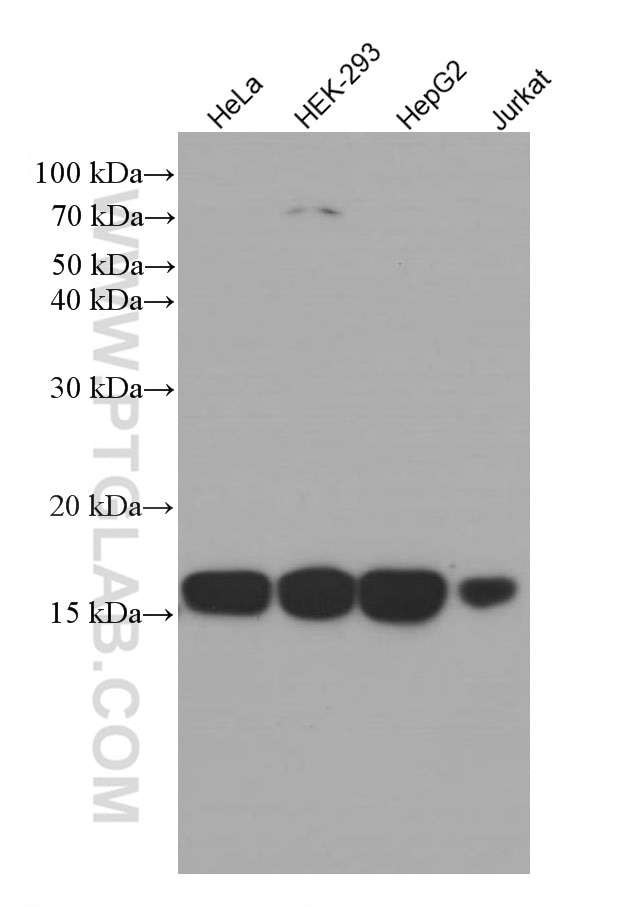
  • Western Blot (WB) analysis of various lysates using COXIV Monoclonal antibody (66110-1-Ig)

    WB analysis using 66110-1-Ig

    Various lysates were subjected to SDS PAGE followed by western blot with 66110-1-Ig (COXIV antibody) at dilution of 1:3000 incubated at room temperature for 1.5 hours.

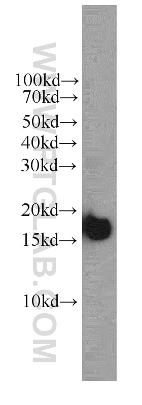
  • Western Blot (WB) analysis of HeLa cells using COXIV Monoclonal antibody (66110-1-Ig)

    WB analysis of HeLa using 66110-1-Ig

    HeLa cells were subjected to SDS PAGE followed by western blot with 66110-1-Ig (COXIV antibody) at dilution of 1:2000 incubated at room temperature for 1.5 hours.

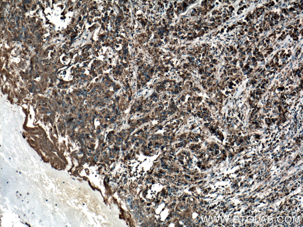
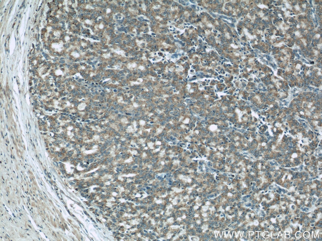
  • Immunohistochemistry (IHC) staining of human prostate cancer tissue using COXIV Monoclonal antibody (66110-1-Ig)

    IHC staining of human prostate cancer using 66110-1-Ig

    Immunohistochemical analysis of paraffin-embedded human prostate cancer tissue slide using 66110-1-Ig (COXIV antibody) at dilution of 1:500 (under 10x lens. Heat mediated antigen retrieval with Tris-EDTA buffer (pH 9.0).

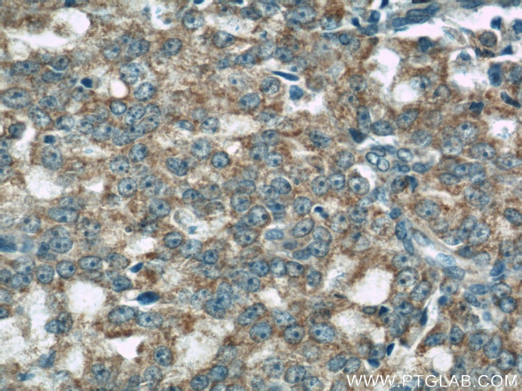
  • Immunohistochemistry (IHC) staining of human prostate cancer tissue using COXIV Monoclonal antibody (66110-1-Ig)

    IHC staining of human prostate cancer using 66110-1-Ig

    Immunohistochemical analysis of paraffin-embedded human prostate cancer tissue slide using 66110-1-Ig (COXIV antibody) at dilution of 1:500 (under 40x lens. Heat mediated antigen retrieval with Tris-EDTA buffer (pH 9.0).

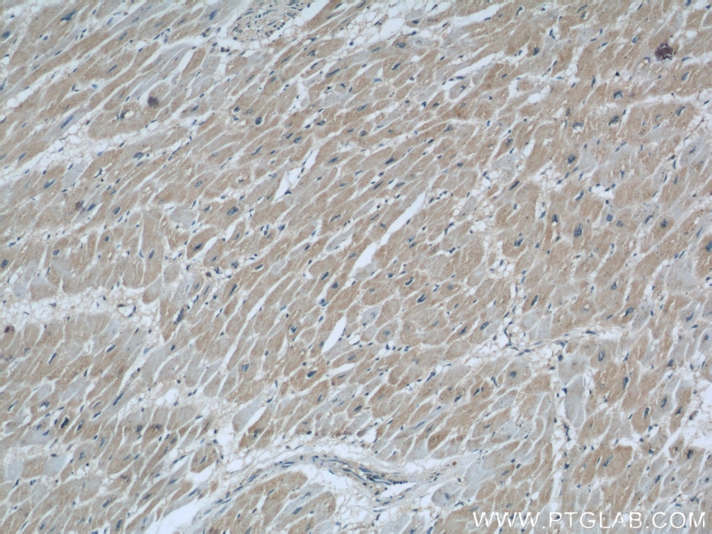
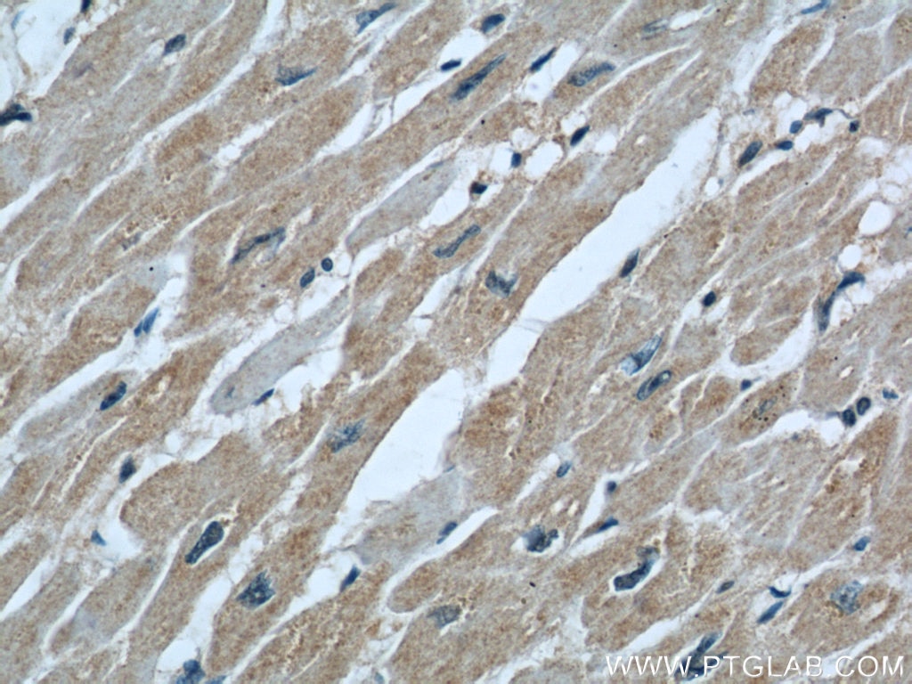
  • Immunohistochemistry (IHC) staining of human heart tissue using COXIV Monoclonal antibody (66110-1-Ig)

    IHC staining of human heart using 66110-1-Ig

    Immunohistochemical analysis of paraffin-embedded human heart using 66110-1-Ig(COXIV antibody) at dilution of 1:200 (under 10x lens).

  • Immunohistochemistry (IHC) staining of human heart tissue using COXIV Monoclonal antibody (66110-1-Ig)

    IHC staining of human heart using 66110-1-Ig

    Immunohistochemical analysis of paraffin-embedded human heart using 66110-1-Ig(COXIV antibody) at dilution of 1:200 (under 40x lens).

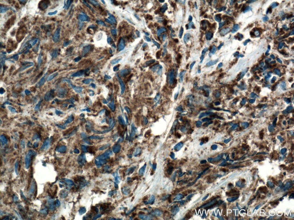
  • Immunohistochemistry (IHC) staining of human prostate cancer tissue using COXIV Monoclonal antibody (66110-1-Ig)

    IHC staining of human prostate cancer using 66110-1-Ig

    Immunohistochemical analysis of paraffin-embedded human prostate cancer using 66110-1-Ig(COXIV antibody) at dilution of 1:200 (under 10x lens).

  • Immunohistochemistry (IHC) staining of human prostate cancer tissue using COXIV Monoclonal antibody (66110-1-Ig)

    IHC staining of human prostate cancer using 66110-1-Ig

    Immunohistochemical analysis of paraffin-embedded human prostate cancer using 66110-1-Ig(COXIV antibody) at dilution of 1:200 (under 40x lens).

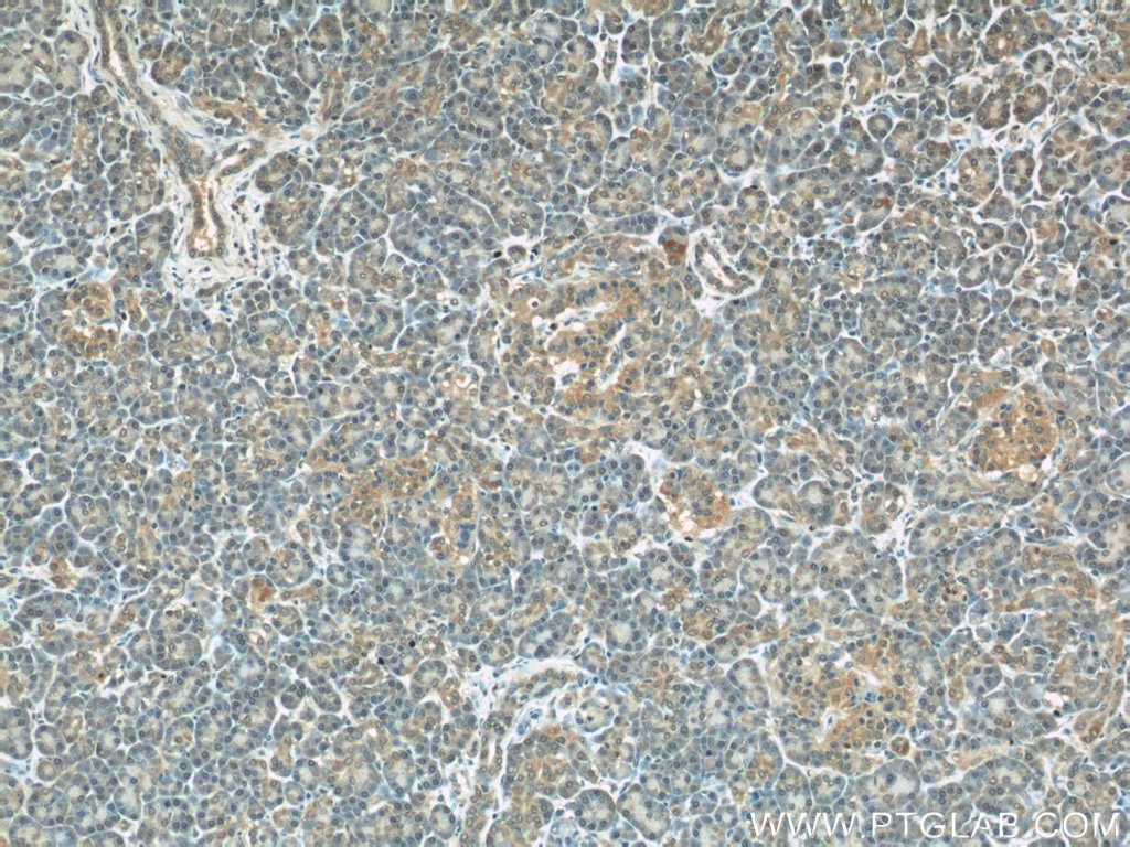
  • Immunohistochemistry (IHC) staining of human pancreas tissue using COXIV Monoclonal antibody (66110-1-Ig)

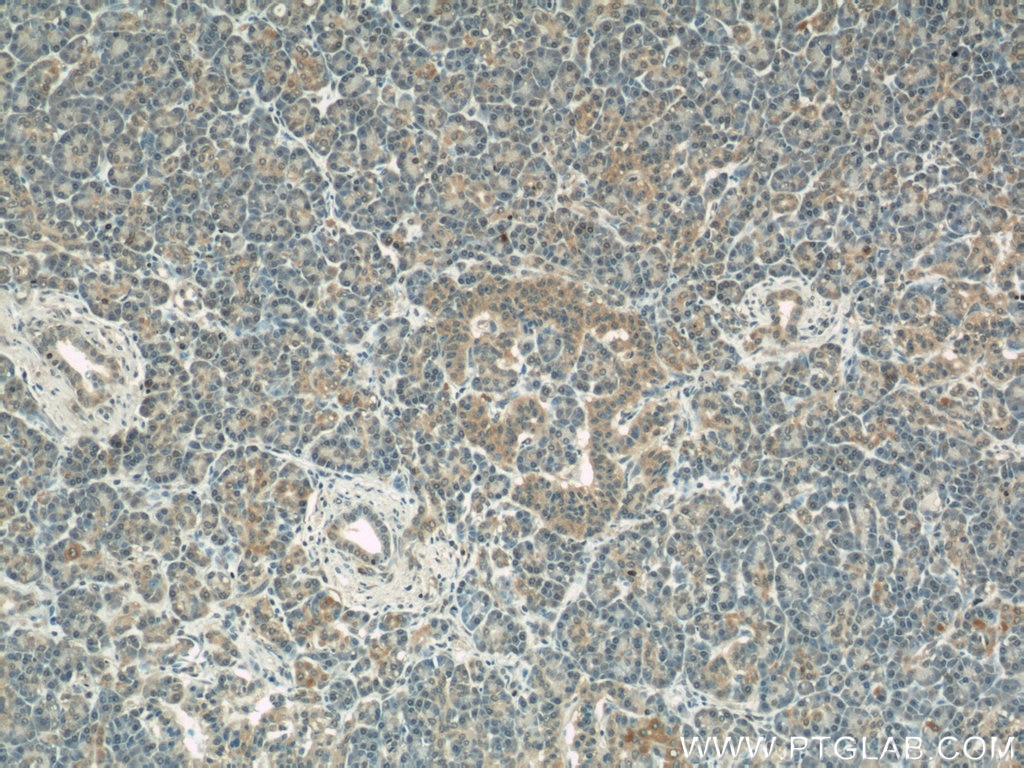
    IHC staining of human pancreas using 66110-1-Ig

    Immunohistochemical analysis of paraffin-embedded human pancreas using 66110-1-Ig(COXIV antibody) at dilution of 1:100 (under 10x lens).

  • Immunohistochemistry (IHC) staining of human pancreas tissue using COXIV Monoclonal antibody (66110-1-Ig)

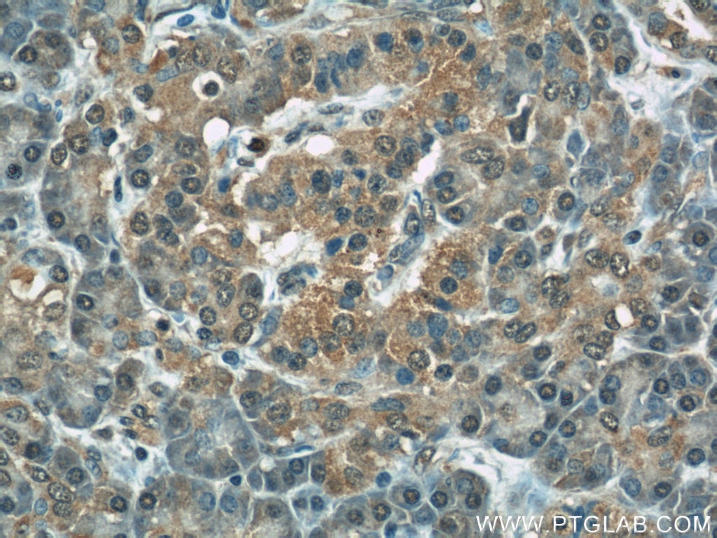
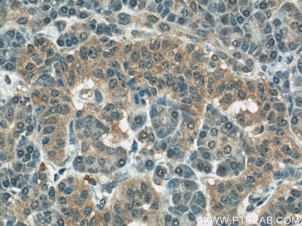
    IHC staining of human pancreas using 66110-1-Ig

    Immunohistochemical analysis of paraffin-embedded human pancreas using 66110-1-Ig(COXIV antibody) at dilution of 1:100 (under 40x lens).

  • Immunohistochemistry (IHC) staining of human pancreas tissue using COXIV Monoclonal antibody (66110-1-Ig)

    IHC staining of human pancreas using 66110-1-Ig

    Immunohistochemical analysis of paraffin-embedded human pancreas using 66110-1-Ig(COXIV antibody) at dilution of 1:200 (under 10x lens).

  • Immunohistochemistry (IHC) staining of human pancreas tissue using COXIV Monoclonal antibody (66110-1-Ig)

    IHC staining of human pancreas using 66110-1-Ig

    Immunohistochemical analysis of paraffin-embedded human pancreas using 66110-1-Ig(COXIV antibody) at dilution of 1:200 (under 40x lens).

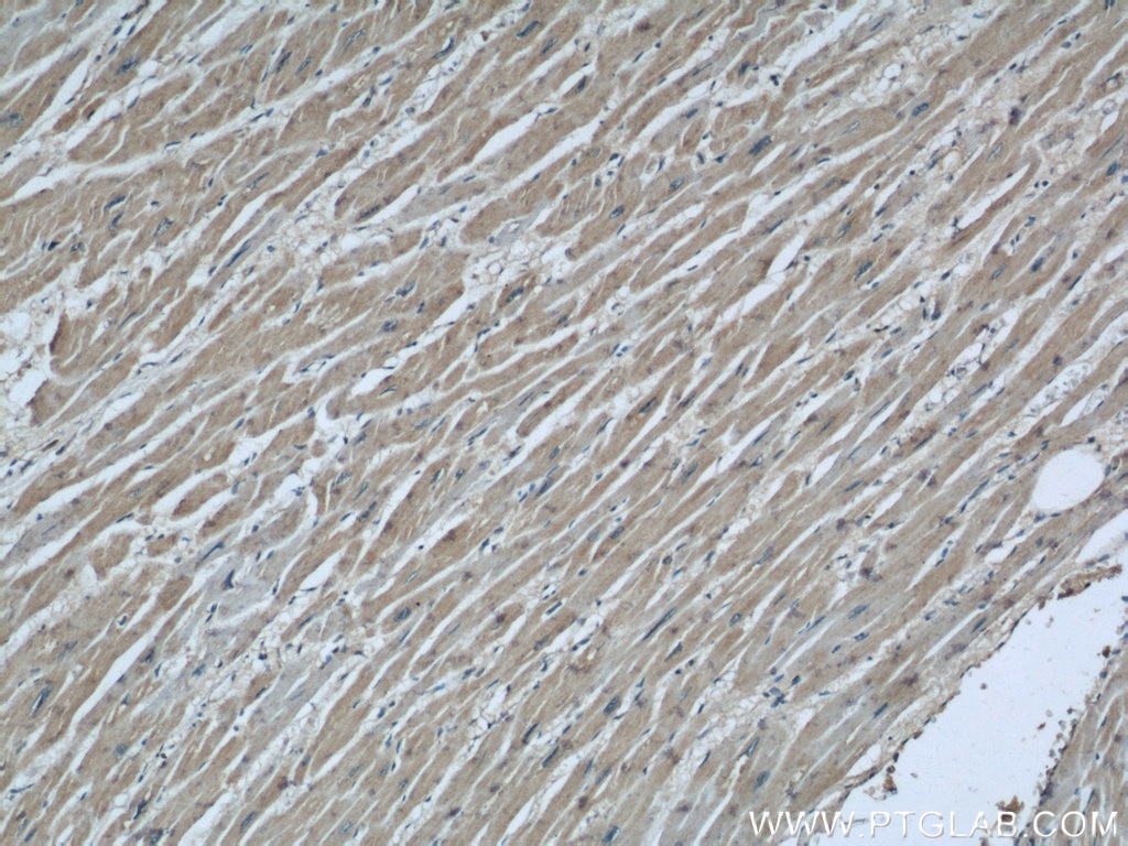
  • Immunohistochemistry (IHC) staining of human heart tissue using COXIV Monoclonal antibody (66110-1-Ig)

    IHC staining of human heart using 66110-1-Ig

    Immunohistochemical analysis of paraffin-embedded human heart using 66110-1-Ig(COXIV antibody) at dilution of 1:100 (under 10x lens).

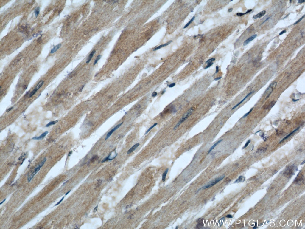
  • Immunohistochemistry (IHC) staining of human heart tissue using COXIV Monoclonal antibody (66110-1-Ig)

    IHC staining of human heart using 66110-1-Ig

    Immunohistochemical analysis of paraffin-embedded human heart using 66110-1-Ig(COXIV antibody) at dilution of 1:100 (under 40x lens).

  • Immunofluorescence (IF) / fluorescent staining of HepG2 cells using COXIV Monoclonal antibody (66110-1-Ig)

    IF Staining of HepG2 using 66110-1-Ig

    Immunofluorescent analysis of (4% PFA) fixed HepG2 cells using 66110-1-Ig (COXIV antibody, 1:100) at dilution of 1:100 and Alexa Fluor 488-Conjugated Goat Anti-Mouse IgG(H+L); Red is the staining with alpha Tubulin antibody (11224-1-AP, 1:100).

"COXIV Antibodies" Comparison

View side-by-side comparison of COXIV antibodies from other vendors to find the one that best suits your research needs.

Tested Applications

Positive WB detected inA431 cells, HeLa cells, HEK-293 cells, HepG2 cells, Jurkat cells, LNCaP cells, K-562 cells
Positive IHC detected inhuman prostate cancer tissue, human pancreas tissue, human heart tissue
Note: suggested antigen retrieval with TE buffer pH 9.0; (*) Alternatively, antigen retrieval may be performed with citrate buffer pH 6.0
Positive IF/ICC detected inHepG2 cells

Recommended dilution

ApplicationDilution
Western Blot (WB)WB : 1:5000-1:50000
Immunohistochemistry (IHC)IHC : 1:250-1:1000
Immunofluorescence (IF)/ICCIF/ICC : 1:50-1:500
It is recommended that this reagent should be titrated in each testing system to obtain optimal results.
Sample-dependent, Check data in validation data gallery.

Product Information

66110-1-Ig targets COXIV in WB, IHC, IF/ICC, ELISA applications and shows reactivity with human samples.

Tested Reactivity human
Cited Reactivityhuman, mouse, rat
Host / Isotype Mouse / IgG1
Class Monoclonal
Type Antibody
Immunogen COXIV fusion protein Ag20551
Full Name cytochrome c oxidase subunit IV isoform 1
Calculated Molecular Weight19.6 kDa
Observed Molecular Weight 17-18 kDa
GenBank Accession NumberBC021236
Gene Symbol COX IV
Gene ID (NCBI) 1327
RRIDAB_2881509
Conjugate Unconjugated
Form Liquid
Purification MethodProtein G purification
Storage Buffer PBS with 0.02% sodium azide and 50% glycerol pH 7.3.
Storage ConditionsStore at -20°C. Stable for one year after shipment. Aliquoting is unnecessary for -20oC storage. 20ul sizes contain 0.1% BSA.

Background Information

COX4I1, also named as COX4 and COXIV-1, belongs to the cytochrome c oxidase IV family. It is one of the nuclear-coded polypeptide chains of cytochrome c oxidase, the terminal oxidase in mitochondrial electron transport. COX4I1 is a marker for mitochondria. It has two isoforms (isoform 1 and 2). Isoform 1(COX4I1) is ubiquitously expressed and isoform 2 is highly expressed in lung tissues. COX4I1 is commonly used as a loading control. This antibody is specific to COX4I1 and do not cross reacts with COX4I2.

Protocols

Product Specific Protocols
WB protocol for COXIV antibody 66110-1-IgDownload protocol
IHC protocol for COXIV antibody 66110-1-IgDownload protocol
IF protocol for COXIV antibody 66110-1-IgDownload protocol
FC protocol for COXIV antibody 66110-1-IgDownload protocol
Standard Protocols
Click here to view our Standard Protocols

Publications

SpeciesApplicationTitle
humanWB

Science

Structural insight into the SAM-mediated assembly of the mitochondrial TOM core complex.

Authors - Qiang Wang
humanWB

ACS Nano

Mitochondria-Targeting Polymer Micelle of Dichloroacetate Induced Pyroptosis to Enhance Osteosarcoma Immunotherapy.

Authors - Jiakang Jin
humanWB

Nat Cell Biol

Mitochondria-localised ZNFX1 functions as a dsRNA sensor to initiate antiviral responses through MAVS.

Authors - Yao Wang
humanWB

Mol Cell

Global mitochondrial protein import proteomics reveal distinct regulation by translation and translocation machinery

Authors - Jasmin Adriana Schafer
humanIF

Nat Commun

PINCH-1 regulates mitochondrial dynamics to promote proline synthesis and tumor growth.

Authors - Ling Guo
humanWB

Elife

Genome-wide CRISPRi screening identifies OCIAD1 as a prohibitin client and regulatory determinant of mitochondrial Complex III assembly in human cells.

Authors - Maxence Le Vasseur

Reviews

The reviews below have been submitted by verified Proteintech customers who received an incentive for providing their feedback.


FH

Bryce (Verified Customer) (02-18-2022)

Decent loading control for mitochondria. I cut the blot too low, but you get the point.

  • Applications: Western Blot
  • Primary Antibody Dilution: 1:1000
  • Cell Tissue Type: Mouse Kidney
COXIV Antibody Western Blot validation (1:1000 dilution) in Mouse Kidney (Cat no:66110-1-Ig)