- Phare
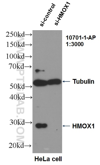
- Validé par KD/KO
Anticorps Polyclonal de lapin anti-HO-1/HMOX1
HO-1/HMOX1 Polyclonal Antibody for WB, IP, IF, IHC, ELISA, FC (Intra)
Hôte / Isotype
Lapin / IgG
Réactivité testée
Humain, rat, souris et plus (5)
Applications
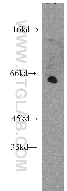
WB, IHC, IF-P, FC (Intra), IP, CoIP, ELISA
Conjugaison
Non conjugué
931
N° de cat : 10701-1-AP
Synonymes
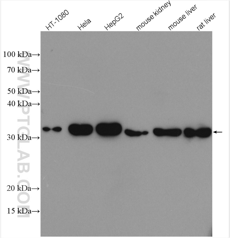
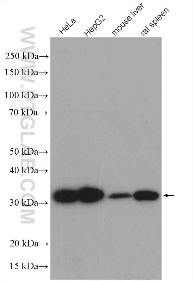
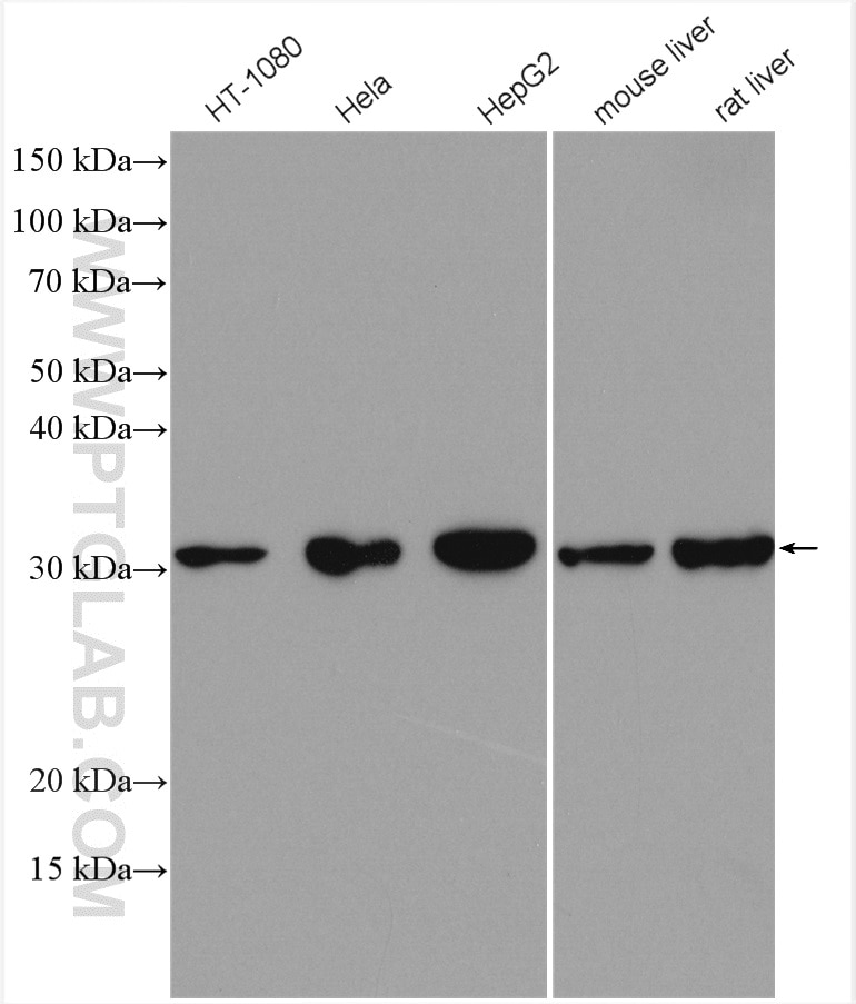
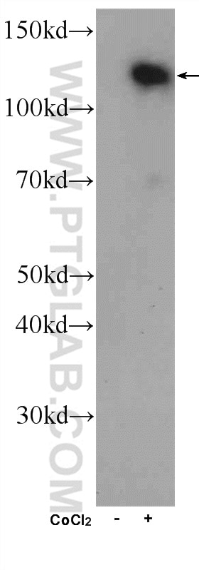
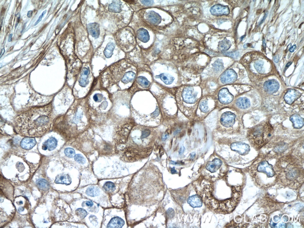
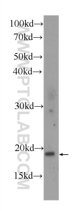
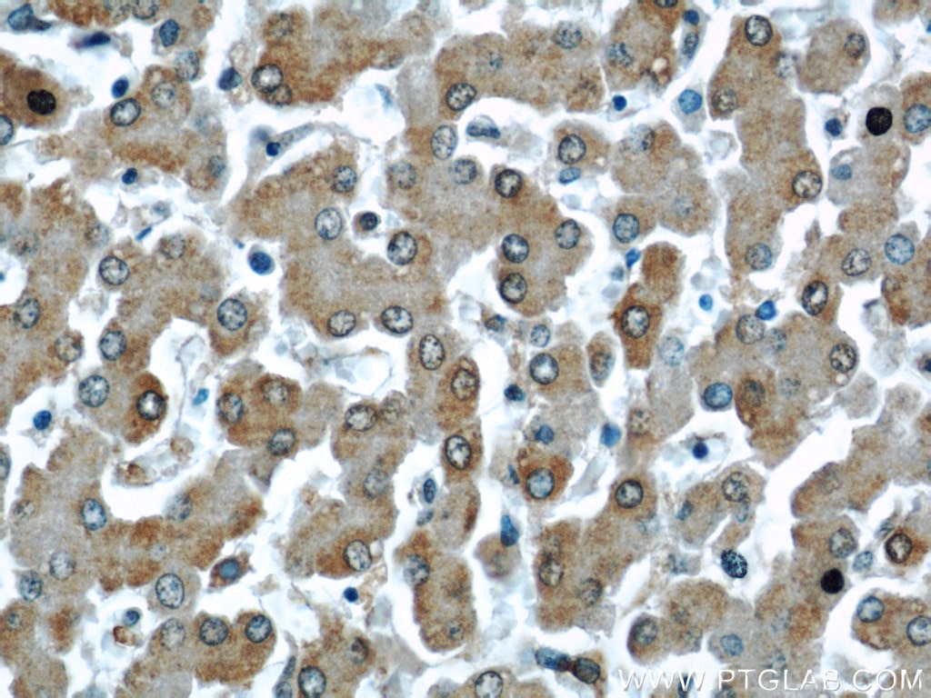
Galerie de données de validation
Applications testées
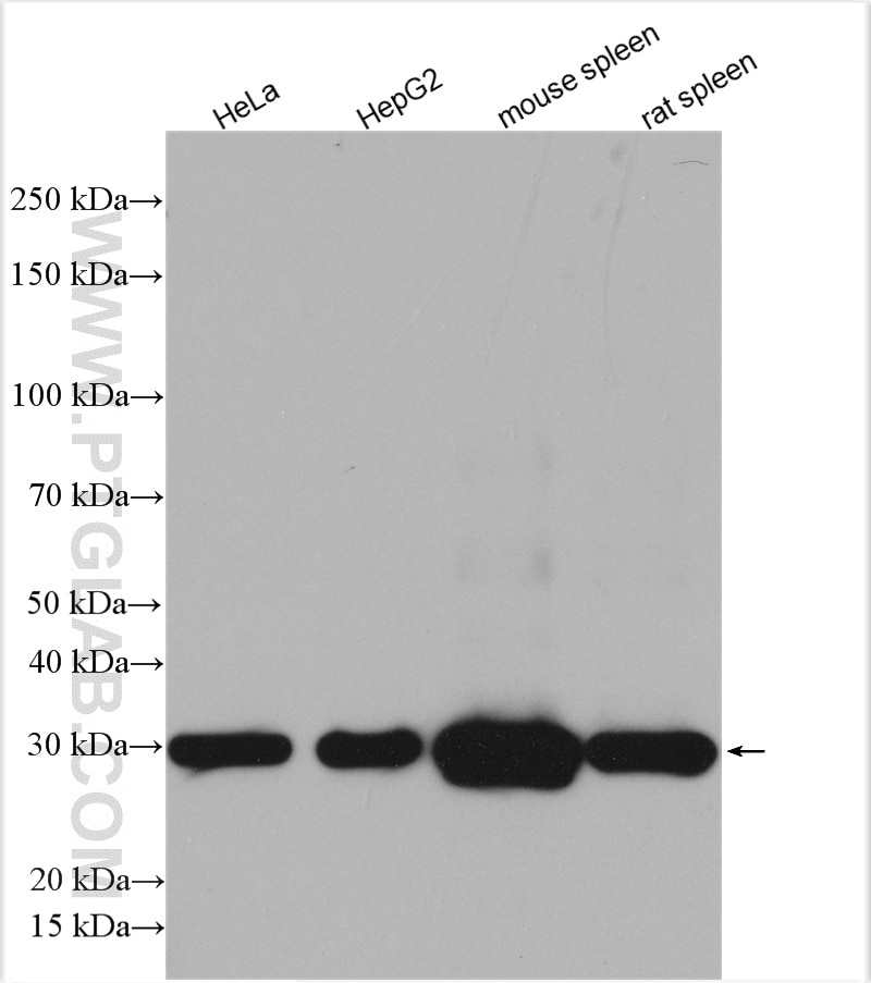
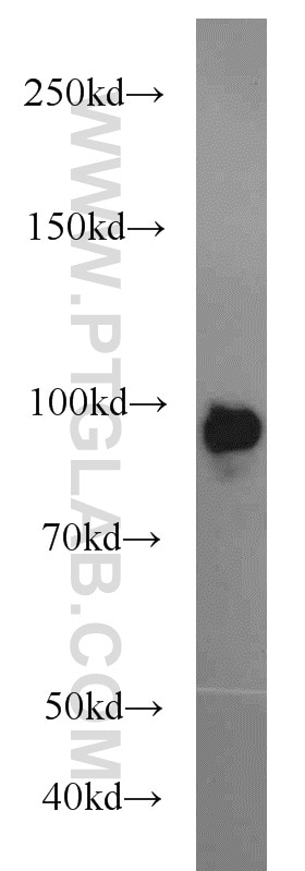
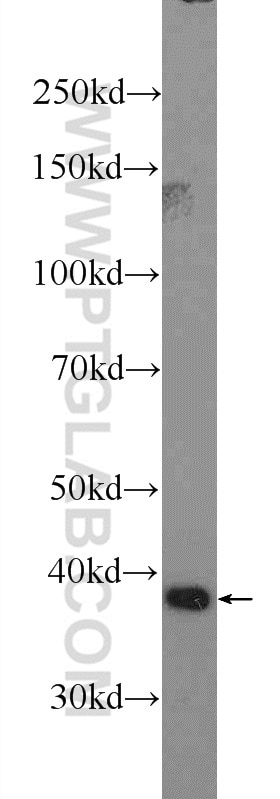
| Résultats positifs en WB | cellules HT-1080, cellules HeLa, cellules HepG2, tissu hépatique de rat, tissu hépatique de souris, tissu rénal de souris, tissu splénique de rat, tissu splénique de souris |
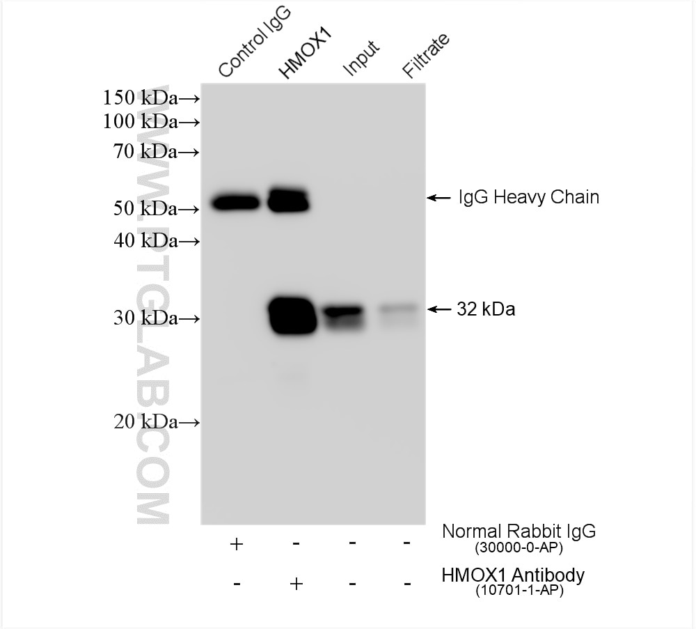
| Résultats positifs en IP | cellules HeLa, |
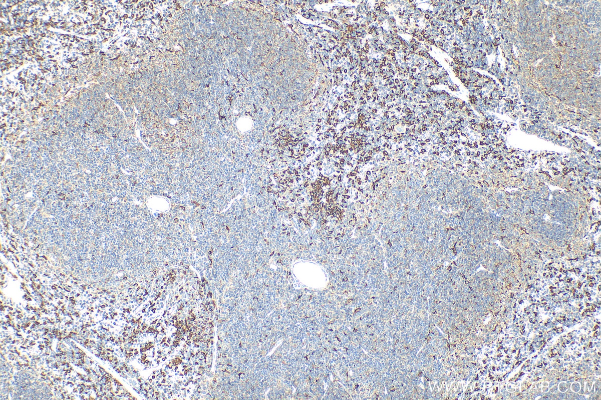
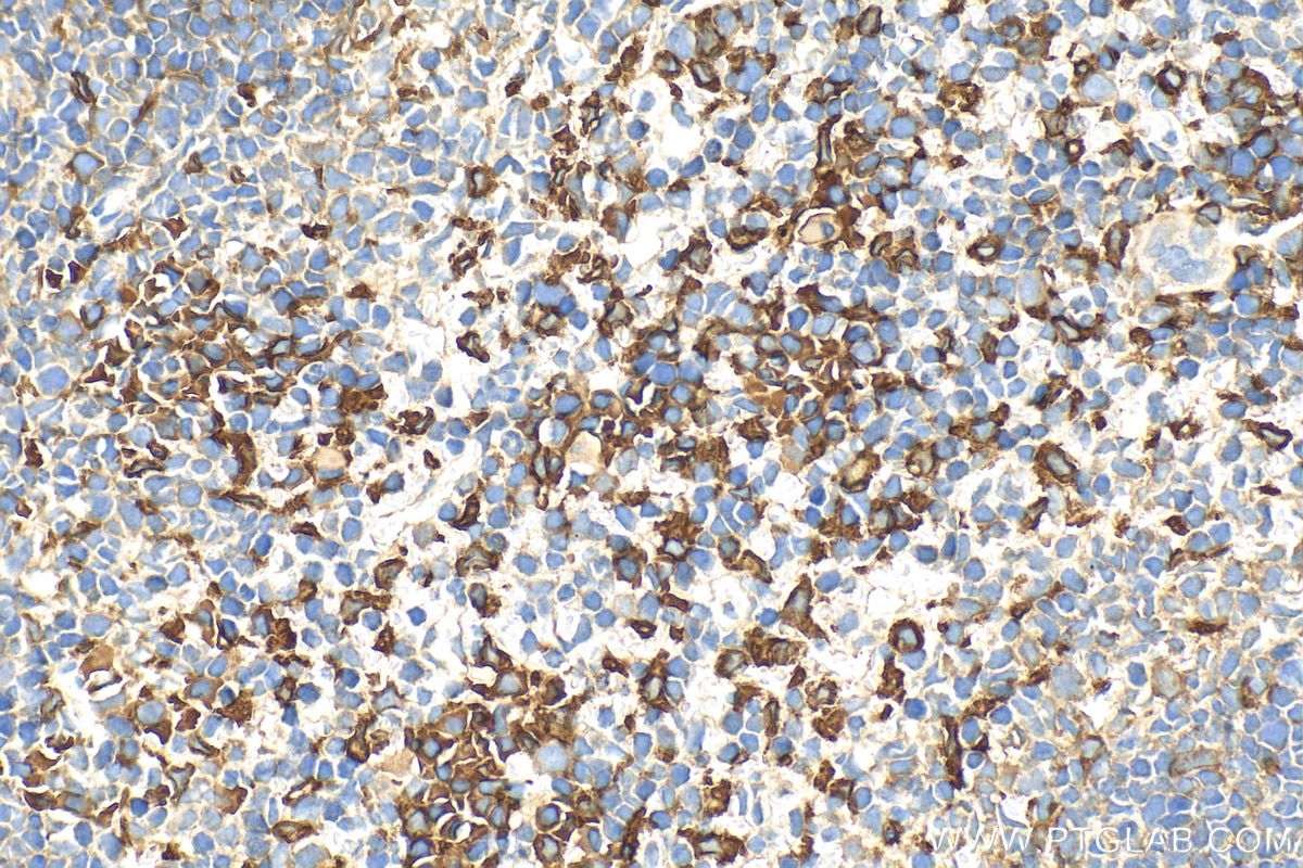
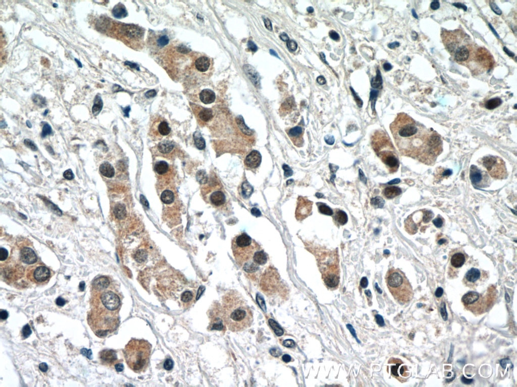
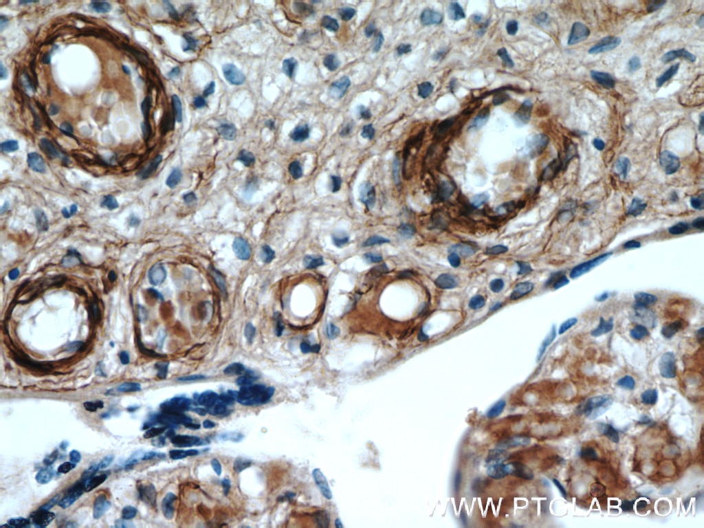
| Résultats positifs en IHC | tissu splénique de souris, il est suggéré de démasquer l'antigène avec un tampon de TE buffer pH 9.0; (*) À défaut, 'le démasquage de l'antigène peut être 'effectué avec un tampon citrate pH 6,0. |
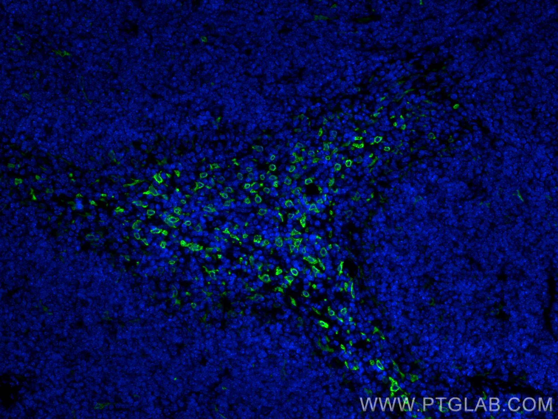
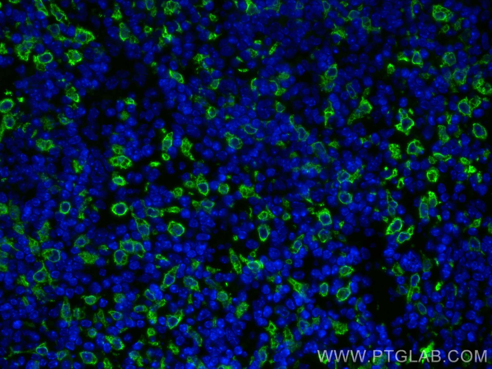
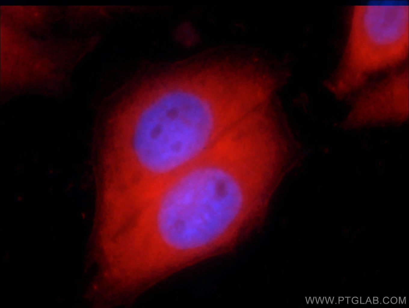
| Résultats positifs en IF-P | tissu splénique de souris, |
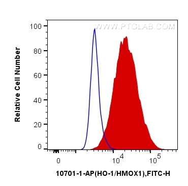
| Résultats positifs en FC (Intra) | cellules HeLa |
| Résultats positifs en cytométrie | cellules HeLa |
Dilution recommandée
| Application | Dilution |
|---|---|
| Western Blot (WB) | WB : 1:1000-1:6000 |
| Immunoprécipitation (IP) | IP : 0.5-4.0 ug for 1.0-3.0 mg of total protein lysate |
| Immunohistochimie (IHC) | IHC : 1:50-1:500 |
| Immunofluorescence (IF)-P | IF-P : 1:50-1:500 |
| Flow Cytometry (FC) (INTRA) | FC (INTRA) : 0.40 ug per 10^6 cells in a 100 µl suspension |
| Flow Cytometry (FC) | FC : 0.40 ug per 10^6 cells in a 100 µl suspension |
| It is recommended that this reagent should be titrated in each testing system to obtain optimal results. | |
| Sample-dependent, check data in validation data gallery | |
Informations sur le produit
10701-1-AP cible HO-1/HMOX1 dans les applications de WB, IHC, IF-P, FC (Intra), IP, CoIP, ELISA et montre une réactivité avec des échantillons Humain, rat, souris
| Réactivité | Humain, rat, souris |
| Réactivité citée | rat, bovin, Chèvre, Humain, porc, poulet, singe, souris |
| Hôte / Isotype | Lapin / IgG |
| Clonalité | Polyclonal |
| Type | Anticorps |
| Immunogène | HO-1/HMOX1 Protéine recombinante Ag1190 |
| Nom complet | heme oxygenase (decycling) 1 |
| Masse moléculaire calculée | 33 kDa |
| Poids moléculaire observé | 28-33 kDa |
| Numéro d’acquisition GenBank | BC001491 |
| Symbole du gène | HO-1 |
| Identification du gène (NCBI) | 3162 |
| Conjugaison | Non conjugué |
| Forme | Liquide |
| Méthode de purification | Purification par affinité contre l'antigène |
| Tampon de stockage | PBS avec azoture de sodium à 0,02 % et glycérol à 50 % pH 7,3 |
| Conditions de stockage | Stocker à -20°C. Stable pendant un an après l'expédition. L'aliquotage n'est pas nécessaire pour le stockage à -20oC Les 20ul contiennent 0,1% de BSA. |
Informations générales
Heme oxygenase (HMOX1) catalyzes the first and rate-limiting step in the degradation of heme to yield equimolar quantities of biliverdin Ixa, carbon monoxide (CO), and iron. It has 3 isoforms: HO-1 is highly inducible, whereas HO-2 and HO-3 are constitutively expressed (PMID:10194478). Heme oxygenase-1 (HO-1) is expressed in many tissues and vascular smooth muscle cells, and endothelial cells (PMID:15451051) and has been identified as an important endogenous protective factor induced in many cell types by various stimulants, such as hemolysis, infiammatory cytokines,oxidative stress, heat shock, heavy metals, and endotoxin (PMID: 11522663). And the full-length HO-1 is very unstable and susceptible to truncation that generates an inactive, soluble form (28 kDa) (James R. Reed, Pharmacology, 535-568).
Protocole
| Product Specific Protocols | |
|---|---|
| WB protocol for HO-1/HMOX1 antibody 10701-1-AP | Download protocol |
| IHC protocol for HO-1/HMOX1 antibody 10701-1-AP | Download protocol |
| IF protocol for HO-1/HMOX1 antibody 10701-1-AP | Download protocol |
| IP protocol for HO-1/HMOX1 antibody 10701-1-AP | Download protocol |
| FC protocol for HO-1/HMOX1 antibody 10701-1-AP | Download protocol |
| Standard Protocols | |
|---|---|
| Click here to view our Standard Protocols |
Publications
| Species | Application | Title |
|---|---|---|
Cell Metab Integrative genetic analysis identifies FLVCR1 as a plasma-membrane choline transporter in mammals | ||
Cell Metab Functional Genomics In Vivo Reveal Metabolic Dependencies of Pancreatic Cancer Cells. | ||
ACS Nano Melatonin-Derived Carbon Dots with Free Radical Scavenging Property for Effective Periodontitis Treatment via the Nrf2/HO-1 Pathway | ||
Sci Transl Med PTEN status determines chemosensitivity to proteasome inhibition in cholangiocarcinoma. |
Avis
The reviews below have been submitted by verified Proteintech customers who received an incentive forproviding their feedback.
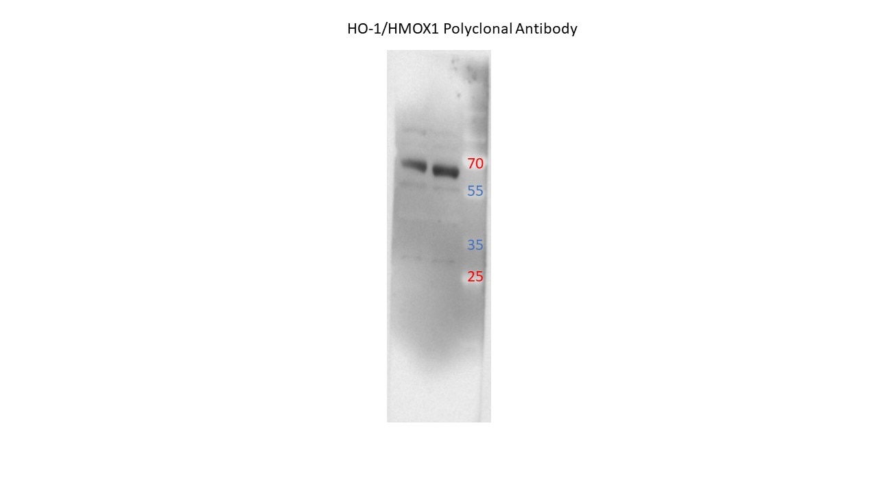
FH Brice-Emmanuel (Verified Customer) (12-18-2023) | Works perfectly. Band a 28-33 kDa
 |
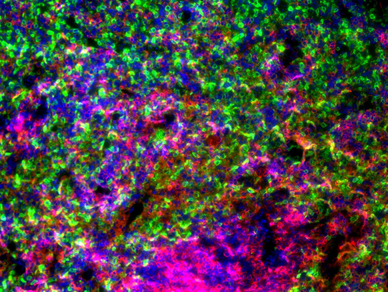
FH James (Verified Customer) (11-18-2022) | Very good antibody! Used it for IF staining of paraffin-embedded spleen tissue. HMOX-1 positive cells are in Green and CD45 in red.
 |
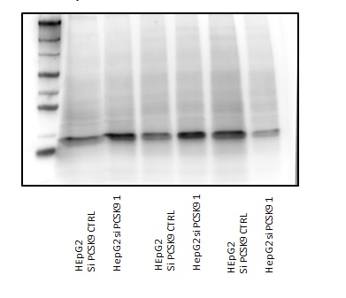
FH Hala (Verified Customer) (07-28-2021) | incubation 1.5 hours at room temperature dilution 1/3000
 |
FH JIM (Verified Customer) (03-26-2019) | This antibody offers a good detection of HO-1 expression in LPS stimulated macrophages. The HO-1 expression can be observed in the cells after 24h stimulation of LPS.
|
