
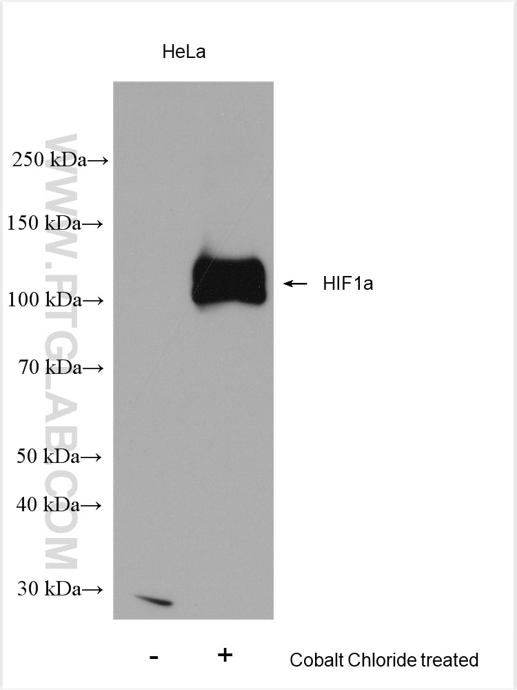
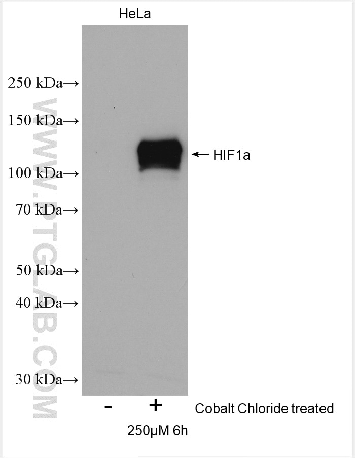
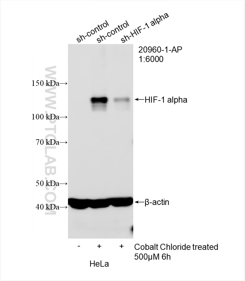
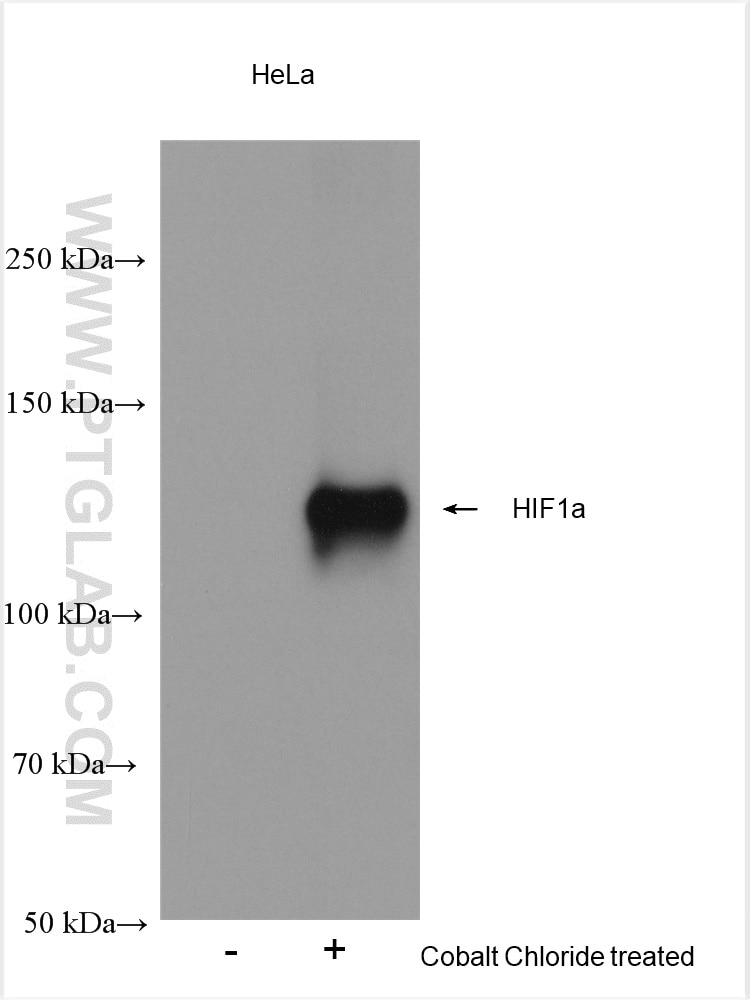

Tested Applications
| Positive WB detected in | Cobalt Chloride treated HeLa cells |
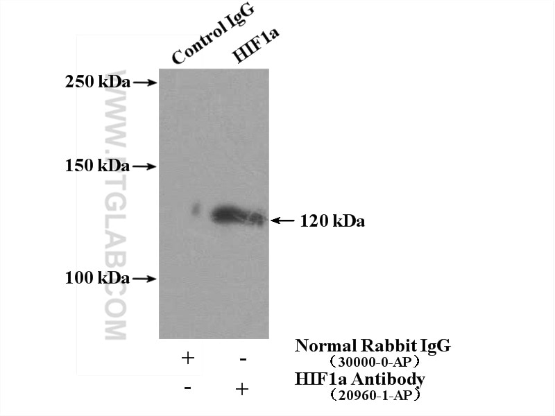
| Positive IP detected in | HeLa cells |
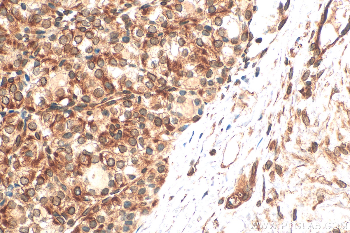
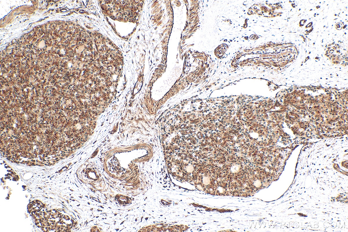
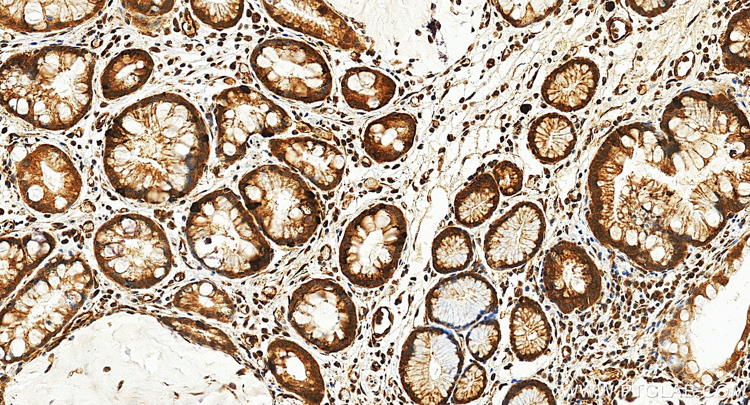
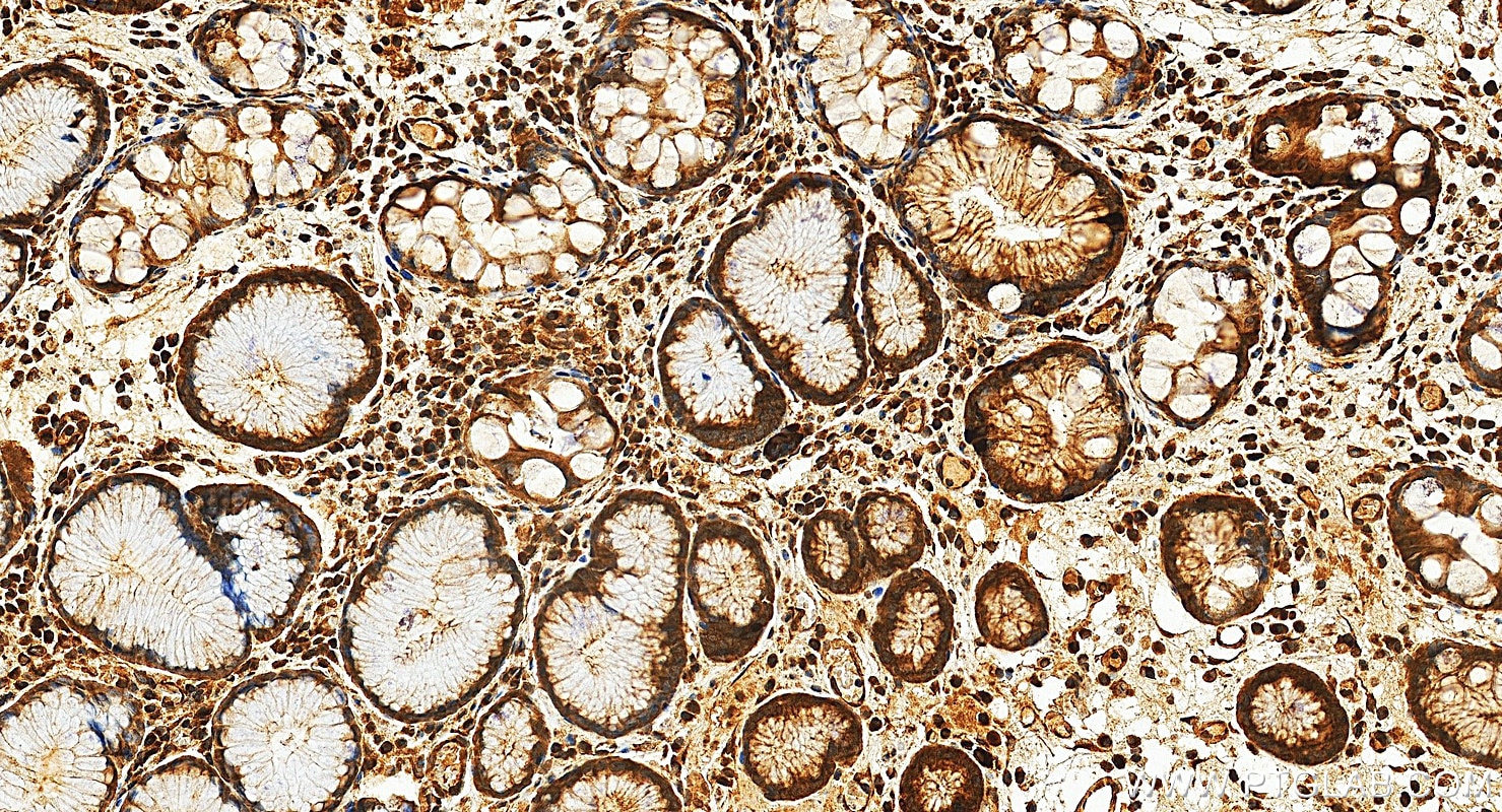
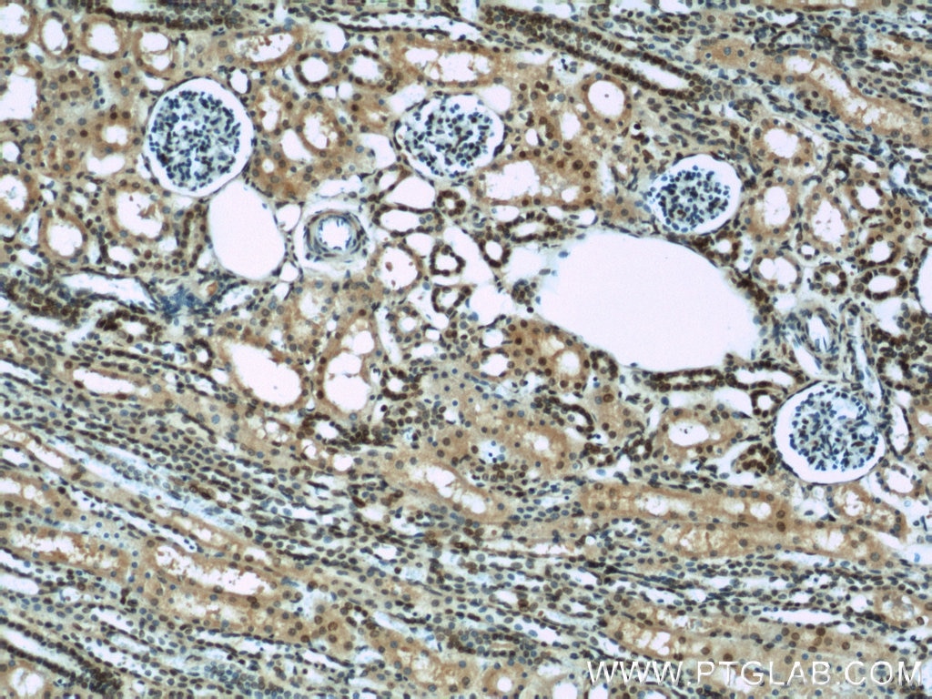
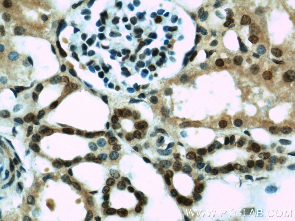
| Positive IHC detected in | human thyroid cancer tissue, human kidney tissue, human stomach cancer tissue Note: suggested antigen retrieval with TE buffer pH 9.0; (*) Alternatively, antigen retrieval may be performed with citrate buffer pH 6.0 |
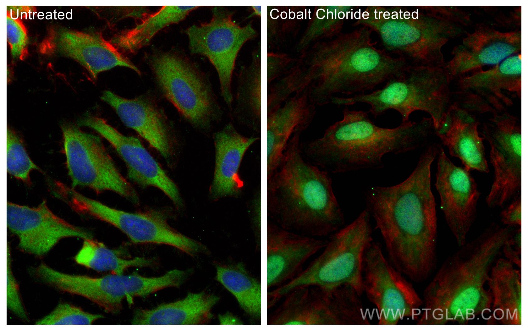
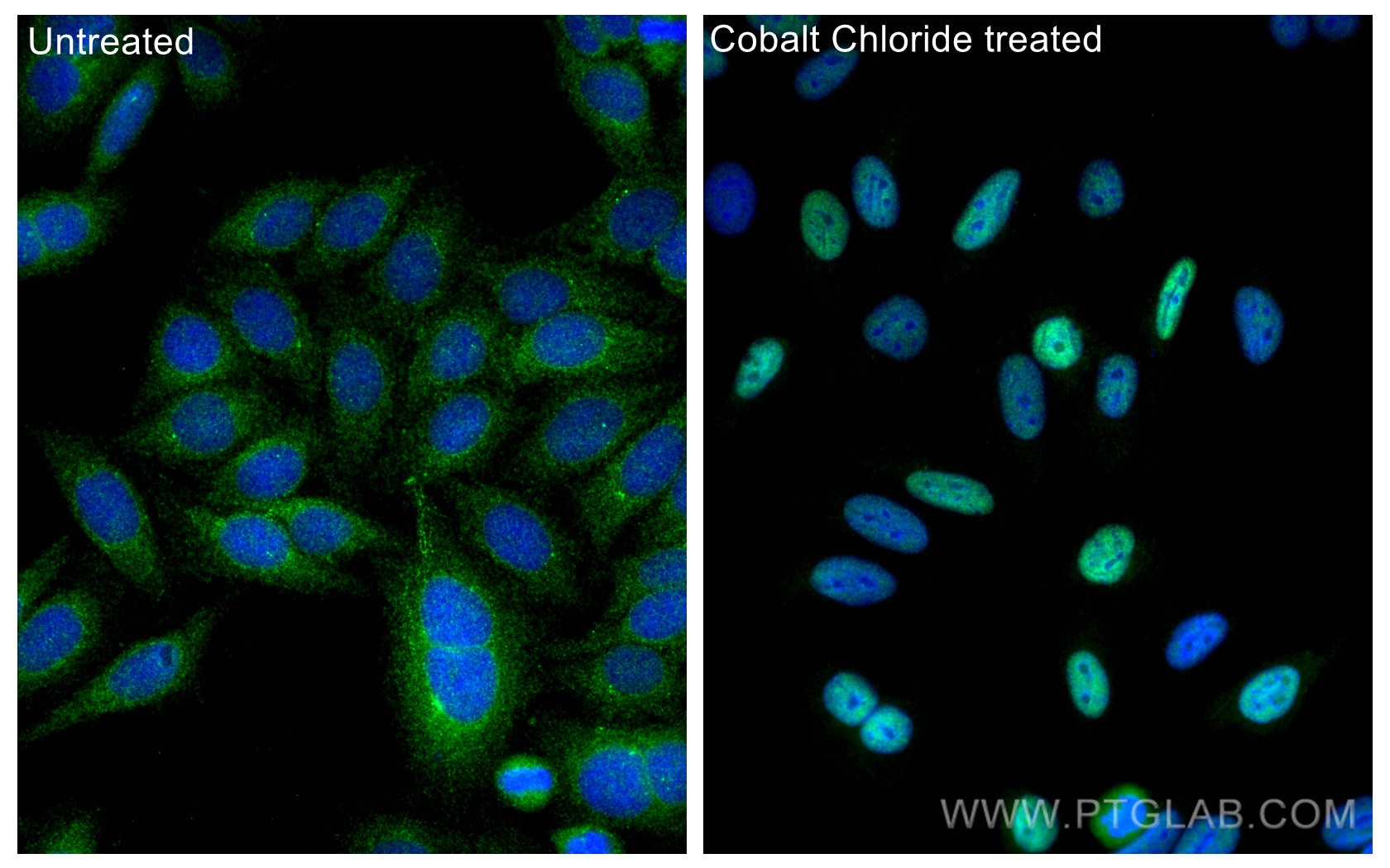
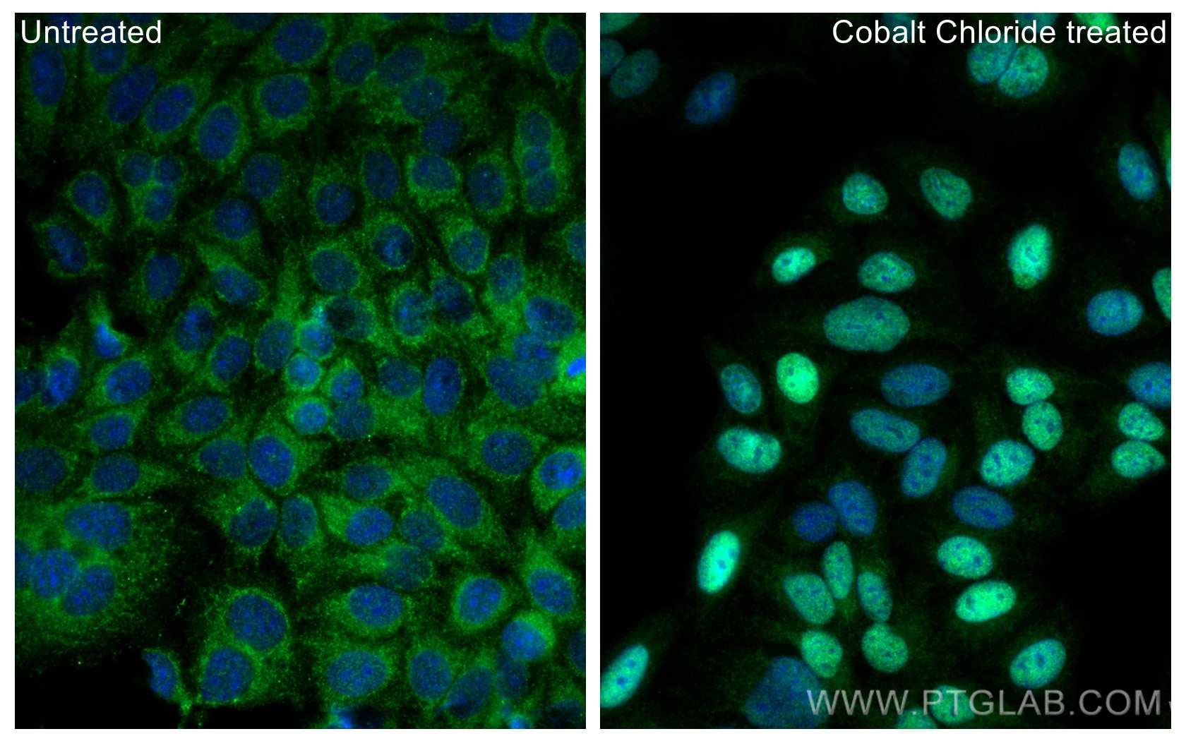
| Positive IF/ICC detected in | Cobalt Chloride treated HeLa cells, Cobalt Chloride treated HepG2 cells |
Samples need to be treated with hypoxia stimulation.
Recommended dilution
| Application | Dilution |
|---|---|
| Western Blot (WB) | WB : 1:2000-1:12000 |
| Immunoprecipitation (IP) | IP : 0.5-4.0 ug for 1.0-3.0 mg of total protein lysate |
| Immunohistochemistry (IHC) | IHC : 1:50-1:500 |
| Immunofluorescence (IF)/ICC | IF/ICC : 1:200-1:800 |
| It is recommended that this reagent should be titrated in each testing system to obtain optimal results. | |
| Sample-dependent, Check data in validation data gallery. | |
Product Information
20960-1-AP targets HIF-1 alpha in WB, IHC, IF/ICC, IP, CoIP, ChIP, RIP, ELISA applications and shows reactivity with human samples.
| Tested Reactivity | human |
| Cited Reactivity | human, pig, rabbit, monkey, chicken, goat |
| Host / Isotype | Rabbit / IgG |
| Class | Polyclonal |
| Type | Antibody |
| Immunogen |
CatNo: Ag15198 Product name: Recombinant human HIF1a protein Source: e coli.-derived, PGEX-4T Tag: GST Domain: 574-799 aa of BC012527 Sequence: LRSFDQLSPLESSSASPESASPQSTVTVFQQTQIQEPTANATTTTATTDELKTVTKDRMEDIKILIASPSPTHIHKETTSATSSPYRDTQSRTASPNRAGKGVIEQTEKSHPRSPNVLSVALSQRTTVPEEELNPKILALQNAQRKRKMEHDGSLFQAVGIGTLLQQPDDHAATTSLSWKRVKGCKSSEQNGMEQKTIILIPSDLACRLLGQSMDESGLPQLTSYD Predict reactive species |
| Full Name | hypoxia inducible factor 1, alpha subunit (basic helix-loop-helix transcription factor) |
| Calculated Molecular Weight | 826 aa, 93 kDa |
| Observed Molecular Weight | 120 kDa |
| GenBank Accession Number | BC012527 |
| Gene Symbol | HIF1A |
| Gene ID (NCBI) | 3091 |
| RRID | AB_10732601 |
| Conjugate | Unconjugated |
| Form | Liquid |
| Purification Method | Antigen affinity purification |
| UNIPROT ID | Q16665 |
| Storage Buffer | PBS with 0.02% sodium azide and 50% glycerol, pH 7.3. |
| Storage Conditions | Store at -20°C. Stable for one year after shipment. Aliquoting is unnecessary for -20oC storage. 20ul sizes contain 0.1% BSA. |
Background Information
HIF1a, the major regulator of the cellular responses to hypoxia, consists of an oxygen-sensitive subunit, HIF1 alpha (HIF1A), and an oxygen-insensitive subunit, HIF1 beta (arylhydrocarbon receptor nuclear transporter [ARNT]). Under normal oxygen conditions, HIF1a is continuously produced and destroyed, in a process involving hydroxylation, interaction with von Hippel-Lindau (VHL) protein, polyubiquitylation and subsequent proteasomal degradation. Under hypoxic conditions, hydroxylation is impaired and HIF1a is stabilized. HIF1a localizes in cytoplasm in normoxia, but it can translocate into nuclear in response to hypoxia. The calculated molecular weight of HIF1a is 93 kDa, but the modified protein HIF1a is about 110-120kDa (PMID: 11698256, .PMID: 7539918). .
Protocols
| Product Specific Protocols | |
|---|---|
| IF protocol for HIF-1 alpha antibody 20960-1-AP | Download protocol |
| IHC protocol for HIF-1 alpha antibody 20960-1-AP | Download protocol |
| IP protocol for HIF-1 alpha antibody 20960-1-AP | Download protocol |
| WB protocol for HIF-1 alpha antibody 20960-1-AP | Download protocol |
| Standard Protocols | |
|---|---|
| Click here to view our Standard Protocols |
Publications
What published studies show
Anti-HIF-1α antibody (Cat# 20960-1-AP) has accumulated 1,032 total citations. Publications appear in high-impact journals including Nature, Cell, Cancer Cell, Nature Communications, Nature Cell Biology, Signal Transduction and Targeted Therapy, Molecular Cell, Cell Death & Disease, Theranostics, Journal of Experimental & Clinical Cancer Research, Biomaterials, ACS Nano, and Advanced Science. Research fields span oncology (tumor metabolism, angiogenesis, ferroptosis, immunotherapy), hypoxia signaling, tissue engineering, bone regeneration, neurology, cardiology, and inflammation.
| Species | Application | Title |
|---|---|---|
Signal Transduct Target Ther Aberrant Notch-signaling promotes tumor angiogenesis in esophageal squamous-cell carcinoma | ||
Signal Transduct Target Ther Hypoxia induced VEGF secretion promotes resistance to bispecific T-cell engagers. | ||
Signal Transduct Target Ther The p-MYH9/USP22/HIF-1α axis promotes lenvatinib resistance and cancer stemness in hepatocellular carcinoma | ||
Signal Transduct Target Ther Hypoxia-inducible factor upregulation by roxadustat attenuates drug reward by altering brain iron homoeostasis | ||
Reviews
What customers say
Most reviewers (5 of 8) rate this HIF-1α antibody highly, praising clean, specific bands in Western blot and immunofluorescence, with reliable performance across WB, IF, and IP applications. Two reviewers reported issues: one observed aspecific binding in cholangiocyte organoids under hypoxic conditions, and another noted significant background and unspecific bands in human chondrocyte and BV2 cell lysates. One reviewer gave a neutral rating with brief positive feedback. Overall, the antibody performs well for hypoxia-related studies but may require optimization for certain cell types or tissues.
The reviews below have been submitted by verified Proteintech customers who received an incentive for providing their feedback.
royruk (Verified Customer) (12-11-2025) | Good HIF-1α antibody with clean, specific bands and reliable performance in both WB and IF. Solid choice for hypoxia-focused experiments.
|
mali (Verified Customer) (12-11-2025) | Reliable HIF-1α antibody that gives clear, specific detection in Western blots and works well in IF/IHC and IP applications, making it a good tool for hypoxia-related studies
|
Vignesh (Verified Customer) (09-15-2025) | The results are excellent.
|
Carlo (Verified Customer) (01-18-2023) | The quality of the band in western blot is well defined
|
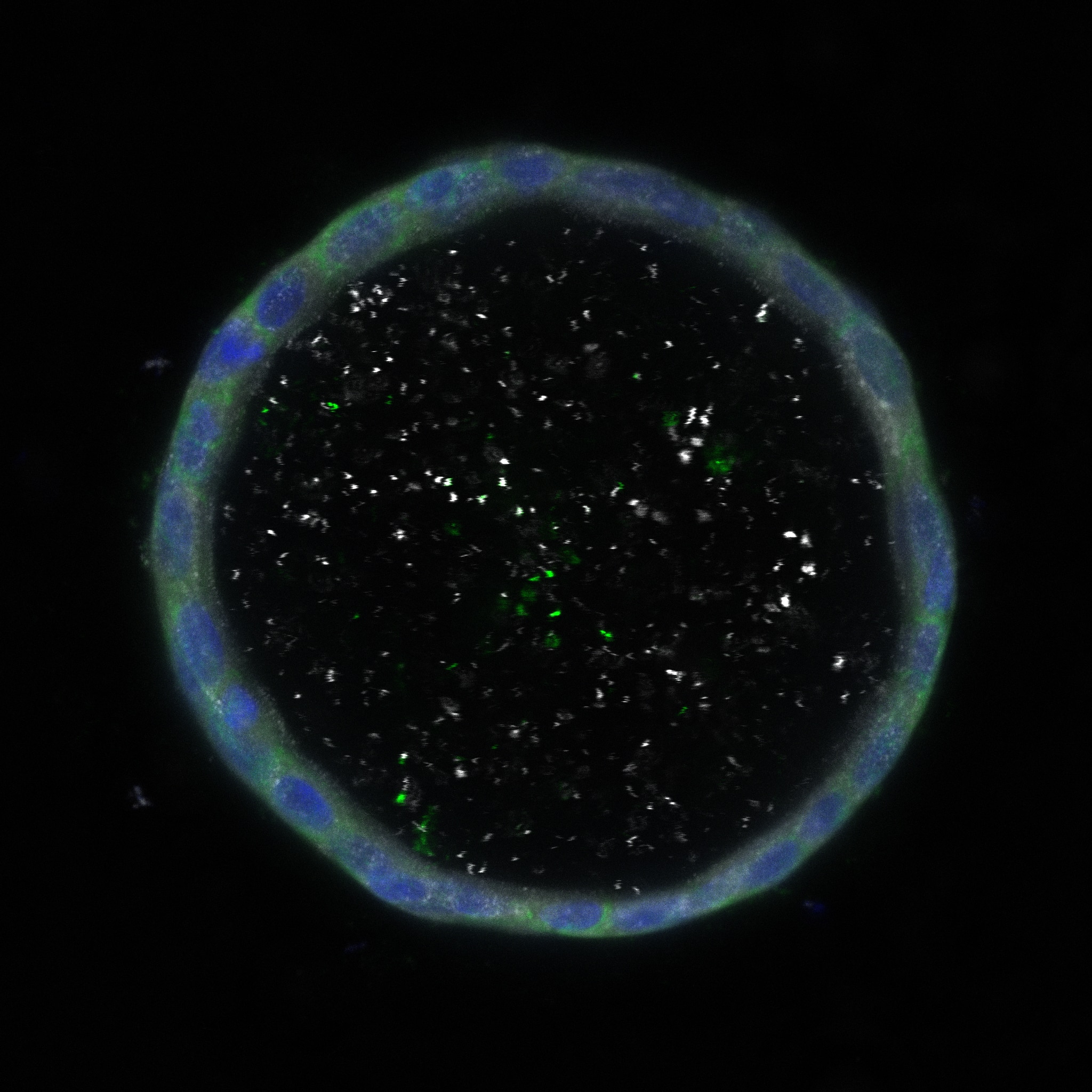
Davide (Verified Customer) (09-20-2022) | The image uploaded refers to a hypoxic condition O2<1%. The antibody signal was aspecific and generated aspecific binding in the middle of the organoid.
 |
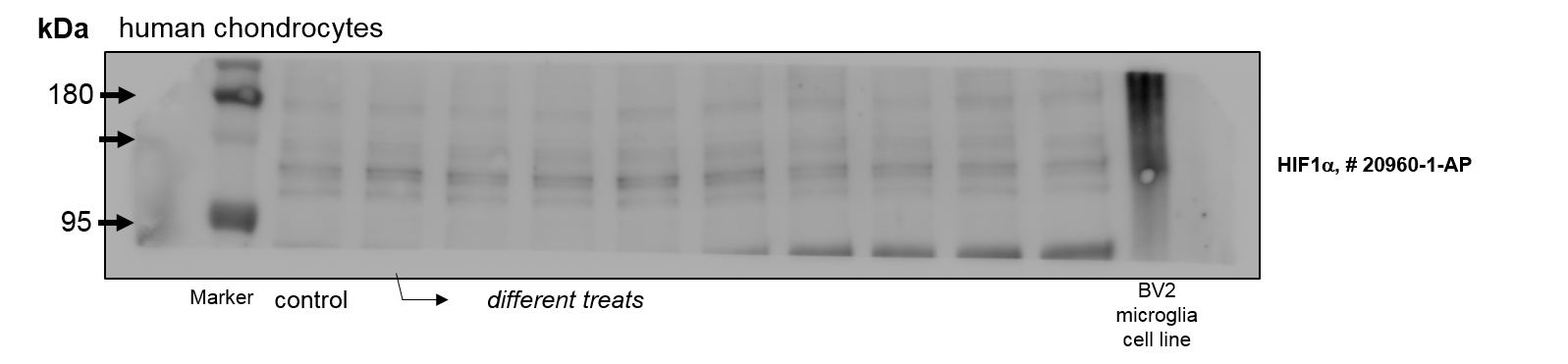
Christian (Verified Customer) (01-07-2022) | Unfortunately much background an many unspecific bands in lysates of human chondrocytes and BV2 microclia cells (less unspecific bands in rat chondrocytes).
 |
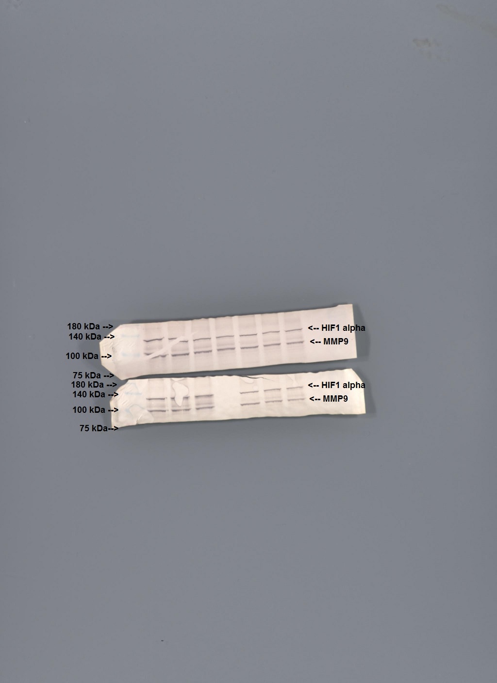
Diane (Verified Customer) (02-17-2021) | Separated on a 7.5% SDS PAGE gel, transferred to nitrocellulose membrane. Block with 5% dry milk/PBST at room temperature for one hour. 1:1000 dilution of the primary antibody in blocking solution. Incubated overnight at 4 degrees C on a rocker platform. Secondary antibody 1:1000 secondary (anti rabbit HRP) incubation for two hours at room temperature. Visualization with the Opti-4CN chromogenic substrate kit (BioRad).
 |
Chun (Verified Customer) (12-05-2019) | A good antibody
|


