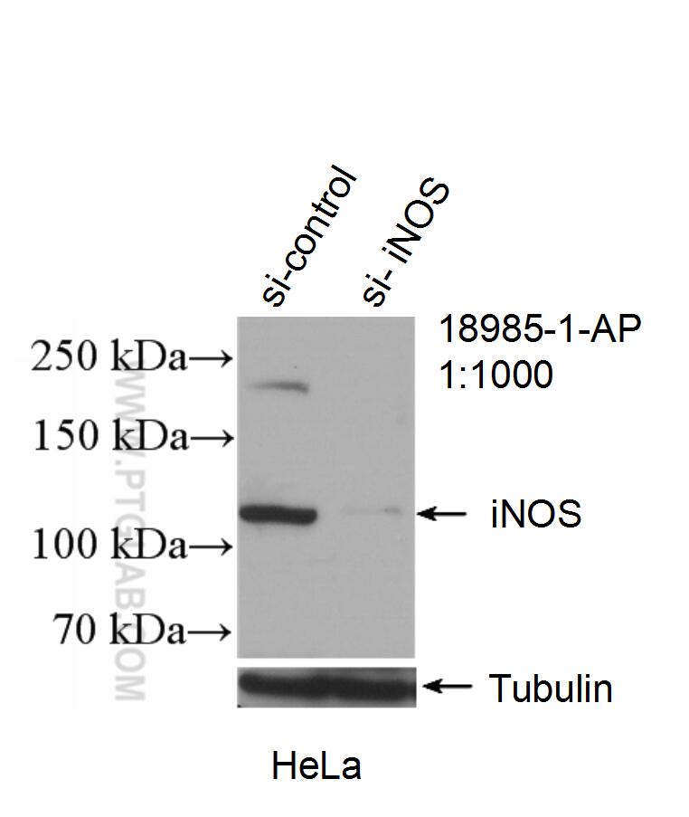
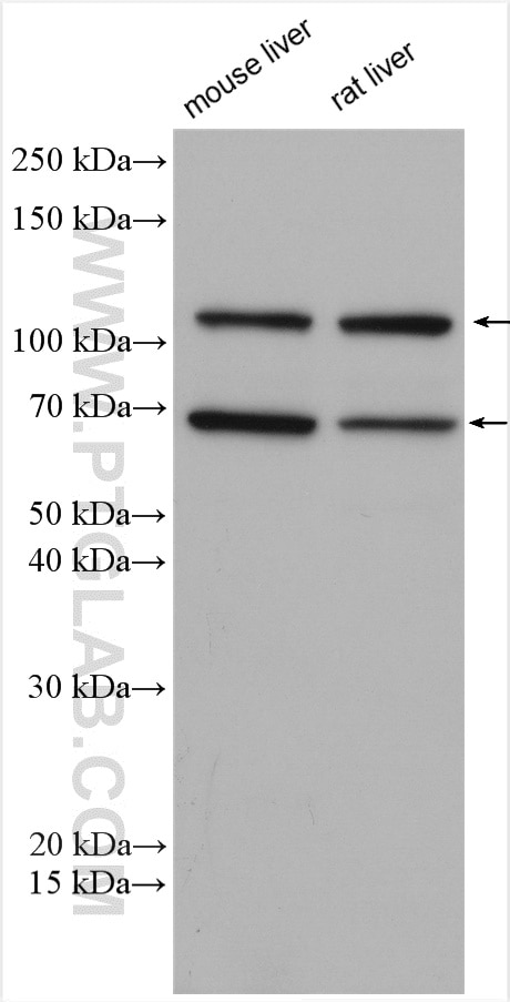
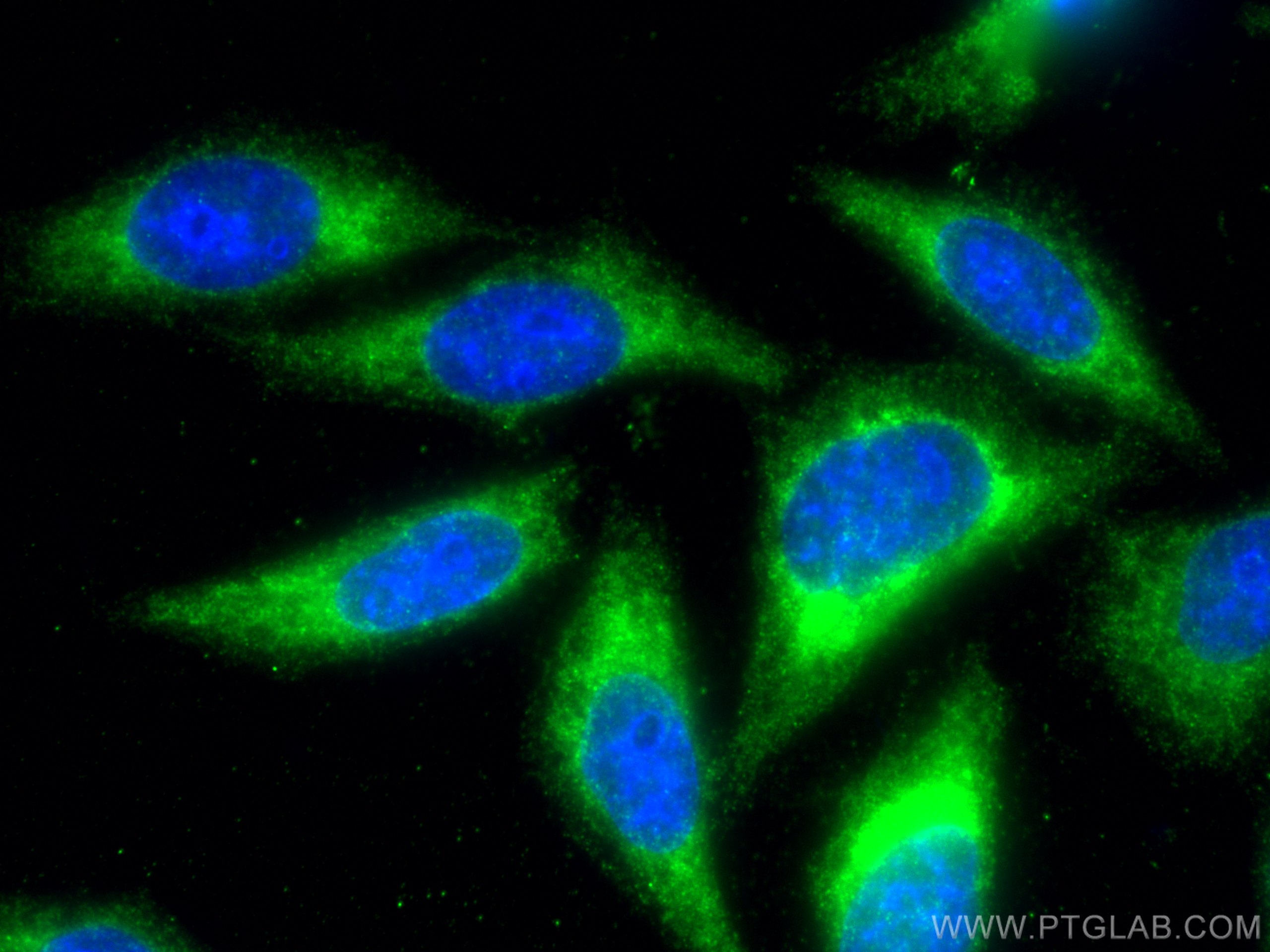
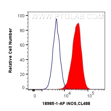
Tested Applications
| Positive WB detected in | mouse liver tissue, rat liver tissue |
| Positive IF/ICC detected in | HepG2 cells |
| Positive FC (Intra) detected in | HepG2 cells |
Recommended dilution
| Application | Dilution |
|---|---|
| Western Blot (WB) | WB : 1:2000-1:10000 |
| Immunofluorescence (IF)/ICC | IF/ICC : 1:50-1:500 |
| Flow Cytometry (FC) (INTRA) | FC (INTRA) : 0.40 ug per 10^6 cells in a 100 µl suspension |
| It is recommended that this reagent should be titrated in each testing system to obtain optimal results. | |
| Sample-dependent, Check data in validation data gallery. | |
Published Applications
| WB | See 720 publications below |
| IF | See 299 publications below |
| IP | See 1 publications below |
| ELISA | See 2 publications below |
Product Information
18985-1-AP targets iNOS in WB, IF/ICC, FC (Intra), IP, ELISA applications and shows reactivity with human, mouse, rat samples.
| Tested Reactivity | human, mouse, rat |
| Cited Reactivity | human, mouse, rat, pig, rabbit, chicken, bovine, goat |
| Host / Isotype | Rabbit / IgG |
| Class | Polyclonal |
| Type | Antibody |
| Immunogen |
Peptide Predict reactive species |
| Full Name | nitric oxide synthase 2, inducible |
| Calculated Molecular Weight | 131 kDa |
| Observed Molecular Weight | 110-130 kDa, 65-70 kDa |
| GenBank Accession Number | NM_000625 |
| Gene Symbol | iNOS |
| Gene ID (NCBI) | 4843 |
| RRID | AB_2782960 |
| Conjugate | Unconjugated |
| Form | Liquid |
| Purification Method | Antigen affinity purification |
| UNIPROT ID | P35228 |
| Storage Buffer | PBS with 0.02% sodium azide and 50% glycerol, pH 7.3. |
| Storage Conditions | Store at -20°C. Stable for one year after shipment. Aliquoting is unnecessary for -20oC storage. 20ul sizes contain 0.1% BSA. |
Background Information
NOS2, also named as iNOS and NOS2A, produces nitric oxide (NO) which is a messenger molecule with diverse functions throughout the body. NO is a reactive free radical which acts as a biologic mediator in several processes, including neurotransmission, antimicrobial and antitumoral activities. NOS2 is a nitric oxide synthase which is expressed in liver and is inducible by a combination of lipopolysaccharide and certain cytokines. iNOS has a very short half-life due to rapid degradation by calpain. iNOS monomer is a direct substrate of calpain I and can be cleaved by calpain I at the canonical CaM-binding site(503-532aa) of iNOS, and then a ~70 kDa band can be detected by western (PMID:11786228). This antibody is specific to NOS2.
Protocols
| Product Specific Protocols | |
|---|---|
| FC protocol for iNOS antibody 18985-1-AP | Download protocol |
| IF protocol for iNOS antibody 18985-1-AP | Download protocol |
| WB protocol for iNOS antibody 18985-1-AP | Download protocol |
| Standard Protocols | |
|---|---|
| Click here to view our Standard Protocols |
Publications
| Species | Application | Title |
|---|---|---|
Adv Mater Biomimetic Immunosuppressive Exosomes that Inhibit Cytokine Storms Contribute to the Alleviation of Sepsis. | ||
Bioact Mater Reprogramming macrophages via immune cell mobilized hydrogel microspheres for osteoarthritis treatments | ||
Adv Sci (Weinh) Reprogramming Lung Redox Homeostasis by NIR Driven Ultra-Small Pd Loaded Covalent Organic Framework Inhibits NF-κB Pathway for Acute Lung Injury Immunotherapy | ||
Adv Sci (Weinh) 3D Printing of a Vascularized Mini-Liver Based on the Size-Dependent Functional Enhancements of Cell Spheroids for Rescue of Liver Failure | ||
J Extracell Vesicles Extracellular vesicle-mediated delivery of circDYM alleviates CUS-induced depressive-like behaviours. | ||
J Exp Clin Cancer Res Chemotherapy-elicited extracellular vesicle CXCL1 from dying cells promotes triple-negative breast cancer metastasis by activating TAM/PD-L1 signaling |
Reviews
The reviews below have been submitted by verified Proteintech customers who received an incentive for providing their feedback.
FH MALLIKARJUNA (Verified Customer) (10-24-2025) | WORKED GOOD FOR IF
|
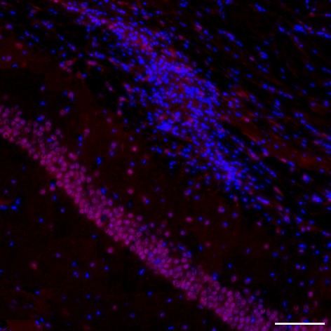
FH Marion (Verified Customer) (11-24-2024) | DAPI (blue) + iNOS (red) We expected iNOS staining on microglia cells. However, only nuclei of CA1 neurons were stained here. After discussion with the support, we came to the joint conclusion that the antibody must surely cross-link with the nNOS protein. We tested dilutions of 1/100, 1/200, 1/400, 1/600, 1/800 and 1/1000.
 |
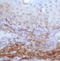
FH Yasuyo (Verified Customer) (03-01-2022) | This is one of the best IHC signaling I ever got for iNOS. Amazing image. I recommend this for iNOS detection by IHC.
 |
FH Xie (Verified Customer) (10-06-2021) | Works nice for WB assay.
 |
FH Prasanth (Verified Customer) (08-19-2019) | This is one of the cleanest and clarity blot I ever got for iNOS. Amazing clarity and no non specific bands. I recommend this for iNOS detection by western blot.
|
FH Elena (Verified Customer) (08-14-2019) | We tried this antibody with 2 cell lines and it worked just fine. We will use this in the future for other cell lines as well. The antibody produced strong bands at the suggested concentration and we diluted it further.
|


