- Phare
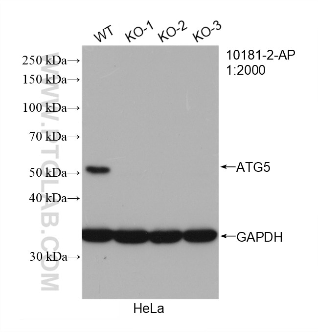
- Validé par KD/KO
Anticorps Polyclonal de lapin anti-ATG5
ATG5 Polyclonal Antibody for WB, IP, IHC, ELISA
Hôte / Isotype
Lapin / IgG
Réactivité testée
Humain, rat, souris et plus (7)
Applications
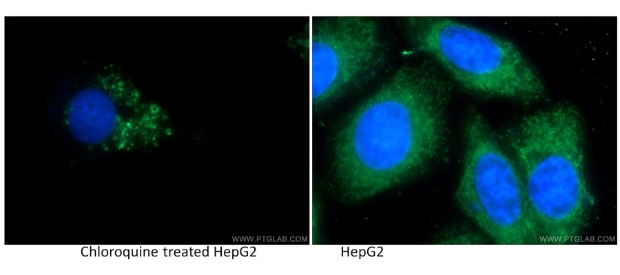
WB, IHC, IP, ELISA, IF
Conjugaison
Non conjugué
295
N° de cat : 10181-2-AP
Synonymes
Galerie de données de validation
Applications testées
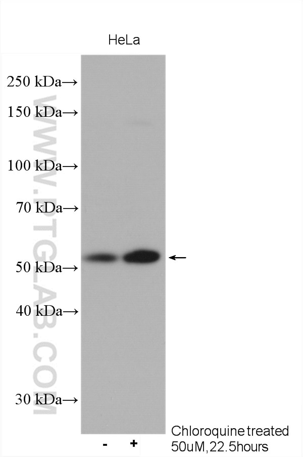
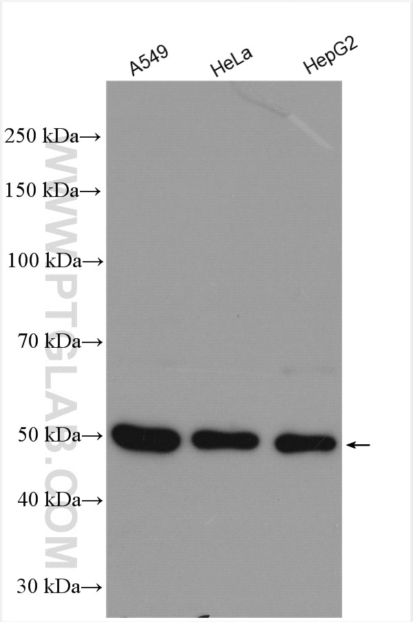
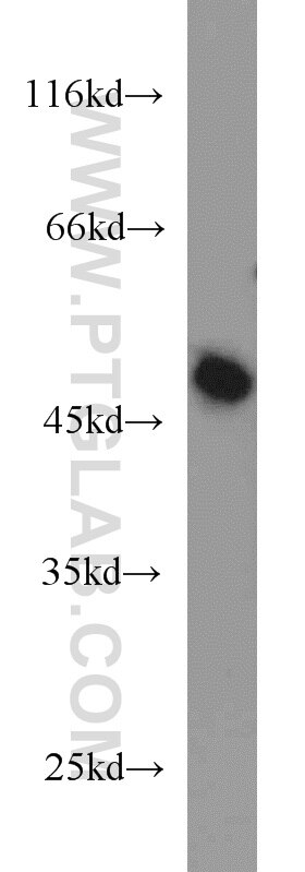
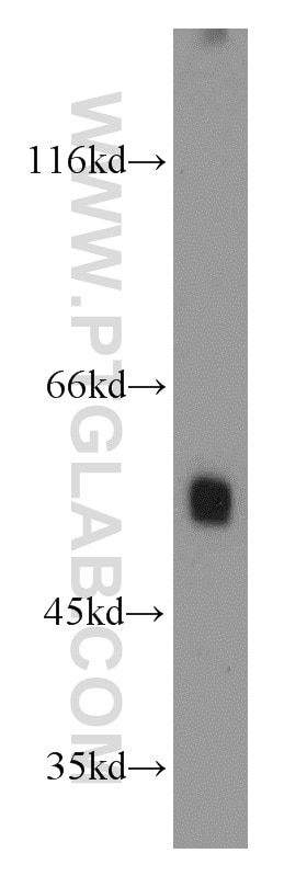
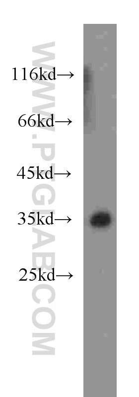
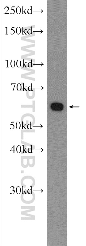
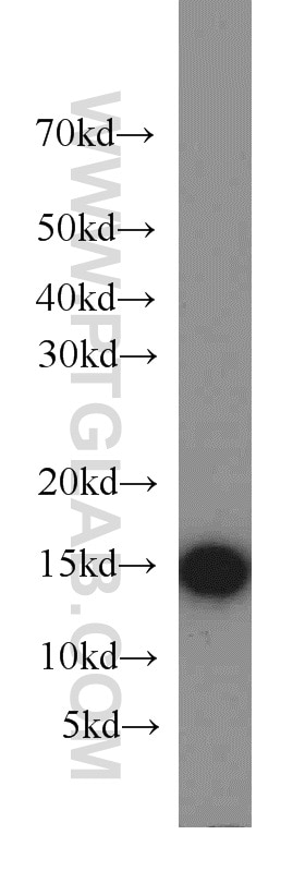
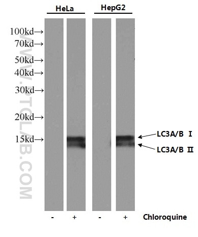
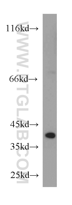
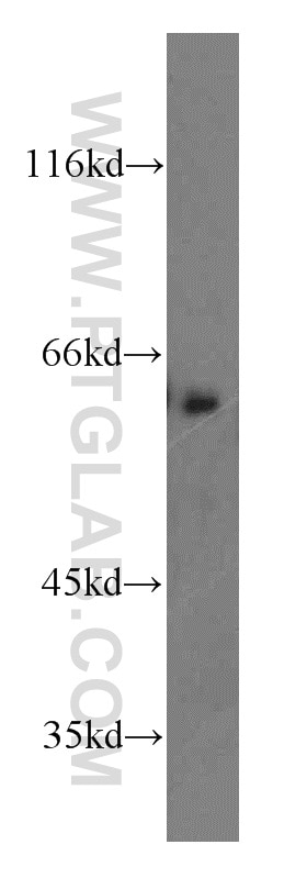
| Résultats positifs en WB | cellules A549, cellules HeLa, cellules HepG2, tissu rénal de souris |
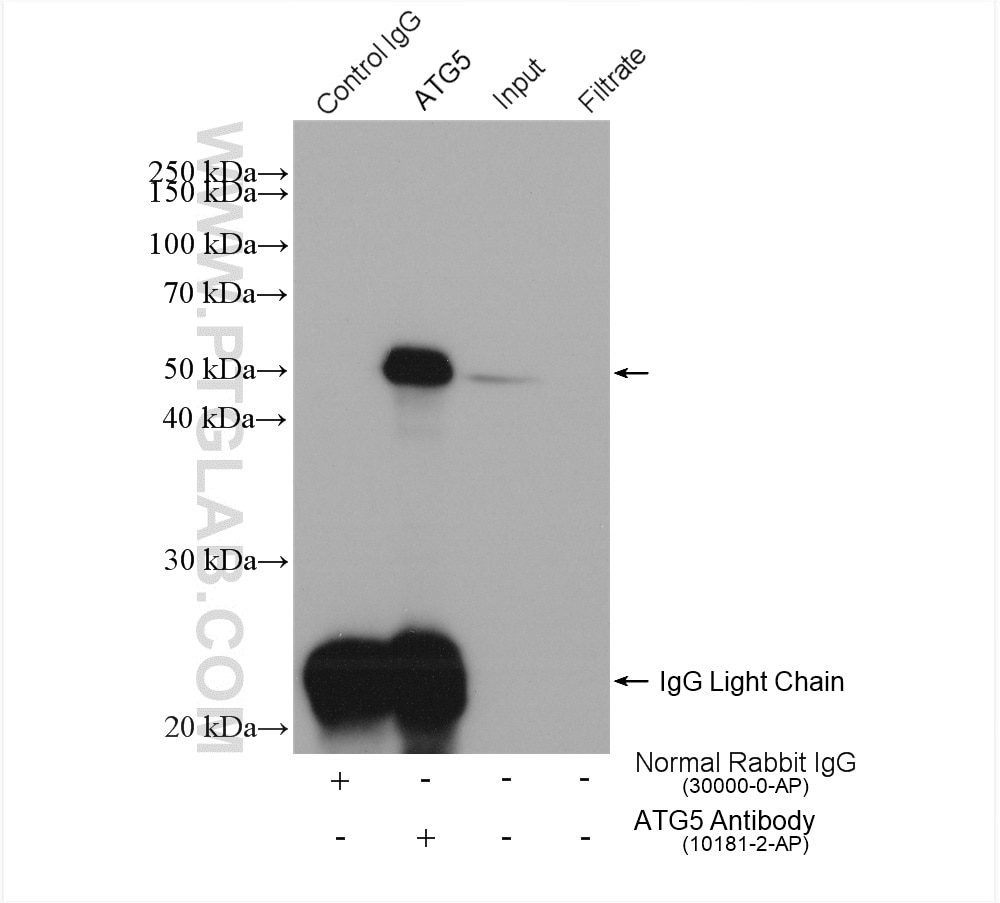
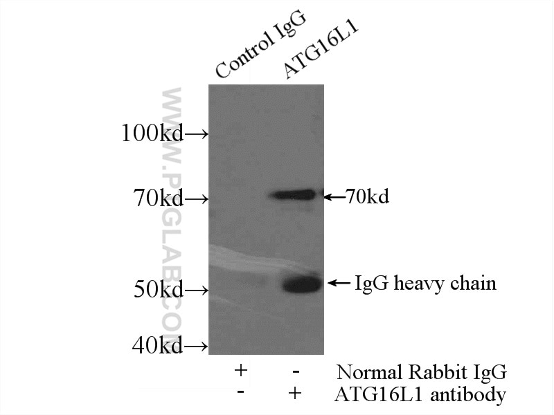
| Résultats positifs en IP | cellules HeLa, |
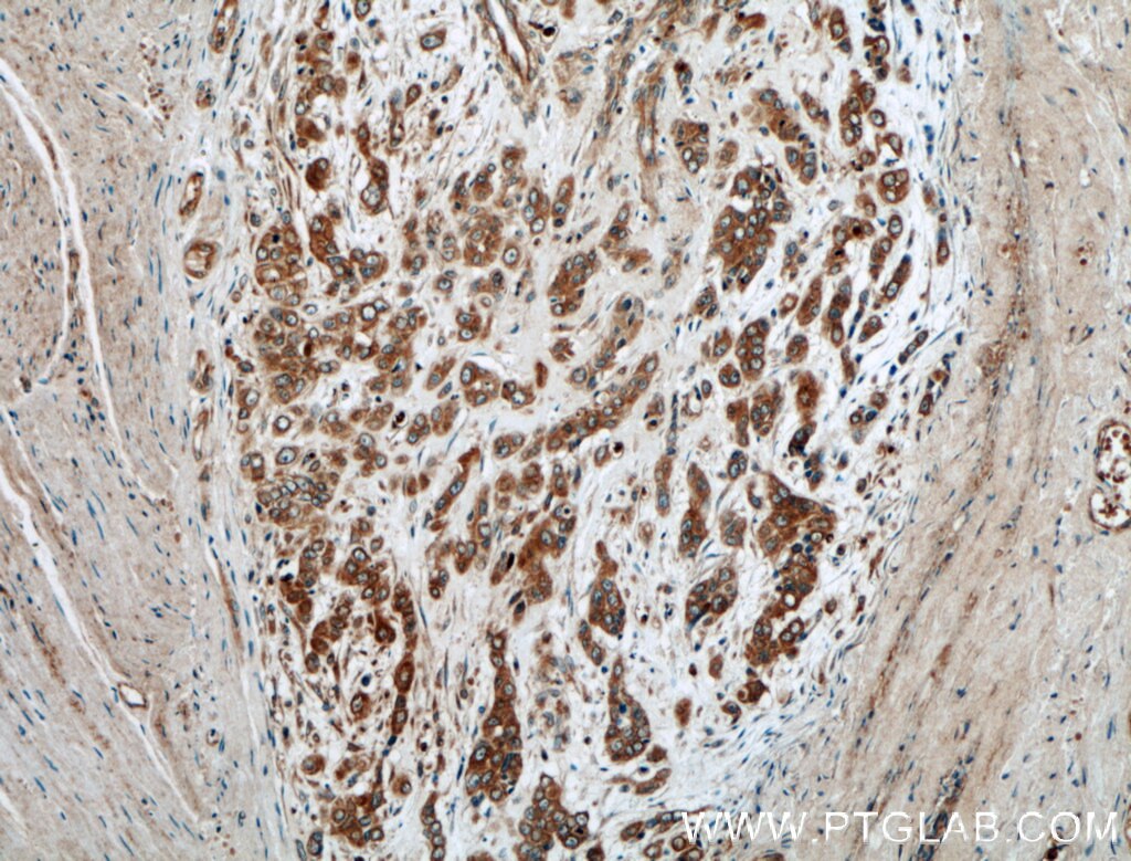
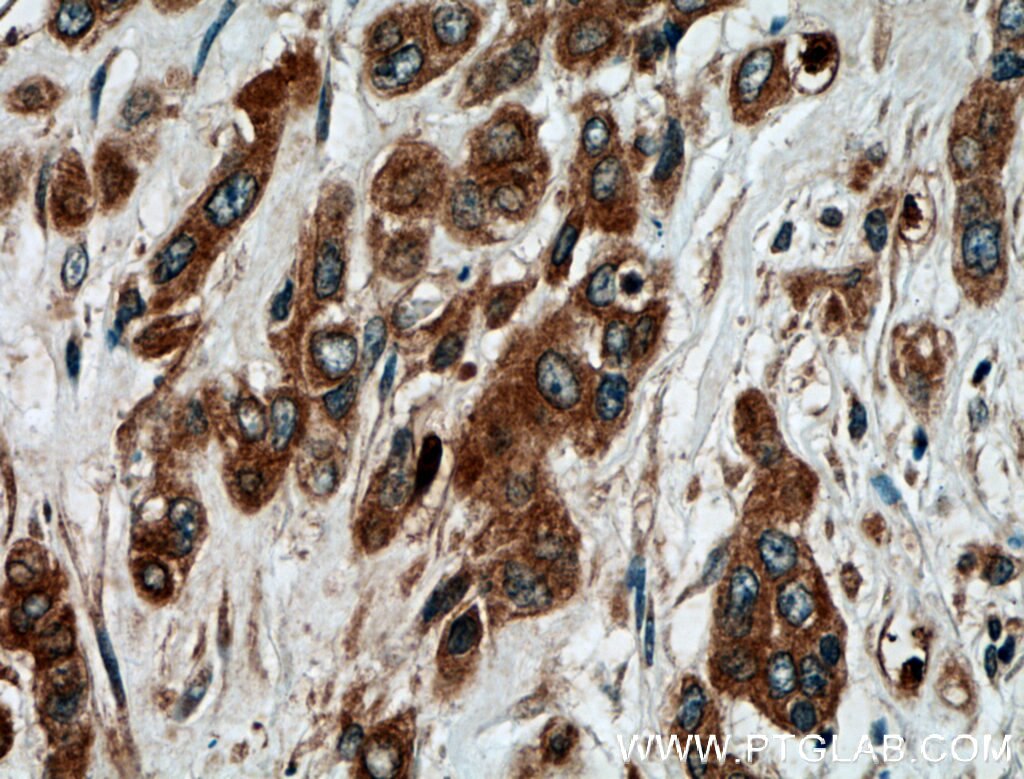
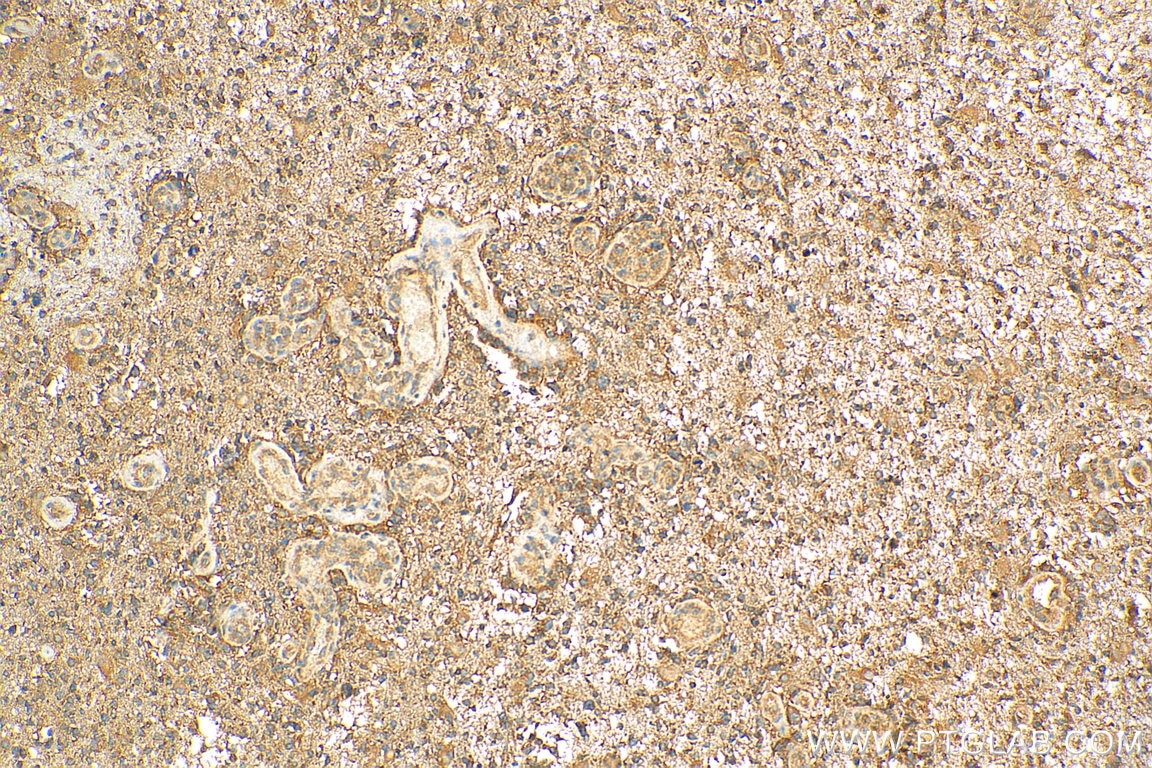
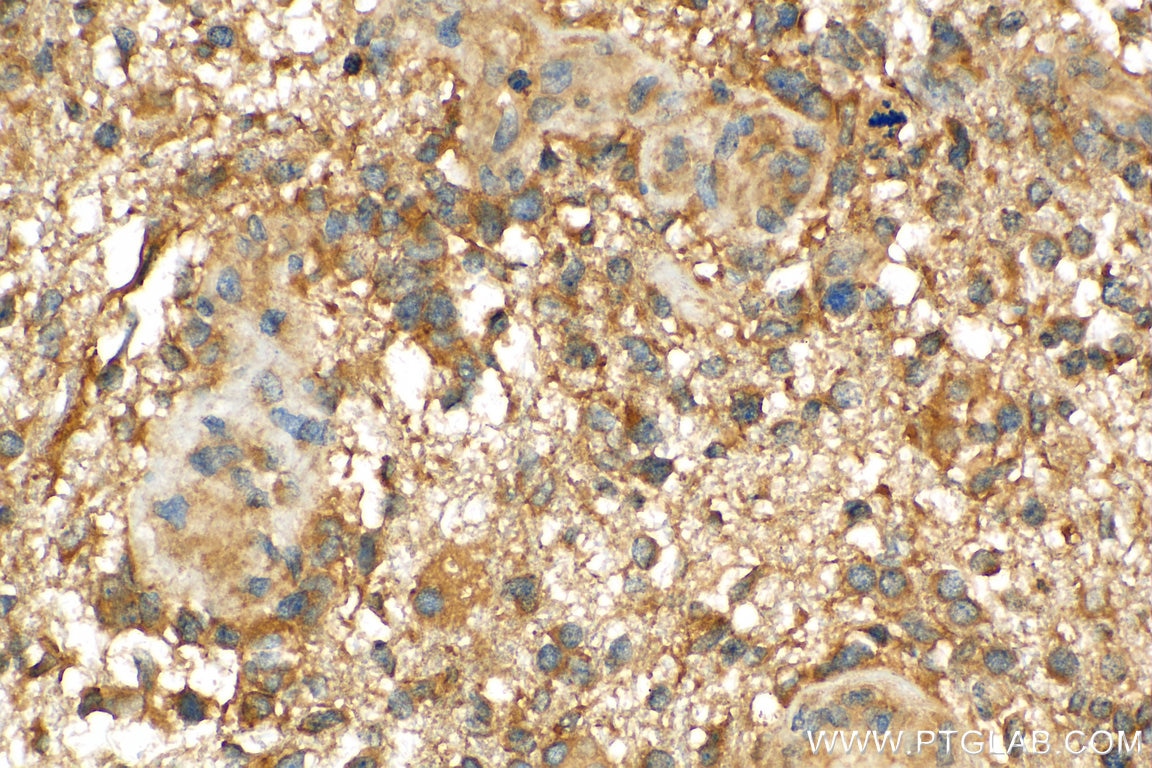
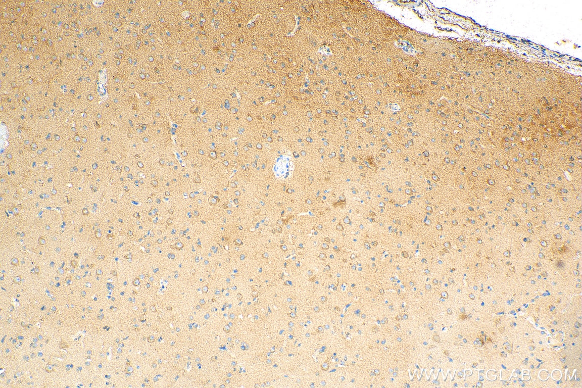
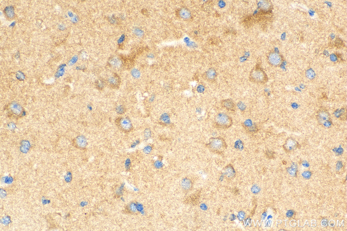
| Résultats positifs en IHC | tissu de cancer du côlon humain, tissu de gliome humain il est suggéré de démasquer l'antigène avec un tampon de TE buffer pH 9.0; (*) À défaut, 'le démasquage de l'antigène peut être 'effectué avec un tampon citrate pH 6,0. |
Dilution recommandée
| Application | Dilution |
|---|---|
| Western Blot (WB) | WB : 1:1000-1:5000 |
| Immunoprécipitation (IP) | IP : 0.5-4.0 ug for 1.0-3.0 mg of total protein lysate |
| Immunohistochimie (IHC) | IHC : 1:50-1:500 |
| It is recommended that this reagent should be titrated in each testing system to obtain optimal results. | |
| Sample-dependent, check data in validation data gallery | |
Applications publiées
| KD/KO | See 42 publications below |
| WB | See 270 publications below |
| IHC | See 25 publications below |
| IF | See 24 publications below |
| FC | See 2 publications below |
Informations sur le produit
10181-2-AP cible ATG5 dans les applications de WB, IHC, IP, ELISA, IF et montre une réactivité avec des échantillons Humain, rat, souris
| Réactivité | Humain, rat, souris |
| Réactivité citée | rat, bovin, Chèvre, Humain, porc, poulet, singe, souris, Hamster, mérou |
| Hôte / Isotype | Lapin / IgG |
| Clonalité | Polyclonal |
| Type | Anticorps |
| Immunogène | ATG5 Protéine recombinante Ag0214 |
| Nom complet | ATG5 autophagy related 5 homolog (S. cerevisiae) |
| Masse moléculaire calculée | 32 kDa |
| Poids moléculaire observé | 32 kDa, 40-45 kDa, 50-55 kDa |
| Numéro d’acquisition GenBank | BC002699 |
| Symbole du gène | ATG5 |
| Identification du gène (NCBI) | 9474 |
| Conjugaison | Non conjugué |
| Forme | Liquide |
| Méthode de purification | Purification par affinité contre l'antigène |
| Tampon de stockage | PBS avec azoture de sodium à 0,02 % et glycérol à 50 % pH 7,3 |
| Conditions de stockage | Stocker à -20°C. Stable pendant un an après l'expédition. L'aliquotage n'est pas nécessaire pour le stockage à -20oC Les 20ul contiennent 0,1% de BSA. |
Informations générales
ATG5, also named as APG5L and ASP, belongs to the ATG5 family. It is required for autophagy. It plays an important role in the apoptotic process, possibly within the modified cytoskeleton. Its expression is a relatively late event in the apoptotic process, occurring downstream of caspase activity. Autophagy is a catabolic process for the autophagosomic-lysosomal degradation of bulk cytoplasmic contents. Formation of the autophagosome involves a ubiquitin-like conjugation system in which Atg12 is covalently bound to Atg5 and targeted to autophagosome vesicles. It mediates autophagosome-independent host protection. This antibody is raised against 28-275 amino acids of human ATG5. It can recognize the ATG5-ATG12 complex (55 kDa) which can be truncated and generate a 40-45 kDa band. 10181-2-AP also recognizes the free ATG5 (32 kDa).
Protocole
| Product Specific Protocols | |
|---|---|
| WB protocol for ATG5 antibody 10181-2-AP | Download protocol |
| IHC protocol for ATG5 antibody 10181-2-AP | Download protocol |
| IP protocol for ATG5 antibody 10181-2-AP | Download protocol |
| Standard Protocols | |
|---|---|
| Click here to view our Standard Protocols |
Publications
| Species | Application | Title |
|---|---|---|
Signal Transduct Target Ther Targeting CRL4 suppresses chemoresistant ovarian cancer growth by inducing mitophagy | ||
Cell Res Degradation of lipid droplets by chimeric autophagy-tethering compounds.
| ||
Mol Cell Syntaxin 13, a Genetic Modifier of Mutant CHMP2B in Frontotemporal Dementia, Is Required for Autophagosome Maturation.
| ||
Adv Sci (Weinh) SETDB1 Methylates MCT1 Promoting Tumor Progression by Enhancing the Lactate Shuttle | ||
Autophagy Autophagy mediates the clearance of oligodendroglial SNCA/alpha-synuclein and TPPP/p25A in multiple system atrophy models | ||
J Clin Invest Acetaldehyde dehydrogenase 2 interactions with LDLR and AMPK regulate foam cell formation. |
Avis
The reviews below have been submitted by verified Proteintech customers who received an incentive forproviding their feedback.
FH Priya (Verified Customer) (08-09-2023) | Used for Caco2 cells and mice colon tissue
|
FH Kamal (Verified Customer) (06-29-2023) | Works well with TBST. Atg5-12 complex (56 kDa) and Atg5 (33kDa) bands came out nice and clear.
|
FH Priya (Verified Customer) (06-21-2023) | Used this antibody for Caco2 cells andmice tissue
|
FH Priya (Verified Customer) (06-21-2023) | Used this antibody for Caco2 cells and mice tissue
|
FH Benjamin (Verified Customer) (01-07-2020) | Works well in western blot in 3% milk TBS-T
|
