
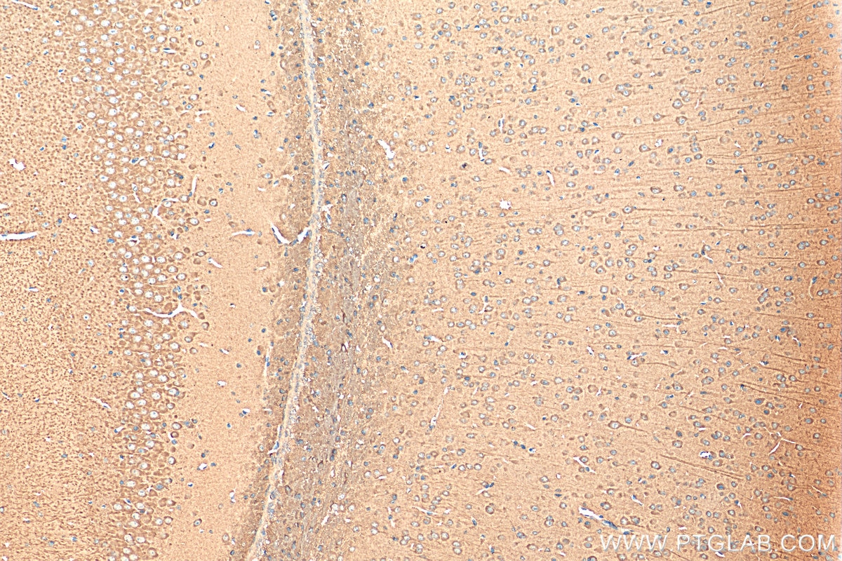
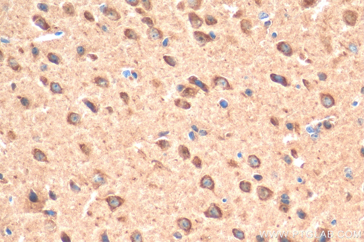
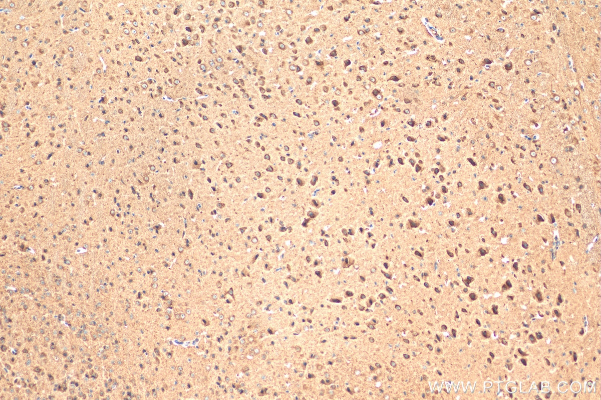
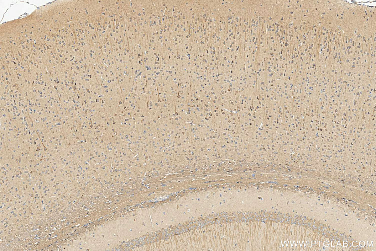

Tested Applications
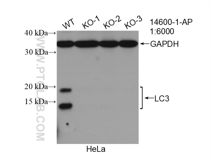
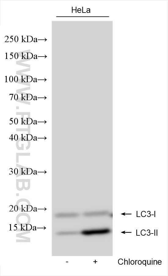
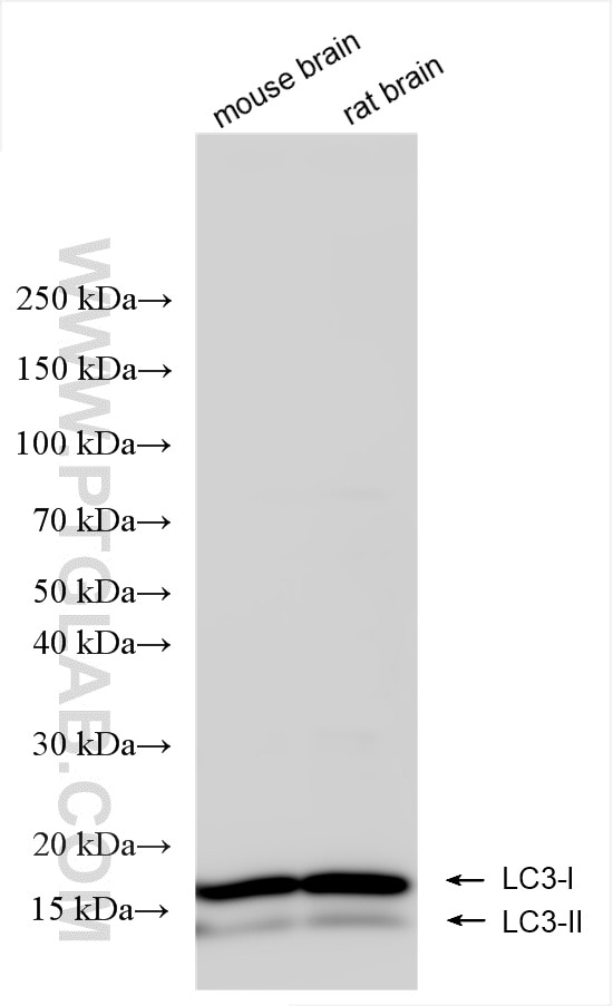
| Positive WB detected in | HeLa cells, mouse brain tissue, Chloroquine treated HeLa cells, rat brain tissue |
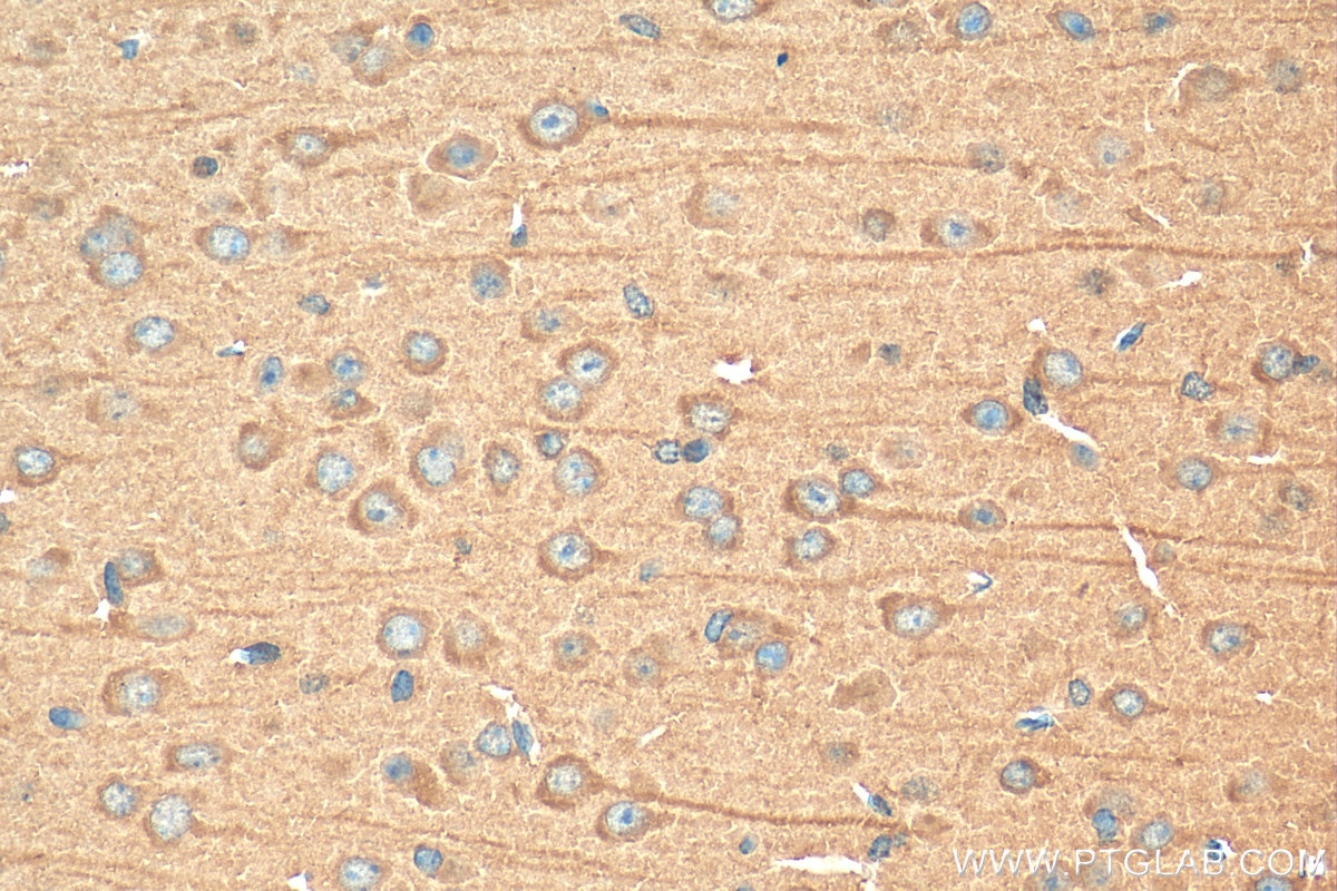
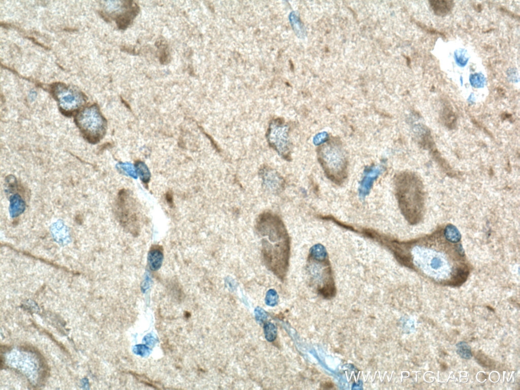
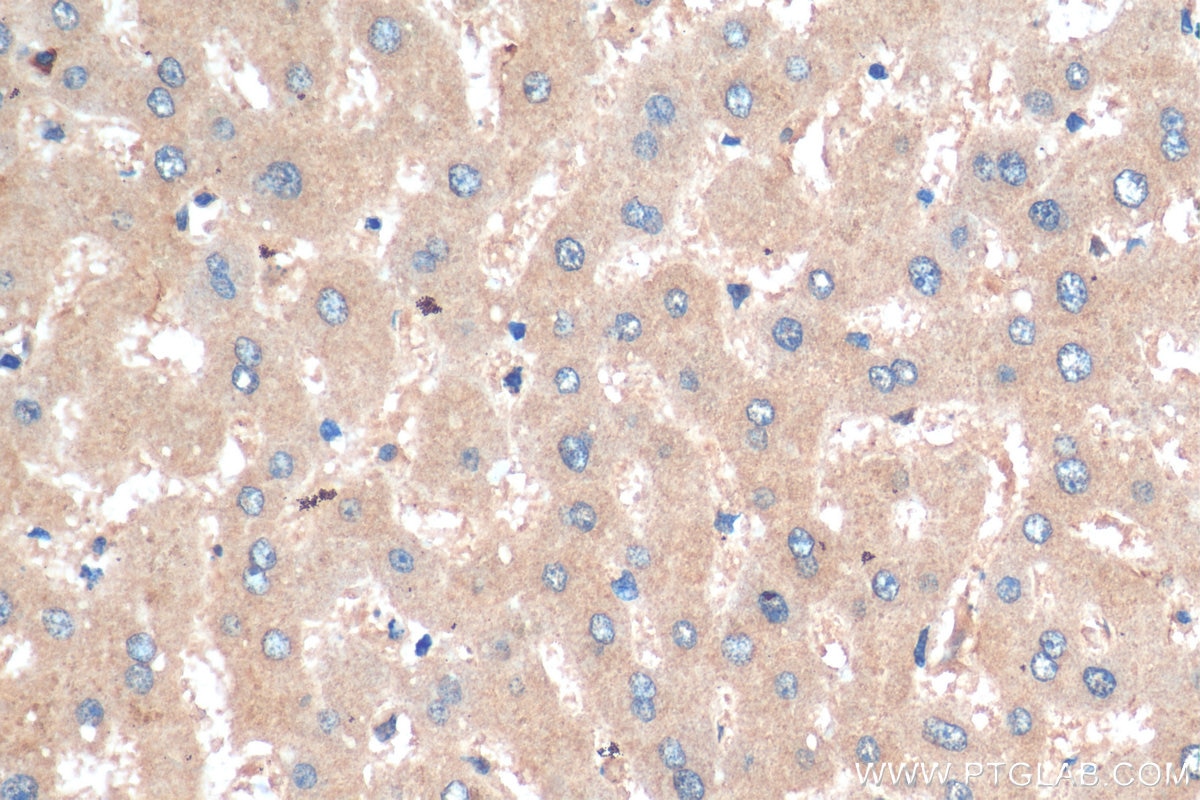
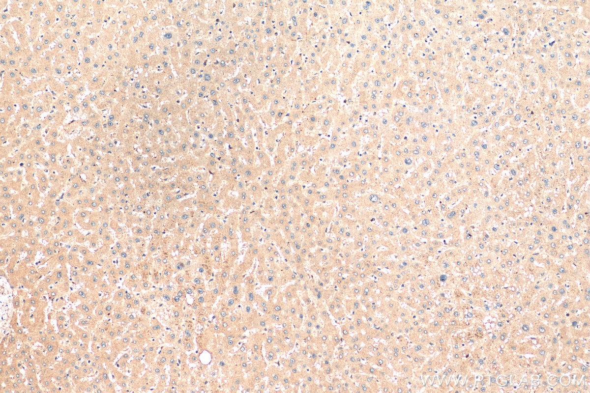
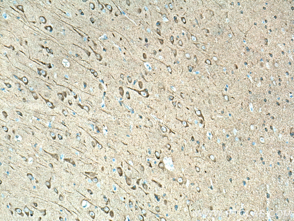
| Positive IHC detected in | human liver tissue, human gliomas tissue, mouse brain tissue Note: suggested antigen retrieval with TE buffer pH 9.0; (*) Alternatively, antigen retrieval may be performed with citrate buffer pH 6.0 |
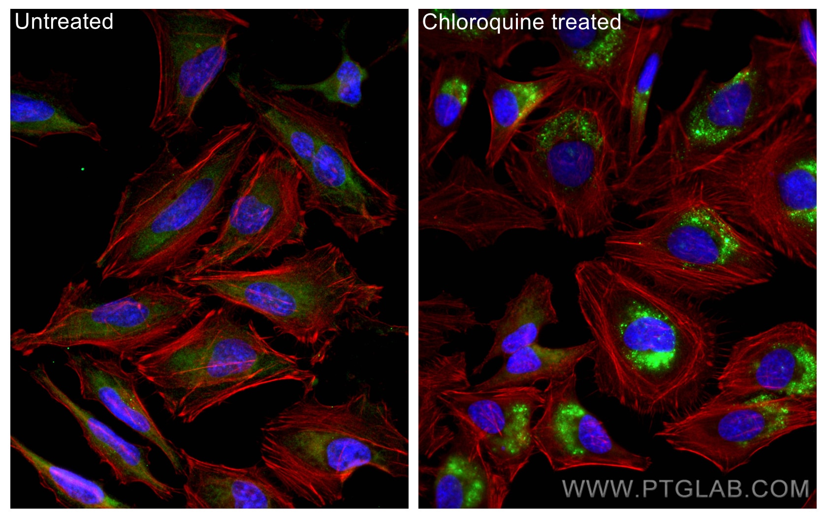
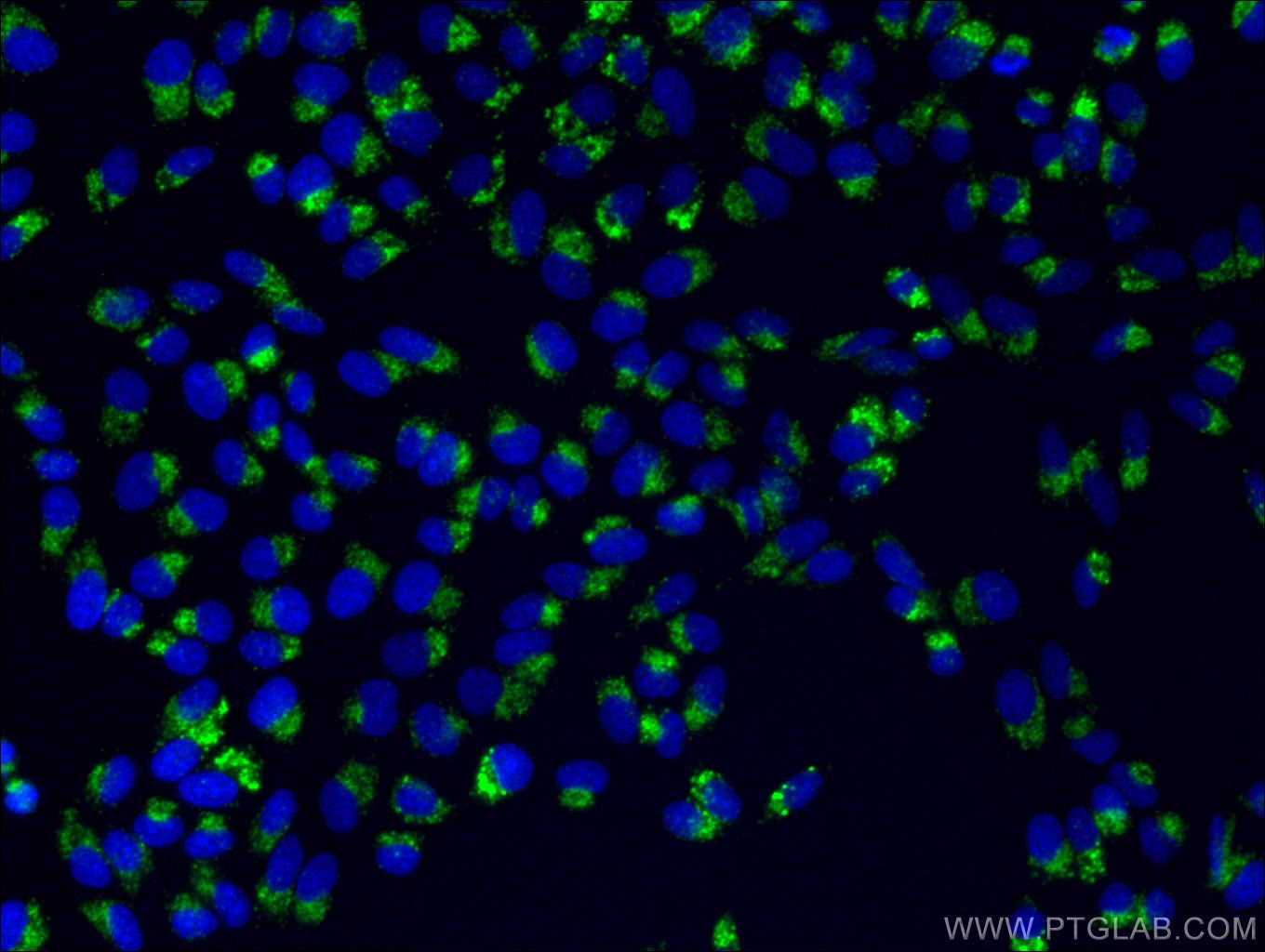
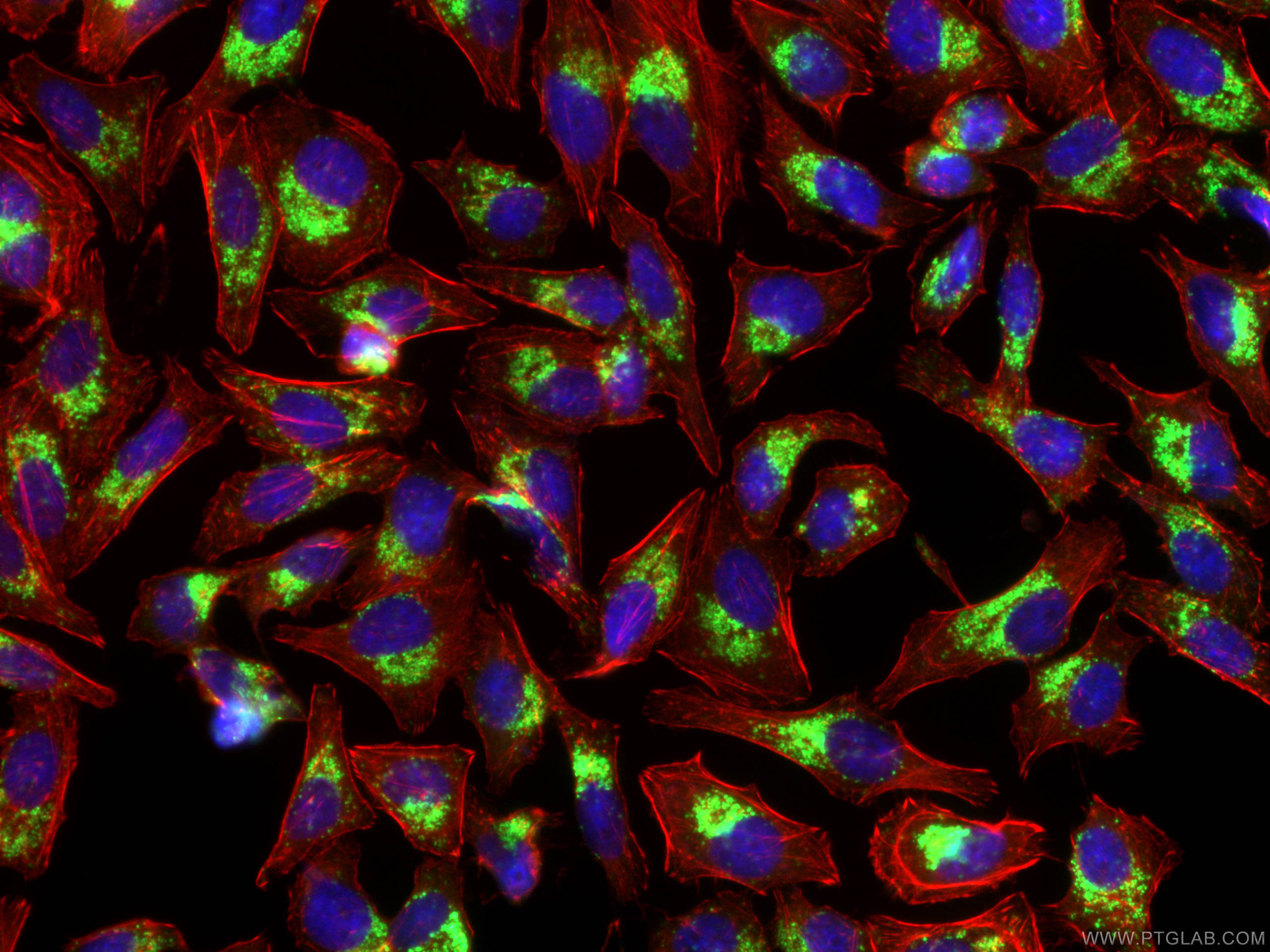
| Positive IF/ICC detected in | Chloroquine treated HeLa cells, Chloroquine treated HepG2 cells |
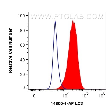
| Positive FC (Intra) detected in | HeLa cells |
Recommended dilution
| Application | Dilution |
|---|---|
| Western Blot (WB) | WB : 1:2000-1:8000 |
| Immunohistochemistry (IHC) | IHC : 1:200-1:1000 |
| Immunofluorescence (IF)/ICC | IF/ICC : 1:250-1:1000 |
| Flow Cytometry (FC) (INTRA) | FC (INTRA) : 0.50 ug per 10^6 cells in a 100 µl suspension |
| It is recommended that this reagent should be titrated in each testing system to obtain optimal results. | |
| Sample-dependent, Check data in validation data gallery. | |
Product Information
14600-1-AP targets LC3 in WB, IHC, IF/ICC, FC (Intra), IP, CoIP, ELISA applications and shows reactivity with human, mouse, rat samples.
| Tested Reactivity | human, mouse, rat |
| Cited Reactivity | human, mouse, pig, monkey, chicken, bovine, hamster, goat, duck |
| Host / Isotype | Rabbit / IgG |
| Class | Polyclonal |
| Type | Antibody |
| Immunogen |
CatNo: Ag6144 Product name: Recombinant human LC3 protein Source: e coli.-derived, PGEX-4T Tag: GST Domain: 1-125 aa of BC067797 Sequence: MPSEKTFKQRRTFEQRVEDVRLIREQHPTKIPVIIERYKGEKQLPVLDKTKFLVPDHVNMGELIKIIRRRLQLNANQAFFLLVNGHSMVSVSTPISEVYESEKDEDGFLYMVYASQETFGMKLSV Predict reactive species |
| Full Name | microtubule-associated protein 1 light chain 3 beta |
| Calculated Molecular Weight | 15 kDa |
| Observed Molecular Weight | 14-18 kDa |
| GenBank Accession Number | BC067797 |
| Gene Symbol | LC3B |
| Gene ID (NCBI) | 81631 |
| ENSEMBL Gene ID | ENSG00000140941 |
| RRID | AB_2137737 |
| Conjugate | Unconjugated |
| Form | Liquid |
| Purification Method | Antigen affinity purification |
| UNIPROT ID | Q9GZQ8 |
| Storage Buffer | PBS with 0.02% sodium azide and 50% glycerol, pH 7.3. |
| Storage Conditions | Store at -20°C. Stable for one year after shipment. Aliquoting is unnecessary for -20oC storage. 20ul sizes contain 0.1% BSA. |
Background Information
Map1LC3, also known as LC3, is the human homolog of yeast Atg8 and is involved in the formation of autophagosomal vacuoles, called autophagosomes. Three human Map1LC3 isoforms, MAP1LC3A, MAP1LC3B, and MAP1LC3C, undergo post-translational modifications during autophagy. And they differ in their post-translation modifications during autophagy. Map1LC3 also exists in two modified forms, an 18 kDa cytoplasmic form that was originally identified as a subunit of the microtubule-associated protein 1, and a 14-16 kDa form that is associated with the autophagosome membrane. This antibody can cross react with MAP1LC3A, MAP1LC3B, and MAP1LC3C.
Protocols
| Product Specific Protocols | |
|---|---|
| FC protocol for LC3 antibody 14600-1-AP | Download protocol |
| IF protocol for LC3 antibody 14600-1-AP | Download protocol |
| IHC protocol for LC3 antibody 14600-1-AP | Download protocol |
| WB protocol for LC3 antibody 14600-1-AP | Download protocol |
| Standard Protocols | |
|---|---|
| Click here to view our Standard Protocols |
Publications
What published studies show
The LC3 Polyclonal antibody (Cat# 14600-1-AP) has accumulated 2,417 citations across high-impact journals including Nature, Science, Cell, Autophagy, Nature Communications, Nature Cell Biology, Signal Transduction and Targeted Therapy, Molecular Cancer, ACS Nano, Theranostics, and Advanced Science. Research fields span autophagy/mitophagy, cancer biology, neurodegeneration, ferroptosis, inflammation, metabolism, vascular disease, bone biology, liver disease, cardiology, nanomedicine, viral infections, stem cell biology, and aging.
| Species | Application | Title |
|---|---|---|
Signal Transduct Target Ther Targeting CRL4 suppresses chemoresistant ovarian cancer growth by inducing mitophagy | ||
Signal Transduct Target Ther Albumosomes formed by cytoplasmic pre-folding albumin maintain mitochondrial homeostasis and inhibit nonalcoholic fatty liver disease | ||
Signal Transduct Target Ther Elaiophylin triggers paraptosis and preferentially kills ovarian cancer drug-resistant cells by inducing MAPK hyperactivation | ||
Signal Transduct Target Ther Nlp promotes autophagy through facilitating the interaction of Rab7 and FYCO1. | ||
Science Liver ALKBH5 regulates glucose and lipid homeostasis independently through GCGR and mTORC1 signaling |
Reviews
What customers say
Users overwhelmingly rate this LC3 antibody positively (avg ~4.3/5 across 13 reviews). It performs well in Western blot, IHC, and IF, producing clear LC3-I and LC3-II bands with minimal background. Several users note it outperforms competing LC3 antibodies from other companies. Common dilutions range from 1:500 to 1:2000 for WB. Tested successfully in diverse samples including HepG2, SH-SY5Y, HCT116, microglia, mouse liver, and zebrafish. One reviewer reported detecting only one LC3 form, but this is the sole negative feedback. Overall, it's regarded as a reliable, cost-effective choice for autophagy studies.
The reviews below have been submitted by verified Proteintech customers who received an incentive for providing their feedback.
Jumm (Verified Customer) (12-11-2025) | Reliable LC3 antibody with clear autophagy-related bands in Western blots and good performance in IF/IHC. Strong signal and minimal background make it a solid choice for LC3/autophagy studies.
|
A (Verified Customer) (07-15-2025) | Used for caco2 cells and animal colon tissue
|
Aditya (Verified Customer) (11-01-2024) | great with ecl
|
David (Verified Customer) (01-02-2024) | Good antibody, single band at the predicted molecular weight, no background.
|
Hala (Verified Customer) (05-09-2023) | works very good in milk5%
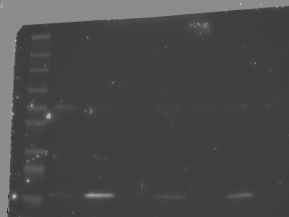 |
Hala (Verified Customer) (04-28-2023) | works very good with milk blocking
|
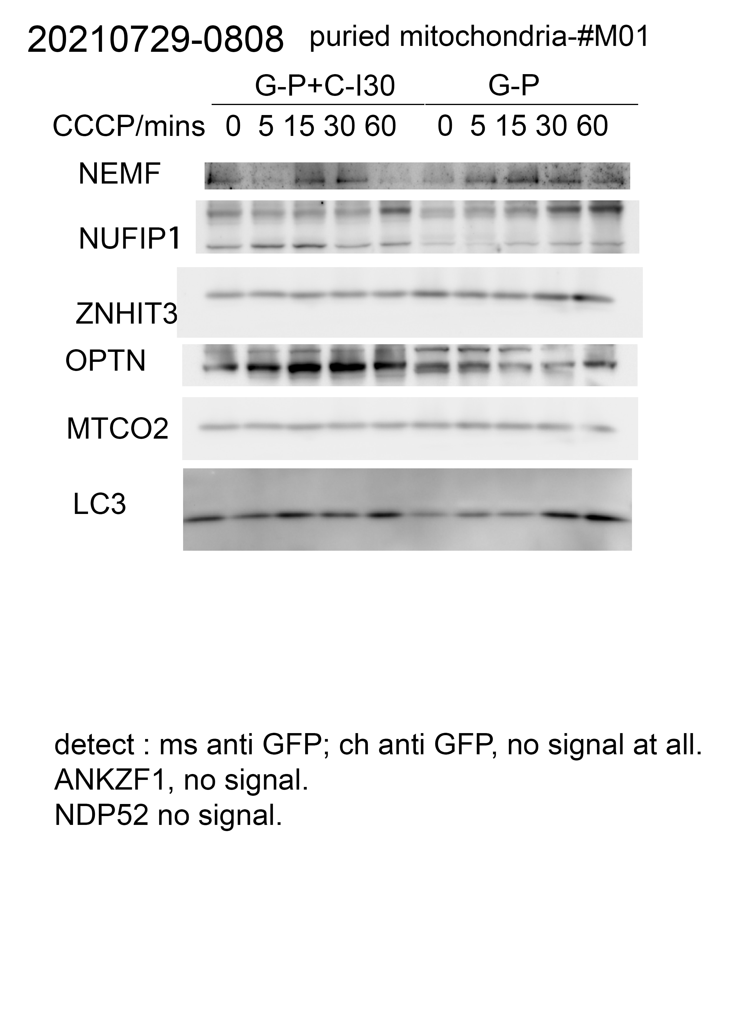
Zhihao (Verified Customer) (07-25-2022) | We could detect one form of LC3 in our cell samples, which should be two.
 |
S (Verified Customer) (12-31-2021) | I have tried three different LC3B antibodies from three different companies so far. This antibody is by far the best (cost efficient as well)
 |
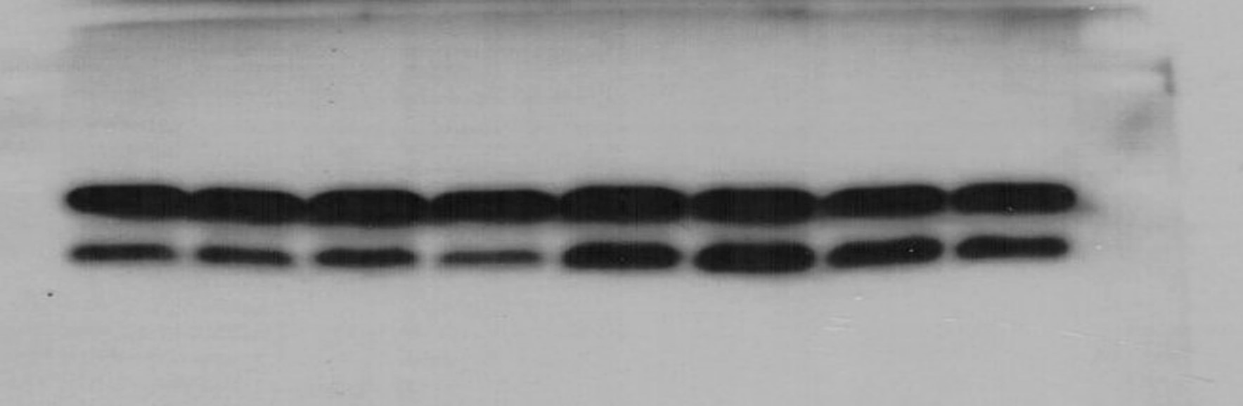
YING (Verified Customer) (09-25-2021) | The antibody works very well. Distinguished LC3-I and LC3-II bands. An increase of LC3-II level in response to treatment with bafilomycin A.
 |
Eric (Verified Customer) (03-13-2021) | Worked very well. I was able to get the LC3A/B bands on western blot using a 12.5% gel and prominent bands on a 8% gel. IF imaging worked fairly well, I used 1:100 which was likely too low so I will repeat at higher but got signal nonetheless. This performed better than some of the other LC3 antibodies we tested.
 |
Elzbieta (Verified Customer) (09-23-2020) | The antibody works very nicely in WB. Clear signal, no aspecific bands. Our lab discarded other products and switched to this antibody.
|
An (Verified Customer) (09-17-2020) | Used for IHC on zebrafish cryosections of eyes. Worked well.
|
David (Verified Customer) (03-30-2020) | Very nice visualisation of LC3-I and LC3-II. Run on 20% gel and significant changes in response to treatment with bafilomycin A and concanamycin A.
|


