at dilution of 1:300 incubated at room temperature for 1.5 hours.")
 at dilution of 1:500 incubated at room temperature for 1.5 hours.")
 at dilution of 1:500 incubated at room temperature for 1.5 hours.")
 at dilution of 1:500 incubated at room temperature for 1.5 hours.")
 at dilution of 1:300 incubated at room temperature for 1.5 hours.")
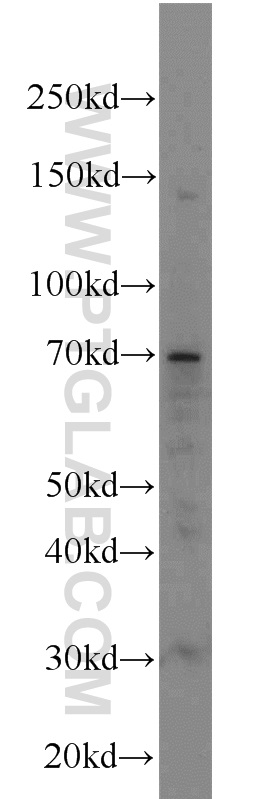
 with MCF-7 cells lysate 2000ug.")
Tested Applications
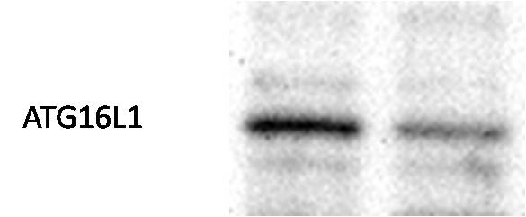
| Positive WB detected in | mouse spleen tissue, MCF-7 cells, Jurkat cells, HEK-293T cells |
| Positive IP detected in | MCF-7 cells |
Recommended dilution
| Application | Dilution |
|---|---|
| Western Blot (WB) | WB : 1:200-1:1000 |
| Immunoprecipitation (IP) | IP : 0.5-4.0 ug for 1.0-3.0 mg of total protein lysate |
| It is recommended that this reagent should be titrated in each testing system to obtain optimal results. | |
| Sample-dependent, Check data in validation data gallery. | |
Published Applications
| WB | See 14 publications below |
| IHC | See 1 publications below |
| IF | See 2 publications below |
| CoIP | See 1 publications below |
Product Information
19812-1-AP targets ATG16L1 in WB, IP, IF, IHC, CoIP, ELISA applications and shows reactivity with human, mouse samples.
| Tested Reactivity | human, mouse |
| Cited Reactivity | human, mouse, rat, pig |
| Host / Isotype | Rabbit / IgG |
| Class | Polyclonal |
| Type | Antibody |
| Immunogen |
CatNo: Ag13844 Product name: Recombinant human ATG16L1 protein Source: e coli.-derived, PGEX-4T Tag: GST Domain: 258-607 aa of BC000061 Sequence: RAISRAATKRLSQPAGGLLDSITNIFGRRSVSSFPVPQDNVDAHPGSGKEVRVPATALCVFDAHDGEVNAVQFSPGSRLLATGGMDRRVKLWEVFGEKCEFKGSLSGSNAGITSIEFDSAGSYLLAASNDFASRIWTVDDYRLRHTLTGHSGKVLSAKFLLDNARIVSGSHDRTLKLWDLRSKVCIKTVFAGSSCNDIVCTEQCVMSRHFDKKIRFWDIRSESIVREMELLGKITALDLNPERTELLSCSRDDLLKVIDLRTNAIKQTFSAPGFKCGSDWTRVVFSPDGSYVAAGSAEGSLYIWSVLTGKVEKVLSKQHSSSINAVAWSPSGSHVVSVDKGCKAVLWAQY Predict reactive species |
| Full Name | ATG16 autophagy related 16-like 1 (S. cerevisiae) |
| Calculated Molecular Weight | 607 aa, 68 kDa |
| Observed Molecular Weight | 63-71 kDa |
| GenBank Accession Number | BC000061 |
| Gene Symbol | ATG16L1 |
| Gene ID (NCBI) | 55054 |
| RRID | AB_10695631 |
| Conjugate | Unconjugated |
| Form | Liquid |
| Purification Method | Antigen affinity purification |
| UNIPROT ID | Q676U5 |
| Storage Buffer | PBS with 0.02% sodium azide and 50% glycerol, pH 7.3. |
| Storage Conditions | Store at -20°C. Stable for one year after shipment. Aliquoting is unnecessary for -20oC storage. 20ul sizes contain 0.1% BSA. |
Background Information
Human ATG16L1 is a 607 amino acid protein (~68 kDa) comprising three major domains: the N‐terminal ATG5 binding domain (ATG5‐BD), the central coiled‐coil domain (CCD) and a predicted C‐terminal WD40‐domain. ATG16L1α and β (Atg16L1α, 63 kDa; and Atg16L1β, 71 kDa) are the major isoforms expressed in intestinal epithelium and macrophages , and all isoforms encode exon 9, which contains Thr 300. Atg16L1 mediates the cellular degradative process of autophagy and is considered a critical regulator of inflammation based on its genetic association with inflammatory bowel disease. ATG16L1 has been implicated in Crohn's disease. (PMID: 24553140, PMID: 22740627,PMID: 28685931)
Publications
| Species | Application | Title |
|---|---|---|
Autophagy ATG4B antagonizes antiviral immunity by GABARAP-directed autophagic degradation of TBK1 | ||
Nat Commun Phase separation of Nur77 mediates celastrol-induced mitophagy by promoting the liquidity of p62/SQSTM1 condensates. | ||
Autophagy AP2M1 mediates autophagy-induced CLDN2 (claudin 2) degradation through endocytosis and interaction with LC3 and reduces intestinal epithelial tight junction permeability. | ||
Curr Biol Cellular mechanotransduction relies on tension-induced and chaperone-assisted autophagy. | ||
Cancer Lett Let-7i-5p promotes a malignant phenotype in nasopharyngeal carcinoma via inhibiting tumor-suppressive autophagy. | ||
Clin Transl Immunology Circular RNA TRAPPC6B inhibits intracellular Mycobacterium tuberculosis growth while inducing autophagy in macrophages by targeting microRNA-874-3p. |
Reviews
The reviews below have been submitted by verified Proteintech customers who received an incentive for providing their feedback.
FH Priya (Verified Customer) (07-31-2023) | Used for Caco2 cells and mice tissue
|
FH Priya (Verified Customer) (06-21-2023) | Used this antibody for Caco2 cells andmice tissue
|
FH Priya (Verified Customer) (04-17-2023) | Used for Caco2 cells
|
FH Priya (Verified Customer) (04-17-2023) | Used for Caco2 cells
|
FH X (Verified Customer) (01-18-2021) | It is OK to use it in WB with human postmortem brain lysate. There is band with MW as expected, although there is other unidentified bands.
 |