Anticorps Polyclonal de lapin anti-VEGFC
VEGFC Polyclonal Antibody for WB, IF, IHC, ELISA
Hôte / Isotype
Lapin / IgG
Réactivité testée
Humain, souris et plus (1)
Applications
WB, IHC, IF/ICC, ELISA
Conjugaison
Non conjugué
N° de cat : 22601-1-AP
Synonymes
Galerie de données de validation
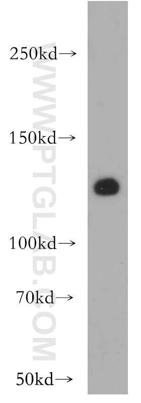
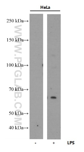
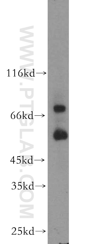
 at dilution of 1:1000 incubated at room temperature for 1.5 hours.")
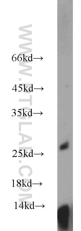
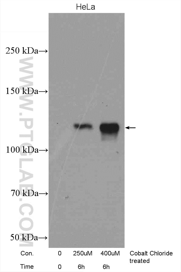
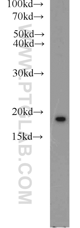
 at dilution of 1:800 incubated at room temperature for 1.5 hours.")
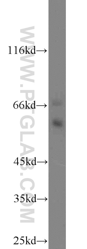
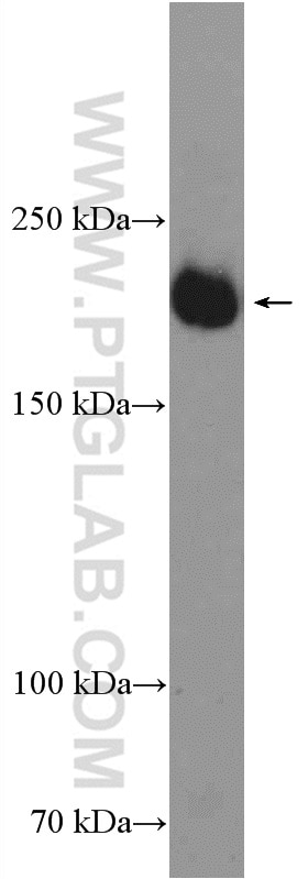
 at dilution of 1:300 incubated at room temperature for 1.5 hours.")
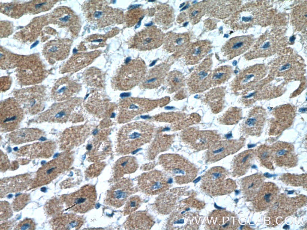
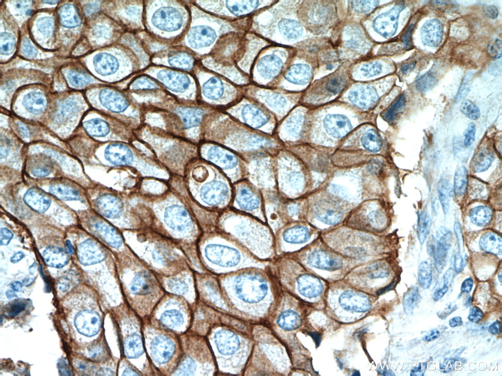
 at dilution of 1:200 (under 10x lens). Heat mediated antigen retrieval with Tris-EDTA buffer (pH 9.0).")
 at dilution of 1:200 (under 40x lens). Heat mediated antigen retrieval with Tris-EDTA buffer (pH 9.0).")
 at dilution of 1:200 (under 10x lens).")
 at dilution of 1:200 (under 40x lens).")
 at dilution of 1:200 (under 10x lens).")
 at dilution of 1:200 (under 40x lens).")
 at dilution of 1:50 and Alexa Fluor 488-conjugated AffiniPure Goat Anti-Rabbit IgG(H+L).")
Applications testées
| Résultats positifs en WB | cellules MCF-7, cellules HCT 116, cellules RAW 264.7, cellules SW480 |
| Résultats positifs en IHC | tissu de cancer de la prostate humain, tissu de cancer du poumon humain il est suggéré de démasquer l'antigène avec un tampon de TE buffer pH 9.0; (*) À défaut, 'le démasquage de l'antigène peut être 'effectué avec un tampon citrate pH 6,0. |
| Résultats positifs en IF/ICC | cellules RAW 264.7 |
Dilution recommandée
| Application | Dilution |
|---|---|
| Western Blot (WB) | WB : 1:500-1:2000 |
| Immunohistochimie (IHC) | IHC : 1:50-1:500 |
| Immunofluorescence (IF)/ICC | IF/ICC : 1:20-1:200 |
| It is recommended that this reagent should be titrated in each testing system to obtain optimal results. | |
| Sample-dependent, check data in validation data gallery | |
Applications publiées
| WB | See 24 publications below |
| IHC | See 13 publications below |
| IF | See 5 publications below |
Informations sur le produit
22601-1-AP cible VEGFC dans les applications de WB, IHC, IF/ICC, ELISA et montre une réactivité avec des échantillons Humain, souris
| Réactivité | Humain, souris |
| Réactivité citée | rat, Humain, souris |
| Hôte / Isotype | Lapin / IgG |
| Clonalité | Polyclonal |
| Type | Anticorps |
| Immunogène | VEGFC Protéine recombinante Ag18182 |
| Nom complet | vascular endothelial growth factor C |
| Masse moléculaire calculée | 419 aa, 47 kDa |
| Poids moléculaire observé | 47 kDa |
| Numéro d’acquisition GenBank | BC035212 |
| Symbole du gène | VEGFC |
| Identification du gène (NCBI) | 7424 |
| Conjugaison | Non conjugué |
| Forme | Liquide |
| Méthode de purification | Purification par affinité contre l'antigène |
| Tampon de stockage | PBS avec azoture de sodium à 0,02 % et glycérol à 50 % pH 7,3 |
| Conditions de stockage | Stocker à -20°C. Stable pendant un an après l'expédition. L'aliquotage n'est pas nécessaire pour le stockage à -20oC Les 20ul contiennent 0,1% de BSA. |
Informations générales
VEGFC is a member of the platelet-derived growth factor / vascular endothelial growth factor (PDGF/VEGF) family. The main function of VEGFC is to promote the growth of lymphatic vessels (lymphangiogenesis). It acts on lymphatic endothelial cells (LECs) primarily via its receptor VEGFR-3 promoting survival, growth, and migration.
Protocole
| Product Specific Protocols | |
|---|---|
| WB protocol for VEGFC antibody 22601-1-AP | Download protocol |
| IHC protocol for VEGFC antibody 22601-1-AP | Download protocol |
| IF protocol for VEGFC antibody 22601-1-AP | Download protocol |
| Standard Protocols | |
|---|---|
| Click here to view our Standard Protocols |
Publications
| Species | Application | Title |
|---|---|---|
J Extracell Vesicles DUSP2 regulates extracellular vesicle-VEGF-C secretion and pancreatic cancer early dissemination. | ||
Clin Transl Med VEGF-C/VEGFR-3 axis protects against pressure-overload induced cardiac dysfunction through regulation of lymphangiogenesis. | ||
Cancer Lett The Hippo-TAZ axis mediates vascular endothelial growth factor C in glioblastoma-derived exosomes to promote angiogenesis. | ||
Oxid Med Cell Longev Angiotensin II Induces Cardiac Edema and Hypertrophic Remodeling through Lymphatic-Dependent Mechanisms. | ||
Front Pharmacol Fenofibrate suppresses corneal neovascularization by regulating lipid metabolism through PPARα signaling pathway | ||
Aging (Albany NY) MicroRNA-330-3p promotes brain metastasis and epithelial-mesenchymal transition via GRIA3 in non-small cell lung cancer. |
Avis
The reviews below have been submitted by verified Proteintech customers who received an incentive forproviding their feedback.
FH Guorong (Verified Customer) (11-22-2022) | No bands were observed. Does not represent the performance in other kind of samples
|
FH hongxuan (Verified Customer) (11-12-2018) | we can detect the obvious signals in the embryo sections by using this primary antibody at a dilution of 1:50. Moreover, the background is very clear. Please forgive me that I cannot upload the files due to it's unpublished data.Thank you very much for you product.
|