A Guide to Immunostaining the Cerebellum
Written by Sophia Leung, PhD, Postdoctoral Fellow at McGill University
Introduction
The cerebellum, also known as the “little brain,” contains half of all the cells in the brain while only accounting for 20% of the brain’s volume. It is classically recognized for its pivotal role in motor coordination. However, recent research has expanded its role to include non-motor cognitive functions. Consequently, there are increasing interest in investigating pathological alterations to the cerebellum in neurodevelopment and neurodegeneration, such as in autism spectrum disorder.
The cerebellar cortex has a uniform cytoarchitecture, consisting of an outer molecular layer and an inner granule cell layer, with Purkinje cell bodies in between. Because of this predictable crystalline organization, histological analysis is one of the most informative assays for revealing pathological changes in the cerebellum, such as alterations in cell arborization, molecular expression patterns, and cellular density.
Proteintech offers a wide range of antibodies targeting various cerebellar cell markers to assist researchers in their investigation of cerebellar features.
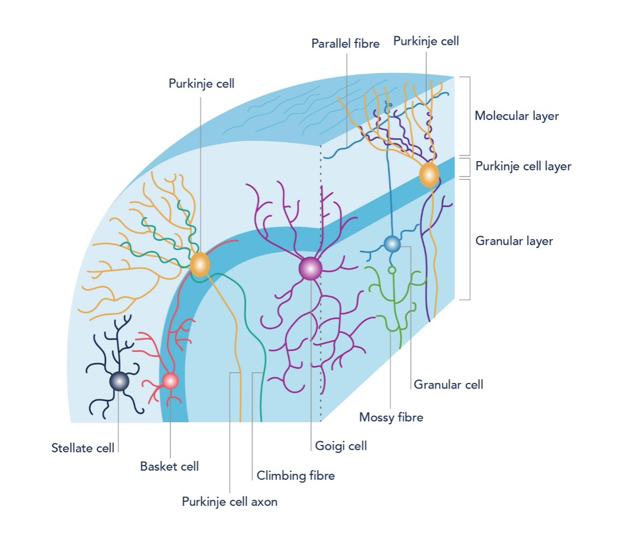
Figure 1: Schematic of cerebellar cortex circuitry and cellular composition
Neuronal Markers:
Calbindin
Calbindin is a well-established marker for Purkinje cells, the principal cerebellar cell type. These cells receive and integrate inputs from hundreds of thousands of other cells within and outside the cerebellum, forming the sole output of the cerebellar cortex. Calbindin is a calcium-binding protein that supports the rapid calcium buffering required for Purkinje cells’ high-frequency firing properties. Immunohistochemistry and immunofluorescence staining against calbindin labels both Purkinje cell bodies and their impressively elaborate dendritic trees.
Proteintech offers conjugated and unconjugated calbindin antibodies suitable for various applications in human, mouse, rat, pig and rabbit tissues.
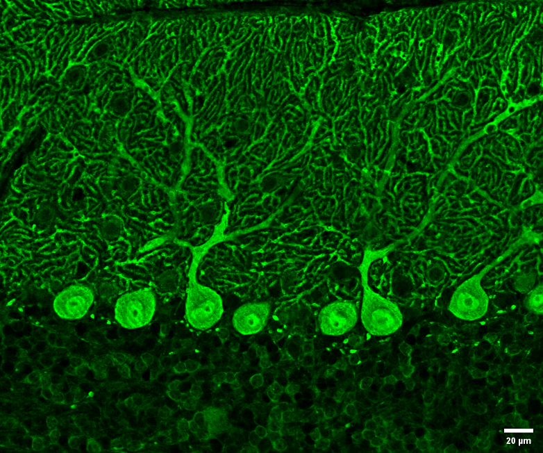
Figure 2: Immunofluorescent analysis of 4% PFA-fixed adult mouse cerebellum tissue using Calbindin-D28k antibody (14479-1-AP) at a dilution of 1:1500 and Alexa Fluor 594 AffiniPure Donkey Anti-Rabbit IgG (H+L).
Parvalbumin
Parvalbumin, another calcium binding protein, is a general marker of cerebellar neurons, labeling a subpopulation of Purkinje cells and molecular layer interneurons. Parvalbumin-positive Purkinje cells have distinct calcium homeostasis and intrinsic firing properties, which may render them selectively vulnerable in diseases. The labeled molecular layer interneurons can be further subcategorized into basket cells and stellate cells. Parvalbumin staining allows visualization of both populations.
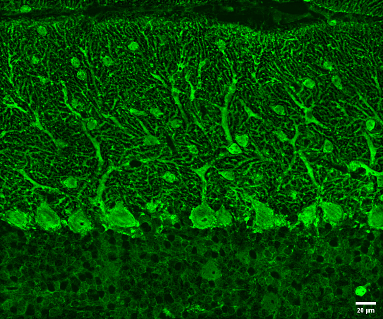
Figure 3: Immunofluorescent analysis of 4% PFA-fixed adult mouse cerebellum tissue using Parvalbumin antibody (29312-1-AP) at a dilution of 1:1500 and Alexa Fluor 594 AffiniPure Donkey Anti-Rabbit IgG (H+L).
Calretinin
Calretinin, a third calcium binding protein in the cerebellum, is commonly used to visualize climbing fibers, granule cells, and granule cell-originating parallel fibers. In certain regions of the cerebellum, it can also be used to label unipolar brush cells in the granule cell layer.
Proteintech offers monoclonal, polyclonal and recombinant calretinin antibodies across wide range of applications.
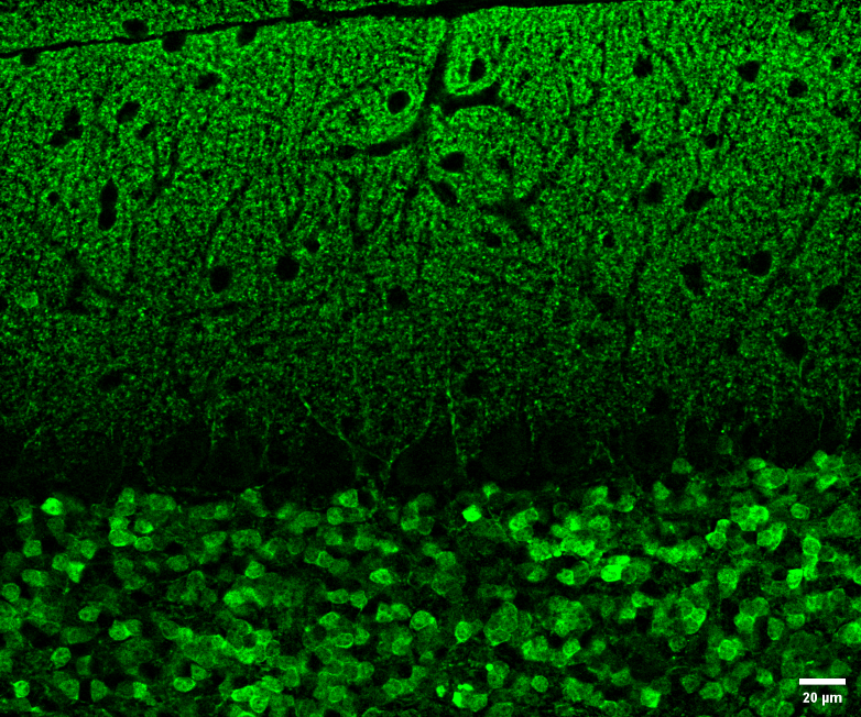
Figure 4: Immunofluorescent analysis of 4% PFA-fixed adult mouse cerebellum tissue using Calretinin antibody (82811-1-RR) at a dilution of 1:500 and Alexa Fluor 594 AffiniPure Donkey Anti-Rabbit IgG (H+L).
VGLUT1/VGLUT2
The vesicular glutamate transporters VGLUT1 and VGLUT2 are commonly used to label parallel fiber and climbing fiber terminals onto Purkinje cells, respectively. These two types of connections represent the main excitatory inputs onto Purkinje cells: parallel fiber inputs are numerous but small, while climbing fiber inputs are fewer but have large amplitudes. Immunostaining against VGLUT1 and VGLUT2 is used to study synaptic abundance and spatial distribution at the parallel fiber and climbing fiber to Purkinje cell synapses, respectively.
View all products for ‘VGLUT1’ and ‘VGLUT2.’
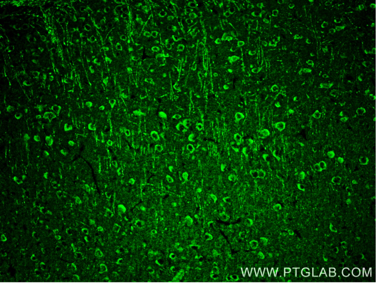
Figure 5: Immunofluorescent analysis of 4% PFA-fixed mouse brain tissue using VGLUT1 antibody (55491-1-AP) at a dilution of 1:200 and CoraLite 488-Conjugated Goat Anti-Rabbit IgG (H+L).
GAD67/GAD65/VGAT
To visualize GABAergic cells in the cerebellum, such as basket cells, stellate cells, Golgi cells, and deep cerebellar nuclei neurons, markers like GAD67 and GAD65 are often used. VGAT is used to label the GABAergic terminals.
View all products for ‘GAD67’, ‘GAD65’, and ‘VGAT.’
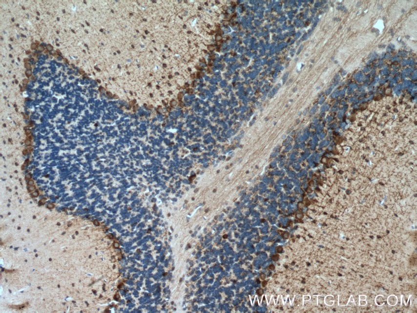
Figure 6: Immunohistochemical analysis of paraffin-embedded mouse brain tissue using GAD65 antibody (21760-1-AP) at a dilution of 1:200.
Aldoc
Despite the seemingly homogenous population of Purkinje cells across the whole cerebellum, they can be classified into many subpopulations based on their molecular identities. Aldolase C (Aldoc), also known as Zebrin, is a widely used marker for categorizing Purkinje cells. The Aldoc identity correlates with intrinsic firing properties and afferent innervation, with significant functional implications.

Figure 7: Immunohistochemical analysis of paraffin-embedded mouse brain tissue using Aldolase C antibody (14884-1-AP) at dilution of 1:200.
Glial Markers
GFAP/S100B
Bergmann glia cells, whose bodies are located in the Purkinje cell layer, extend processes to enwrap Purkinje cell synapses. GFAP is commonly used to stain Bergmann glia. In some pathological conditions, these glia become overactive and express higher level of S100B, which is used to stain activated Bergmann glia.
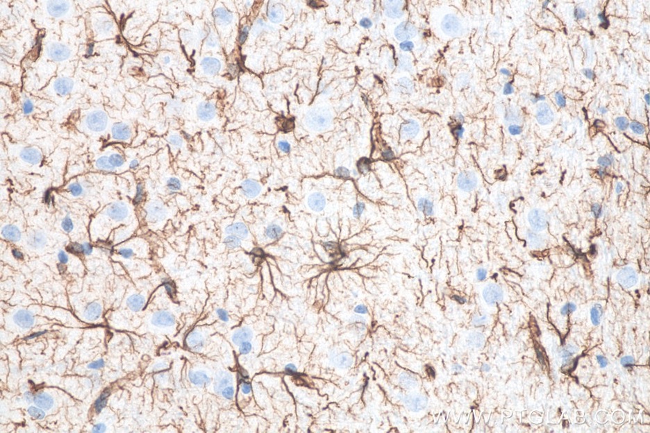
Figure 8: Immunohistochemical analysis of paraffin-embedded rat brain tissue using GFAP IHC Kit (KHC0002).
IBA1
Microglia, which exhibit regional heterogeneity in morphology, density and molecular identity, play key roles in cerebellar circuit development. IBA1 is used to stain microglial populations in the cerebellum.
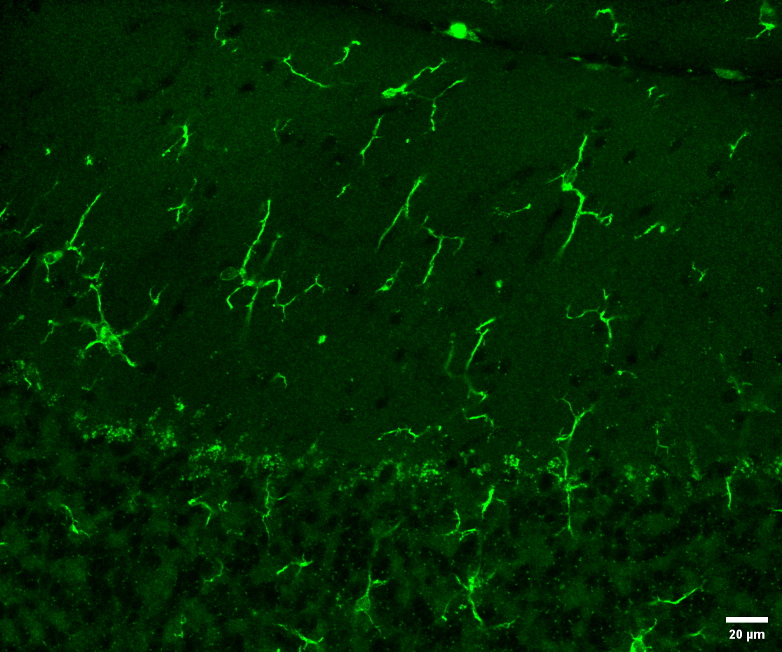
Figure 9: Immunofluorescent analysis of 4% PFA-fixed adult mouse cerebellum tissue using IBA1 antibody (81728-1-RR) at a dilution of 1:500 and Alexa Fluor 594 AffiniPure Donkey Anti-Rabbit IgG (H+L). Heat mediated antigen retrieval with PBS buffer (pH 7.4).
EAAT1/GLAST
Glial cells play a crucial role in preventing excitotoxicity by removing glutamate from the extracellular space. In the cerebellum, Bergmann glia remove glutamate through the excitatory amino acid transporter 1 (EAAT1/GLAST), making it a wildly used marker for glutamate signalling.
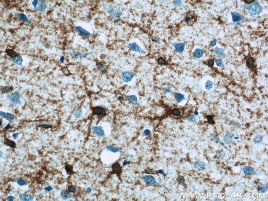
Figure 10: Immunohistochemical analysis of paraffin-embedded rat brain tissue using GLAST antibody (20786-1-AP) at a dilution of 1:50.
Discovery of new cell-specific markers
Recent advances in single-cell sequencing technology have enabled the molecular delineation of cerebellar cells across developmental milestones, leading to the discovery of new cell-type-specific markers. Proteintech’s portfolio continues to expand to meet the growing demand for new cerebellar markers.
Emerging cerebellar markers
|
Cell type |
Specific Markers |
Cat No. |
|
Purkinje cell |
GLUD2 ITPKA |
|
|
Molecular layer interneurons |
Kcna2 (basket cell specific) |
|
|
Granule cells |
ZIC1 |
Reference
Brandenburg, C., Smith, L. A., Kilander, M. B., Bridi, M. S., Lin, Y., Huang, S., & Blatt, G. J. (2021). Parvalbumin subtypes of cerebellar Purkinje cells contribute to differential intrinsic firing properties. Molecular and Cellular Neuroscience, 115, 103650. https://doi.org/10.1016/j.mcn.2021.103650
Nakayama, H., Abe, M., Morimoto, C., Iida, T., Okabe, S., Sakimura, K., & Hashimoto, K. (2018). Microglia permit climbing fiber elimination by promoting GABAergic inhibition in the developing cerebellum. Nature Communications, 9(1). https://doi.org/10.1038/s41467-018-05100-z
Tam, W. Y., Wang, X., Cheng, A. S. K., & Cheung, K. (2021). In search of molecular markers for cerebellar neurons. International Journal of Molecular Sciences, 22(4), 1850. https://doi.org/10.3390/ijms22041850
Related Content
The Complete Guide to Optimizing Immunofluorescence Staining
7 Tips to successfully culture primary rodent neurons
FlexAble: The Latest Technology in Antibody Labeling
How to choose the right secondary antibody?
Support
Newsletter Signup
Stay up-to-date with our latest news and events. New to Proteintech? Get 10% off your first order when you sign up.

