- Phare
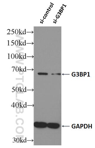
- Validé par KD/KO
Anticorps Polyclonal de lapin anti-G3BP1
G3BP1 Polyclonal Antibody for WB, IP, IF, IHC, ELISA, FC (Intra)
Hôte / Isotype
Lapin / IgG
Réactivité testée
Humain, rat, souris et plus (3)
Applications
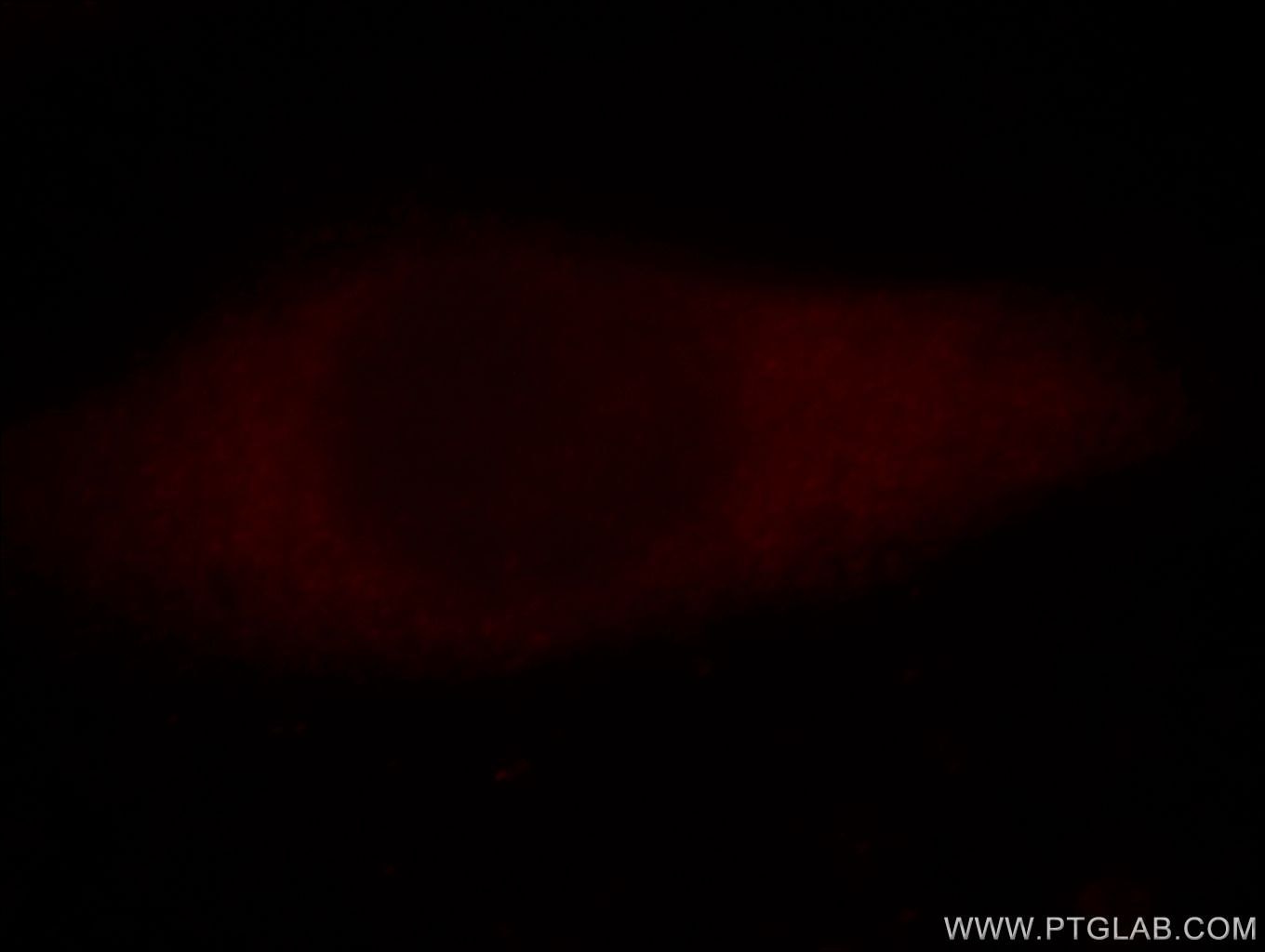
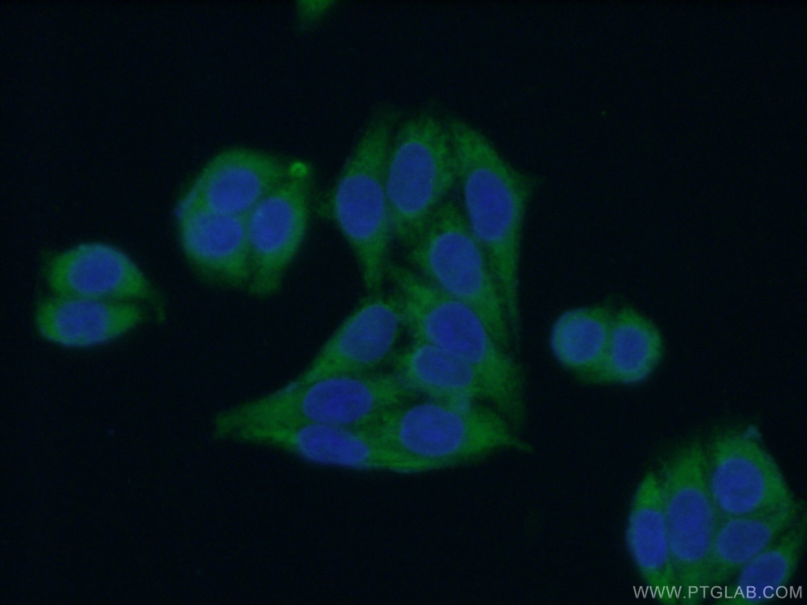
WB, IHC, IF/ICC, FC (Intra), IP, CoIP, RIP, ELISA
Conjugaison
Non conjugué
167
N° de cat : 13057-2-AP
Synonymes
Galerie de données de validation
Applications testées
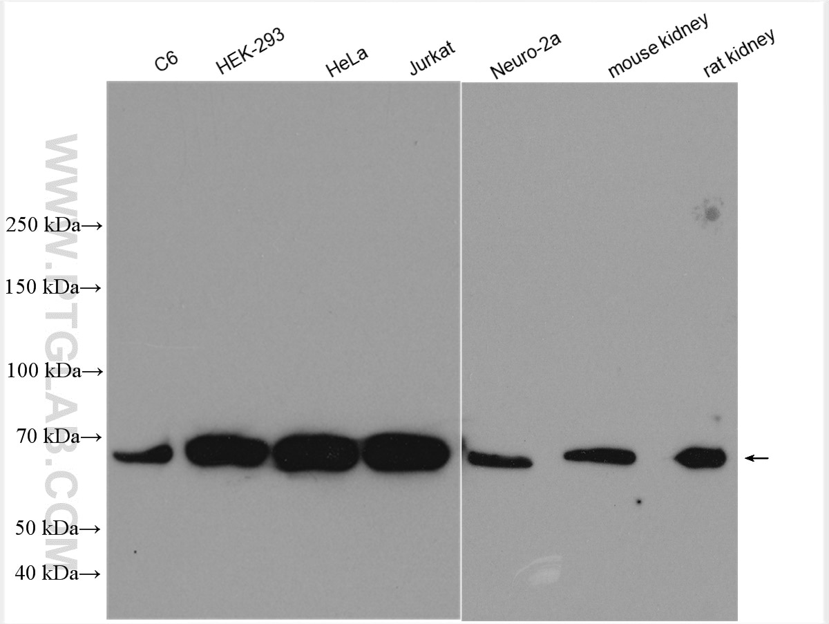
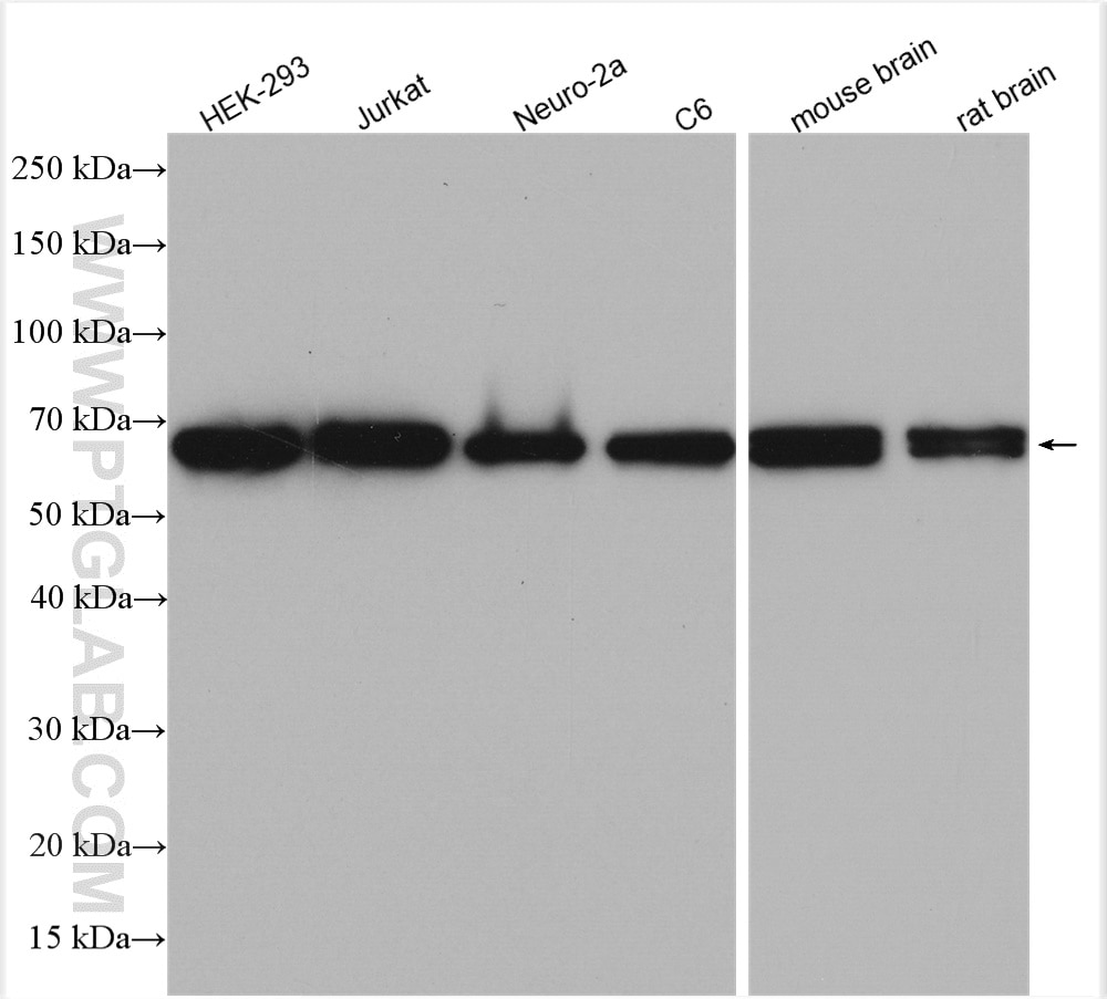
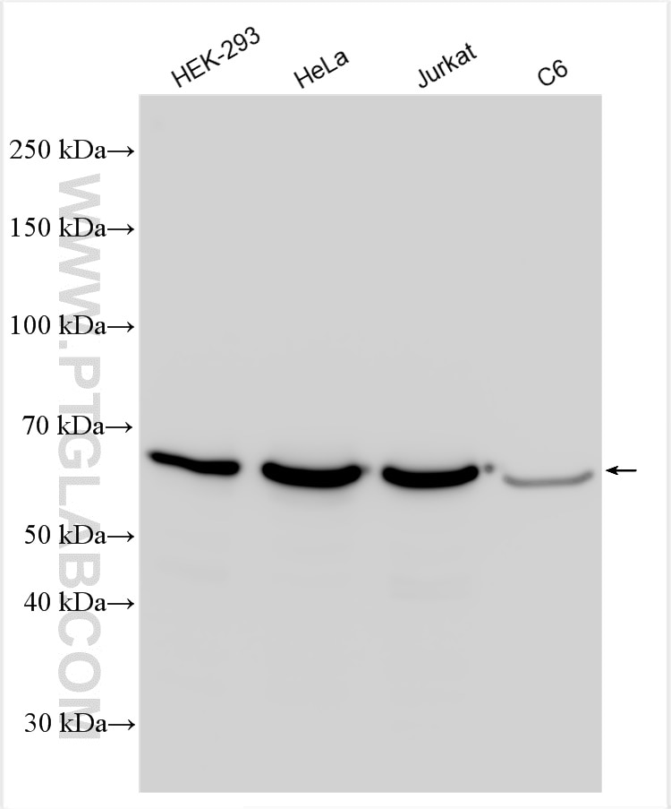
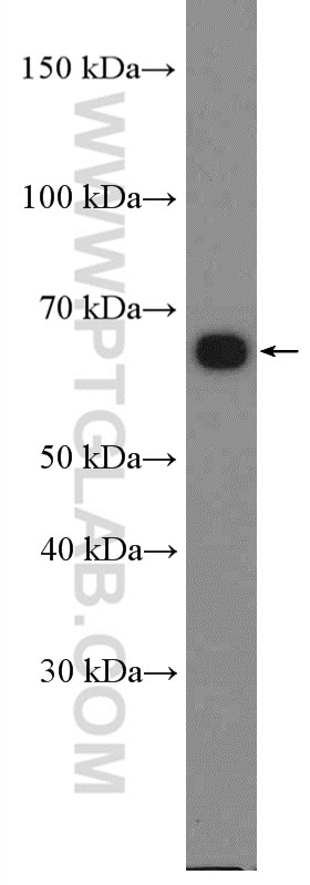
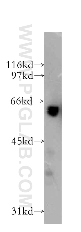
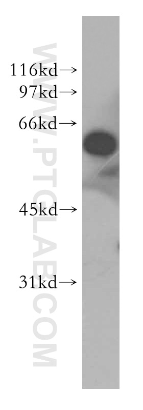
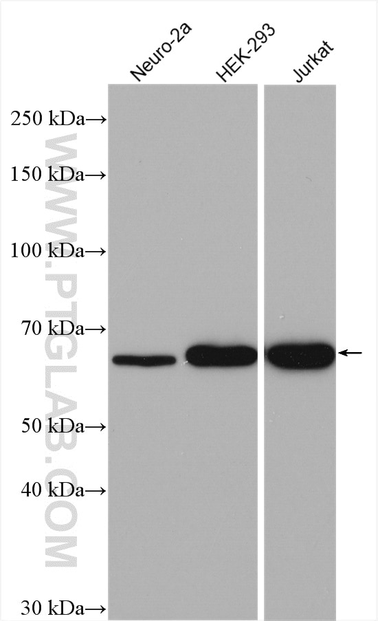
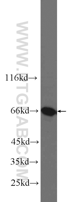
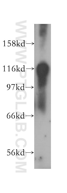
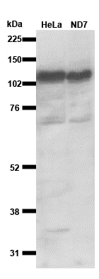
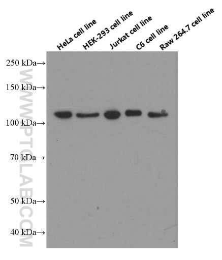
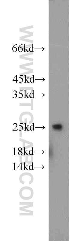
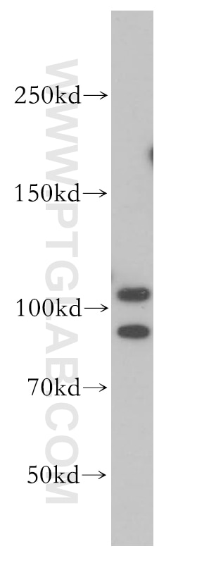
| Résultats positifs en WB | cellules C6, cellules HEK-293, cellules HeLa, cellules HepG2, cellules Jurkat, cellules MCF-7, cellules Neuro-2a, tissu cérébral de rat, tissu cérébral de souris, tissu cérébral humain, tissu rénal de rat, tissu rénal de souris |
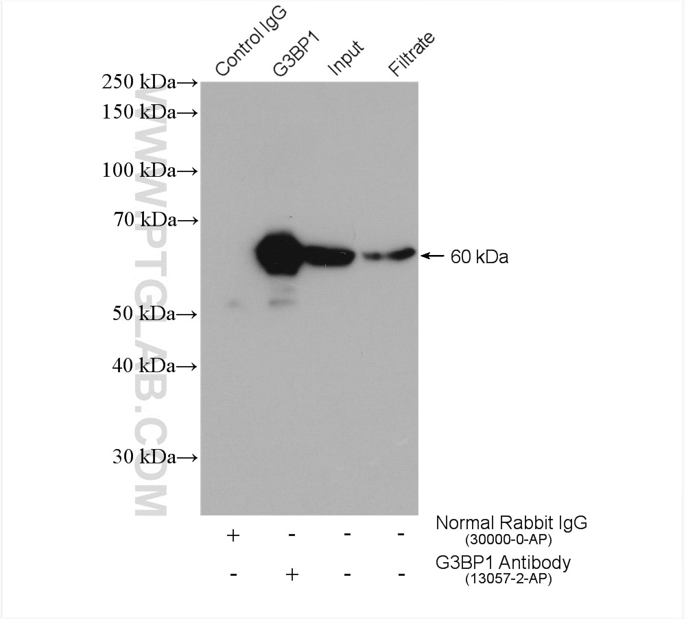
| Résultats positifs en IP | cellules HEK-293, |
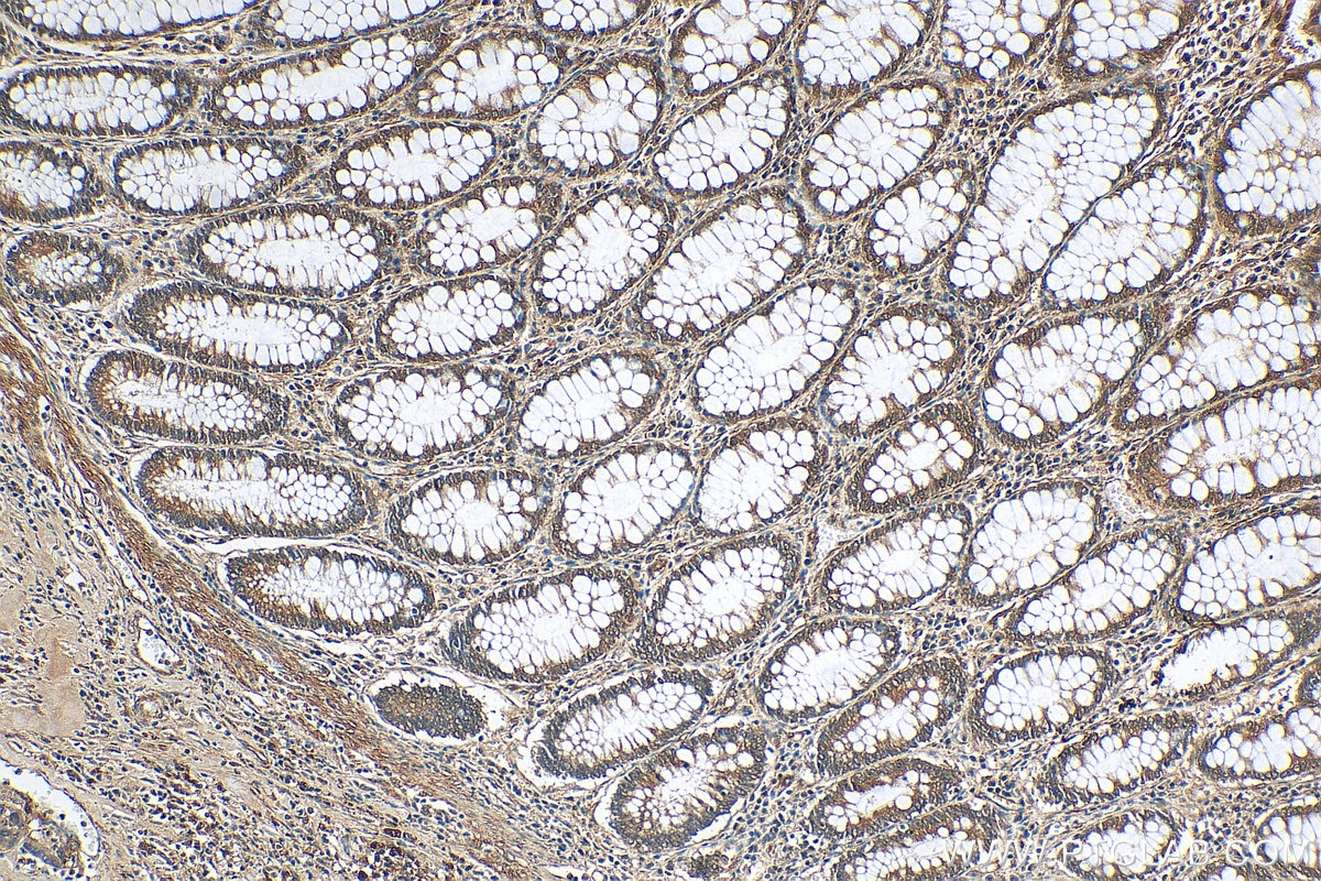
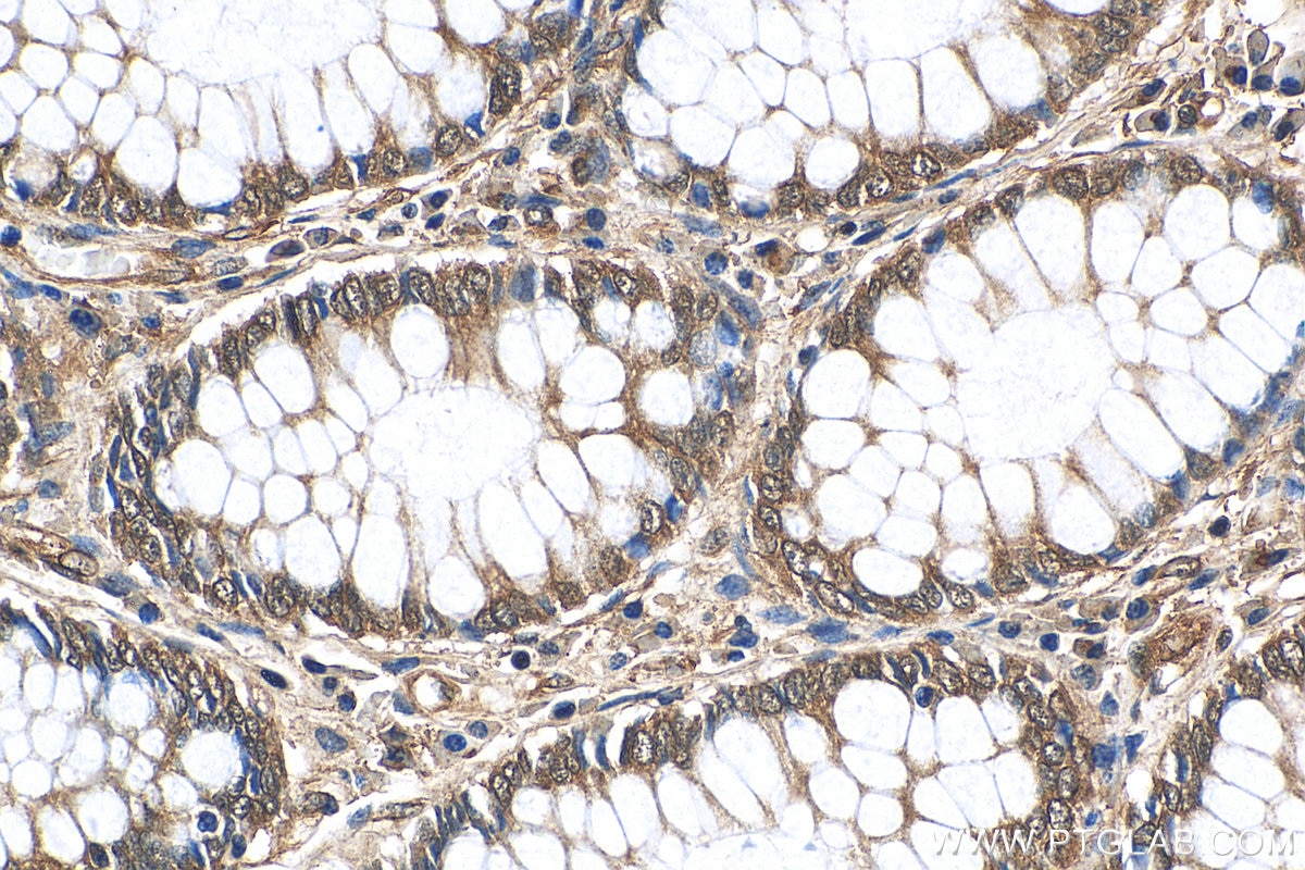
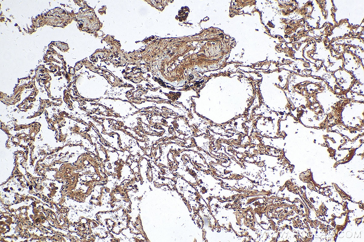
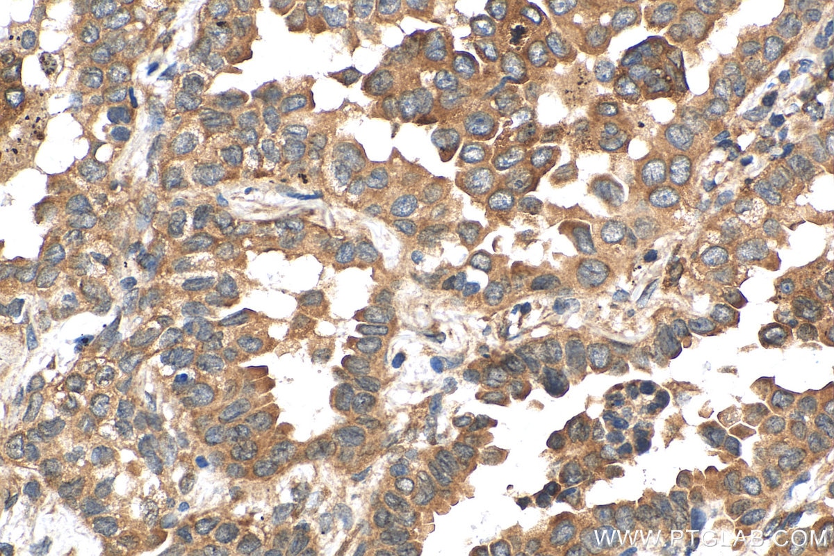
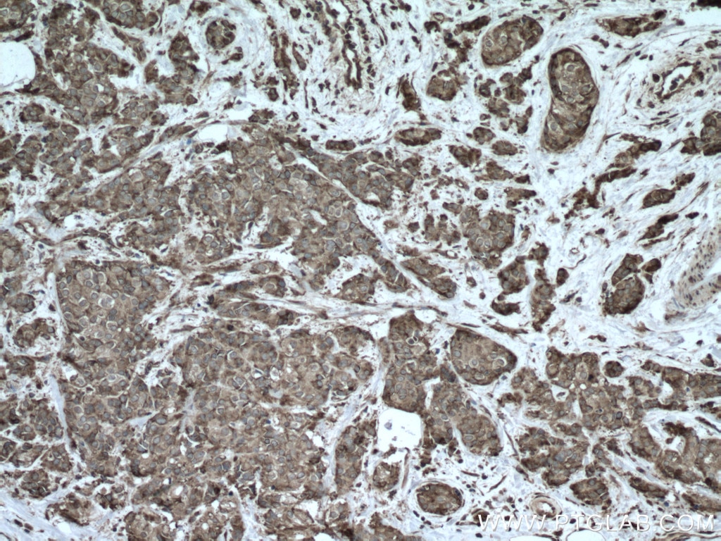
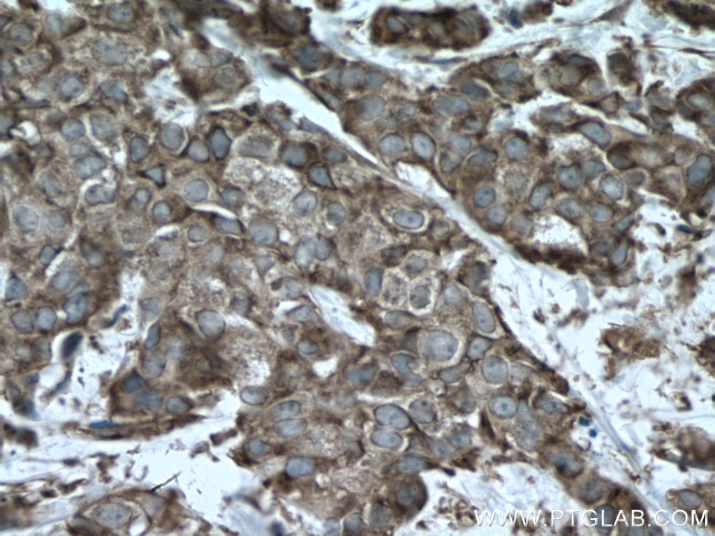
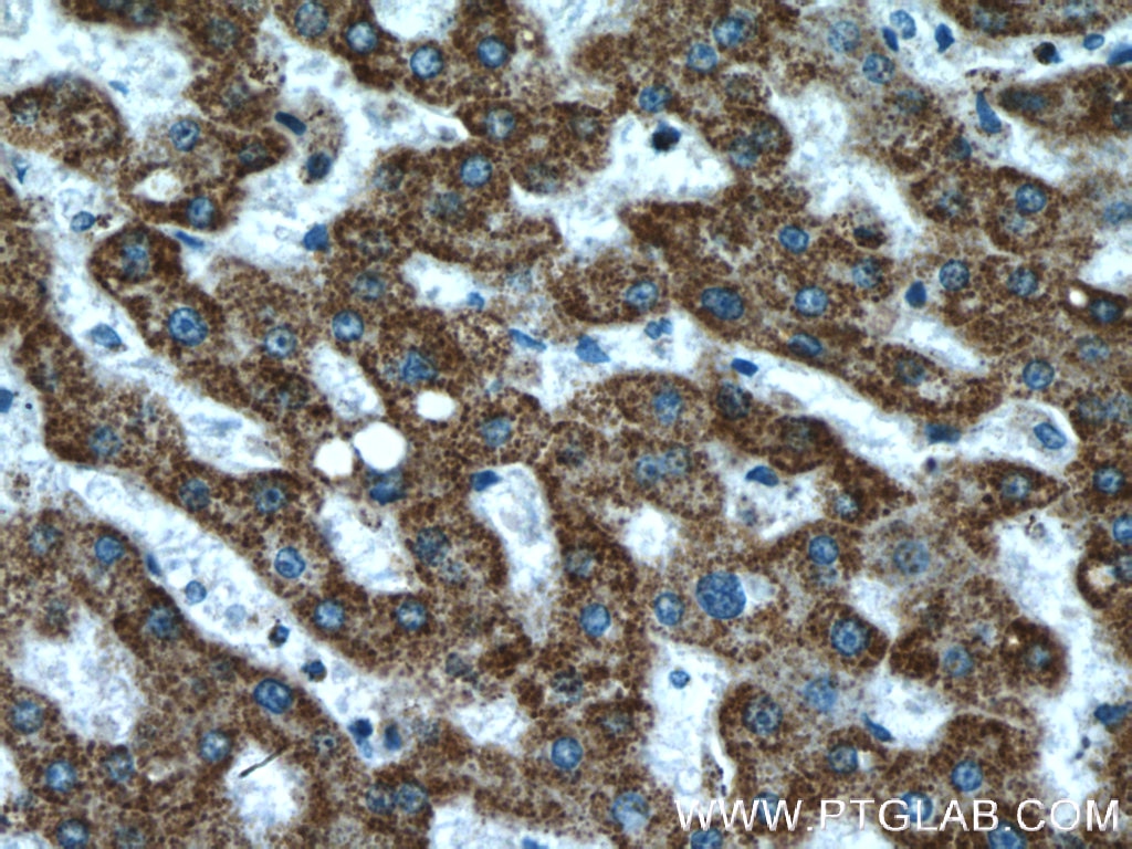
| Résultats positifs en IHC | tissu de cancer du côlon humain, tissu de cancer du poumon humain, tissu de cancer du sein humain il est suggéré de démasquer l'antigène avec un tampon de TE buffer pH 9.0; (*) À défaut, 'le démasquage de l'antigène peut être 'effectué avec un tampon citrate pH 6,0. |
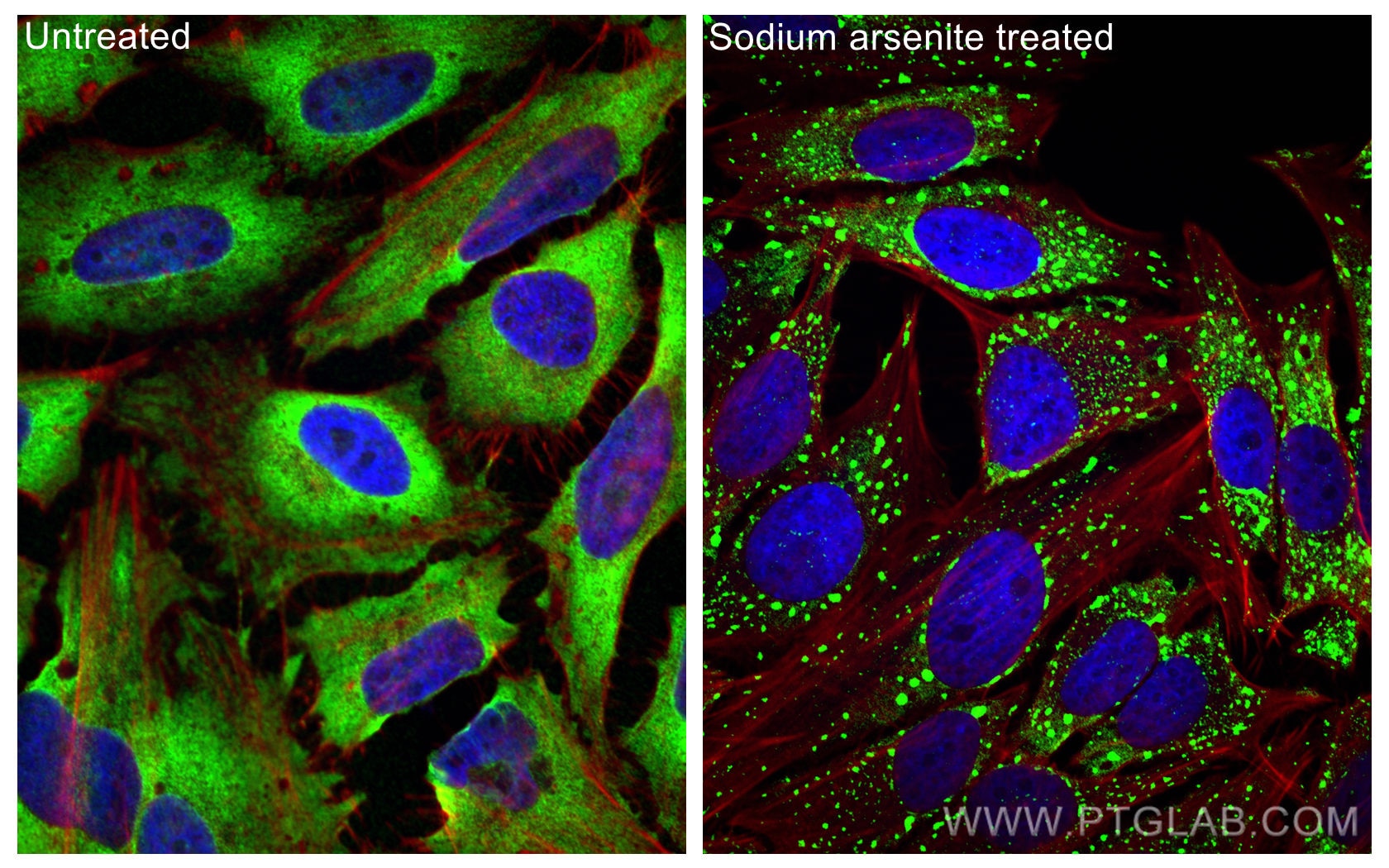
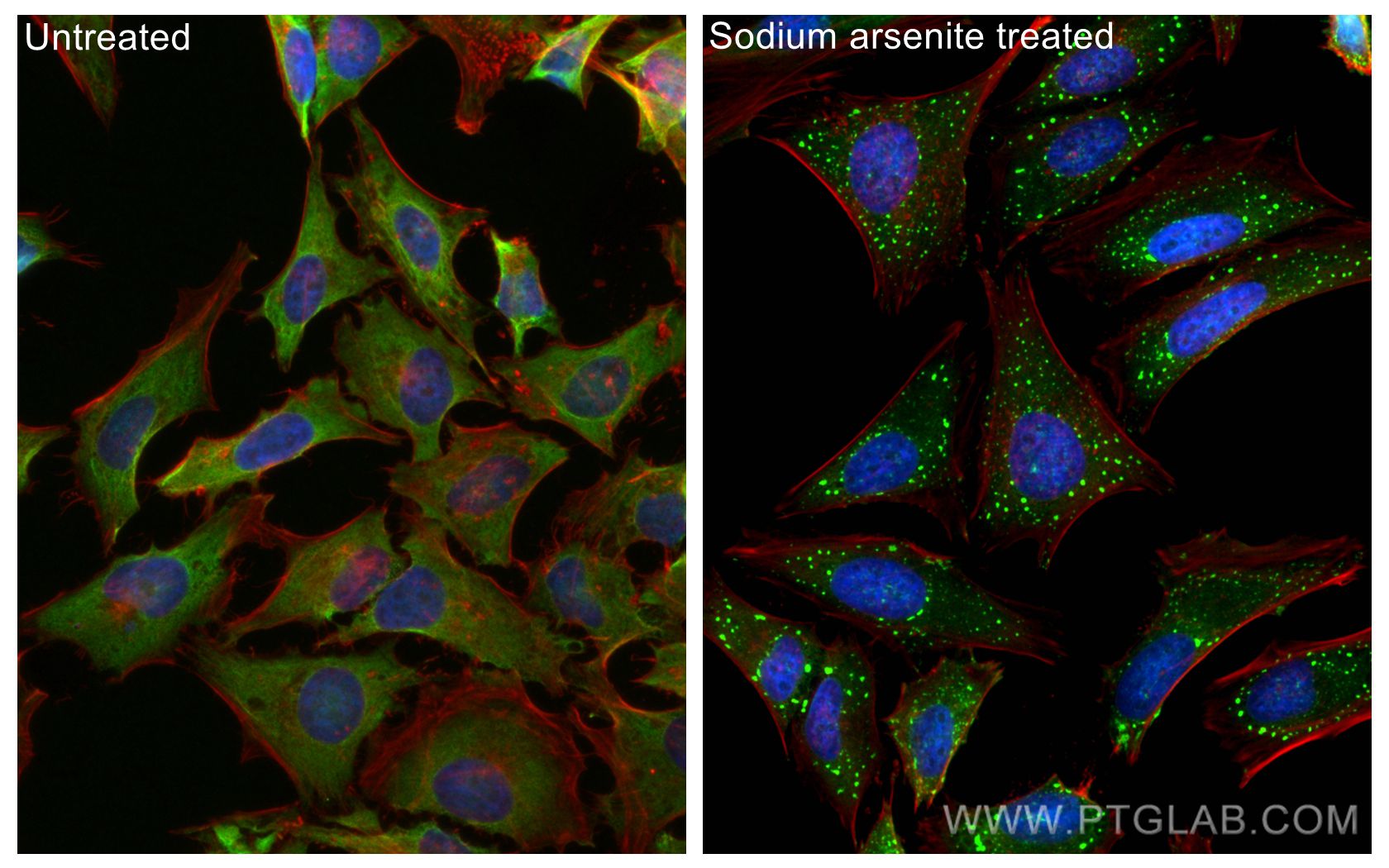
| Résultats positifs en IF/ICC | sodium arsenite treated HeLa cells, |
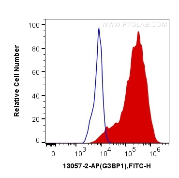
| Résultats positifs en FC (Intra) | cellules HeLa |
| Résultats positifs en cytométrie | cellules HeLa |
Dilution recommandée
| Application | Dilution |
|---|---|
| Western Blot (WB) | WB : 1:2000-1:16000 |
| Immunoprécipitation (IP) | IP : 0.5-4.0 ug for 1.0-3.0 mg of total protein lysate |
| Immunohistochimie (IHC) | IHC : 1:200-1:800 |
| Immunofluorescence (IF)/ICC | IF/ICC : 1:1000-1:4000 |
| Flow Cytometry (FC) (INTRA) | FC (INTRA) : 0.20 ug per 10^6 cells in a 100 µl suspension |
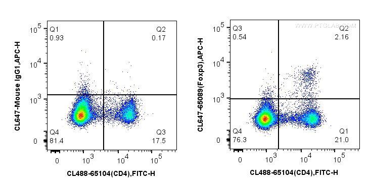
| Flow Cytometry (FC) | FC : 0.20 ug per 10^6 cells in a 100 µl suspension |
| It is recommended that this reagent should be titrated in each testing system to obtain optimal results. | |
| Sample-dependent, check data in validation data gallery | |
Informations sur le produit
13057-2-AP cible G3BP1 dans les applications de WB, IHC, IF/ICC, FC (Intra), IP, CoIP, RIP, ELISA et montre une réactivité avec des échantillons Humain, rat, souris
| Réactivité | Humain, rat, souris |
| Réactivité citée | rat, Humain, porc, poulet, singe, souris |
| Hôte / Isotype | Lapin / IgG |
| Clonalité | Polyclonal |
| Type | Anticorps |
| Immunogène | G3BP1 Protéine recombinante Ag3728 |
| Nom complet | GTPase activating protein (SH3 domain) binding protein 1 |
| Masse moléculaire calculée | 466 aa, 52 kDa |
| Poids moléculaire observé | 68 kDa |
| Numéro d’acquisition GenBank | BC006997 |
| Symbole du gène | G3BP1 |
| Identification du gène (NCBI) | 10146 |
| Conjugaison | Non conjugué |
| Forme | Liquide |
| Méthode de purification | Purification par affinité contre l'antigène |
| Tampon de stockage | PBS avec azoture de sodium à 0,02 % et glycérol à 50 % pH 7,3 |
| Conditions de stockage | Stocker à -20°C. Stable pendant un an après l'expédition. L'aliquotage n'est pas nécessaire pour le stockage à -20oC Les 20ul contiennent 0,1% de BSA. |
Informations générales
GAP SH3 Binding Protein 1 (G3BP1), also named as G3BP, is an effector of stress granule (SG) assembly. SG biology plays an important role in the pathophysiology of TDP-43 in ALS and FTLD-U. G3BP1 can be used as a marker of SG. It has been shown to function downstream of Ras and play a role in RNA metabolism, signal transduction, and proliferation. G3BP1 is a ubiquitously expressed protein that localizes to the cytoplasm in proliferating cells and to the nucleus in non-proliferating cells. G3BP1 has recently been implicated in cancer biology.
Protocole
| Product Specific Protocols | |
|---|---|
| WB protocol for G3BP1 antibody 13057-2-AP | Download protocol |
| IHC protocol for G3BP1 antibody 13057-2-AP | Download protocol |
| IF protocol for G3BP1 antibody 13057-2-AP | Download protocol |
| IP protocol for G3BP1 antibody 13057-2-AP | Download protocol |
| FC protocol for G3BP1 antibody 13057-2-AP | Download protocol |
| Standard Protocols | |
|---|---|
| Click here to view our Standard Protocols |
Publications
| Species | Application | Title |
|---|---|---|
Cell Diverse CMT2 neuropathies are linked to aberrant G3BP interactions in stress granules | ||
Science Ubiquitination of G3BP1 mediates stress granule disassembly in a context-specific manner. | ||
Cell ELAVL4, splicing, and glutamatergic dysfunction precede neuron loss in MAPT mutation cerebral organoids. | ||
Cell RNA Granules Hitchhike on Lysosomes for Long-Distance Transport, Using Annexin A11 as a Molecular Tether. | ||
Cell Phase Separation of FUS Is Suppressed by Its Nuclear Import Receptor and Arginine Methylation. |
Avis
The reviews below have been submitted by verified Proteintech customers who received an incentive forproviding their feedback.
FH Roy (Verified Customer) (06-12-2024) | Works great on WB (1/1000 - Overnight 4°C) - Very beautiful IF staining in non stressed (diffuse staining) and Sodium Arsenite-mediated Stress induction (Punctate staining corresponding to stress granules) in HeLa cells (1/250 - 1h at RT).
|
FH Vinny (Verified Customer) (02-20-2024) | Good product.
|
FH Andrea (Verified Customer) (10-05-2023) | Good and strong signal.
|
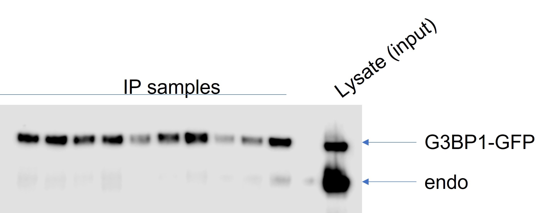
FH Tatyana (Verified Customer) (01-21-2023) | Used for IP and WB of GFP-tagged overexpressed human G3BP1 (HEK293 cells). Lysates were subjected to IP using GFP-Trap beads. WB was done using semi-dry transfer. Antibody was used in 4% milk in TBST at 1:1,000 dilution. It could be reused up to 3 times. As the image shows, the antibody can successfully detect both endogenous and OE protein.
 |
FH Tobias (Verified Customer) (10-25-2022) |
|
FH Peter (Verified Customer) (10-24-2022) | Best G3BP1 antibody I've used for Western, IF and IP
|
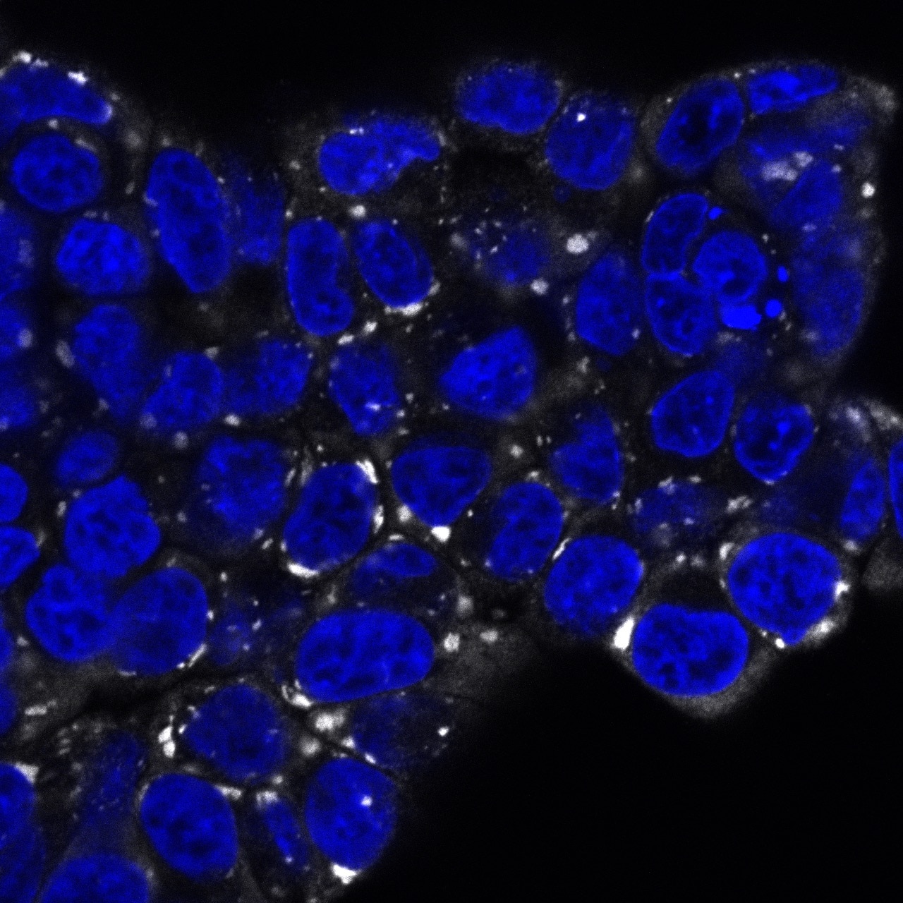
FH Patryk (Verified Customer) (03-18-2021) | I used the antibody for immunofluorescence imaging to label stress granules induced by treating the cells with with 50µM sodium arsenite. Used the antibody at a dilution 1:100 overnight at 4°C. Worked perfectly well, strong and specific signal. I am very satisfied of this antibody and strongly recommend if for immunofluorescence.
 |
FH Kun (Verified Customer) (03-23-2020) | Very specific and sensitive
|
FH Biao (Verified Customer) (03-11-2020) | This antibody is very specific and good quality.
|
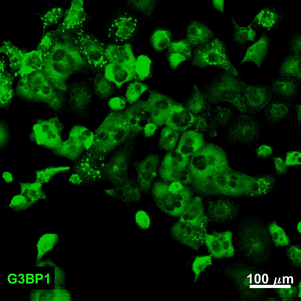
FH Joshua (Verified Customer) (12-28-2019) | PANC1 cells fixed in 4% paraformaldehdye. Bright localization to stress granules.
 |
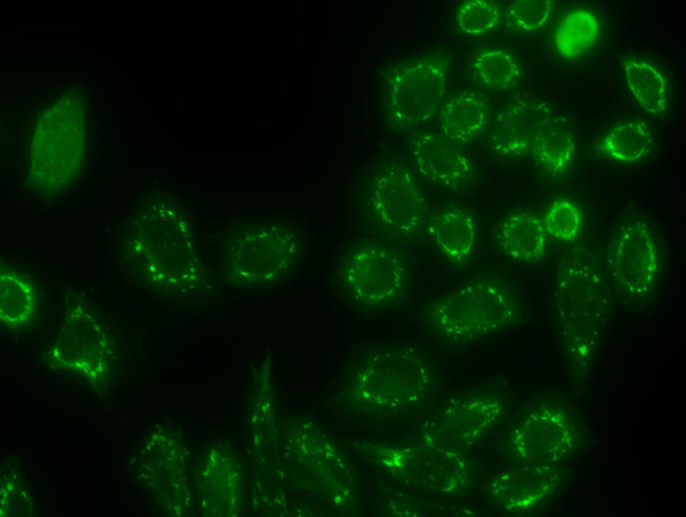
FH Yuan (Verified Customer) (11-02-2019) | Very bright staining for stress granule on NaAsO2 treated Hela cells. 1:500 should be sufficient for IF staining.
 |
FH Zeinab (Verified Customer) (08-19-2019) | It worked great
|
FH Erica (Verified Customer) (05-15-2019) | Our lab has been using this antibody for IP, WB and IF for many years and it always worked well. I highly recommend this antibody, especially for IP for stress granules.
|
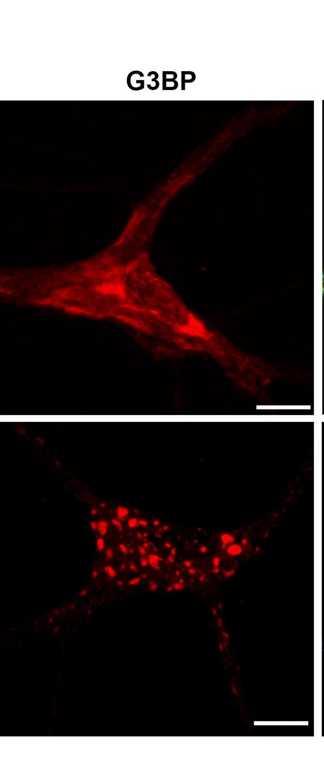
FH Karthik (Verified Customer) (04-24-2019) | Upon induction of sodium arsenite stress in neurons G3BP1 positive stress granules formed in 90 minutes.Cells permeabilized with 0.2% triton for 10 minutes
 |
FH Tian (Verified Customer) (01-23-2019) | I used it for ICC and it worked Great.
|
