Actin Chromobody: Visualization of actin dynamics – NOT alteration
The ChromoTek Actin Chromobody is the only probe that does not interfere with actin filament dynamics.
Everybody really wants to use a detection probe that does not influence its target. For measurement of actin and actin-related functions there are a number of actin visualization probes and techniques available that researchers can select. Hence, one should only apply such actin visualization probes, which do not alter actin dynamics. How do the actin probes for visualization perform? Which probes do interfere with function and which probes do not?
Robert Grosse and his team from the Institute of Pharmacology at the Biochemical-Pharmacological Center (BPC), University of Marburg, Germany took the effort and systematically looked into detection technologies of actin filaments in order to avoid potential pitfalls. In the recently published mini review/poster “Actin Visualization at a Glance” (Melak et al. 2017) they have discussed the pros and cons of current probes to visualize actin filaments, i.e. Phalloidin, LifeAct, Utrophin, F-tractin, SiR-actin, GFP-actin, tagged actin, and Actin Chromobody® (see table 1 and figure 1 below). Grosse’s lab considers only the Actin Chromobody to have minimal effect on actin dynamics. All other presented probes are suspected to alter actin dynamics. Obviously genetic constructs, i.e. actin fusion proteins don’t disturb actin dynamics neither, but have other disadvantages. Hence, if actin probes are used, which do actually interfere with polymerization and depolymerization of the actin skeleton, they should be handled with great care
Table 1: Overview of stte-of-the-art technologies/probes to visualize actin filaments
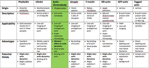
Adopted and modified from Melak et al. (2017). Product names may be covered by registered trademarks of corresponding corporations.
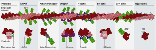
Figure 1: Schematics of actin binding/visualization sites by probe/technology (from Melak et al. (2017)
The Actin Chromobody is an intracellular anti-actin single chain antibody (VHH or so-called “nanobody” from alpaca), which is genetically fused to green or red fluorescent proteins, for real time analysis of endogenous actin in live cells. Carefully balanced on and off rates of this internal anti-actin VHH, its binding and dissociation from the actin epitope, prevent from interfering with actin dynamics. These make the Actin Chromobody an ideal tool for live cell imaging respectively in vivo analysis of endogenous proteins, i.e. actin.
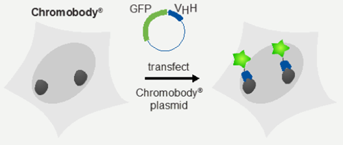
Two types of Actin Chromobodies are available:
one to visualize cytosolic actin filaments conjugated to TagGFP2 or TagRFP, Actin-Chromobody® plasmid (TagGFP2) and Actin-Chromobody® plasmid (TagRFP), and one to visualize nuclear actin filaments, Nuclear Actin-Chromobody® plasmid (TagGFP).
Selected Literature:
Melak, M., Plessner, M. and Grosse, R. 2017. ‘Actin Visualization at a glance’, J Cell Sci 130: 525-530: doi:10.1242/jcs.189068.
Danzl, Johann G., Sven C. Sidenstein, Carola Gregor, Nicolai T. Urban, Peter Ilgen, Stefan Jakobs, and Stefan W. Hell. 2016. 'Coordinate-targeted fluorescence nanoscopy with multiple off states', Nat Photon, 10: 122-28.
Panza, P., J. Maier, C. Schmees, U. Rothbauer, and C. Sollner. 2015. 'Live imaging of endogenous protein dynamics in zebrafish using chromobodies', Development, 142: 1879-84.
Rocchetti, A., C. Hawes, and V. Kriechbaumer. 2014. 'Fluorescent labelling of the actin cytoskeleton in plants using a cameloid antibody', Plant Methods, 10: 12.
Related Content
Nanobody-based reagents by ChromoTek
Intracellular Nanobodies for live-cell analysis in real-time
Chromobodies for live cell imaging
Actin Chromobody for live-cell super-resolution imaging
Visualize Histones in live cells: Histone-Chromobody
Visualize the onset of metastasis with Chromobodies
Novel cell cycle modulators identified using the Cell Cycle Chromobody Technology

Support
Newsletter Signup
Stay up-to-date with our latest news and events. New to Proteintech? Get 10% off your first order when you sign up.