Anticorps Polyclonal de lapin anti-Synaptophysin
Synaptophysin Polyclonal Antibody for WB, IHC, IF/ICC, IF-P, IP, ELISA
Hôte / Isotype
Lapin / IgG
Réactivité testée
Humain, rat, souris et plus (2)
Applications
WB, IHC, IF/ICC, IF-P, IP, ELISA
Conjugaison
Non conjugué
173
N° de cat : 17785-1-AP
Synonymes
Galerie de données de validation
Applications testées
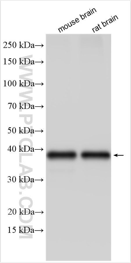
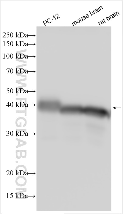
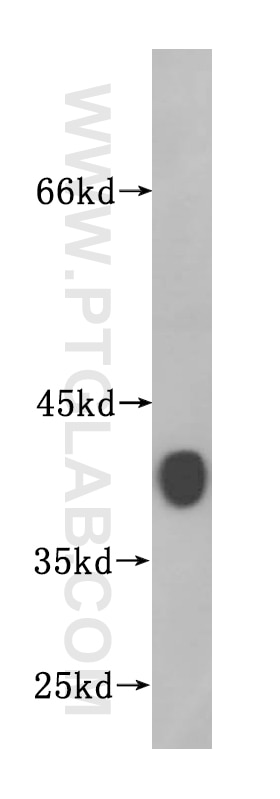
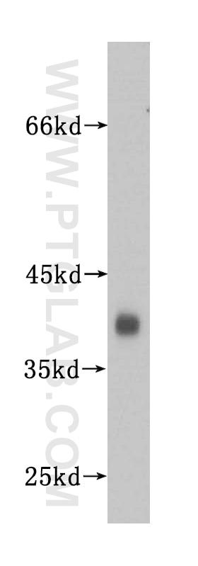
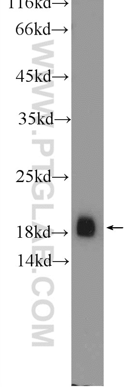
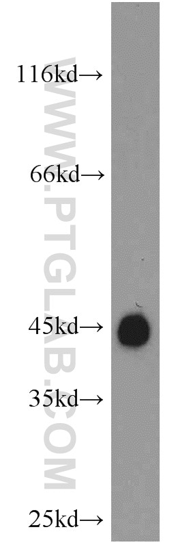
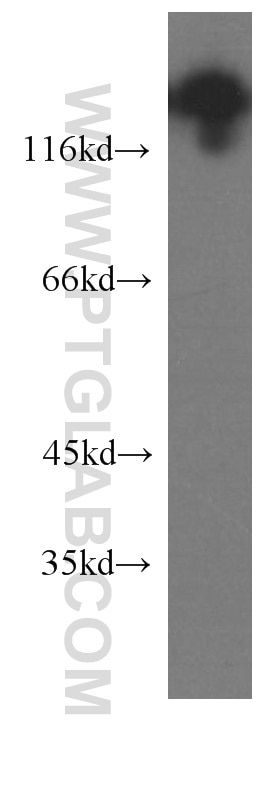
| Résultats positifs en WB | tissu cérébral de souris, cellules PC-12, tissu cérébral de rat, tissu cérébral humain, tissu de cervelet de souris |
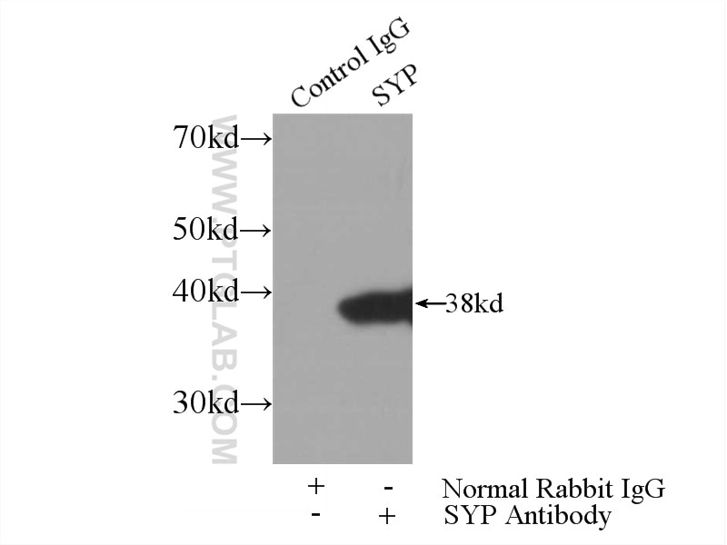
| Résultats positifs en IP | tissu cérébral de souris |
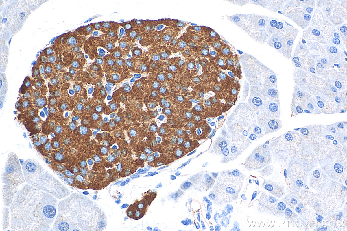
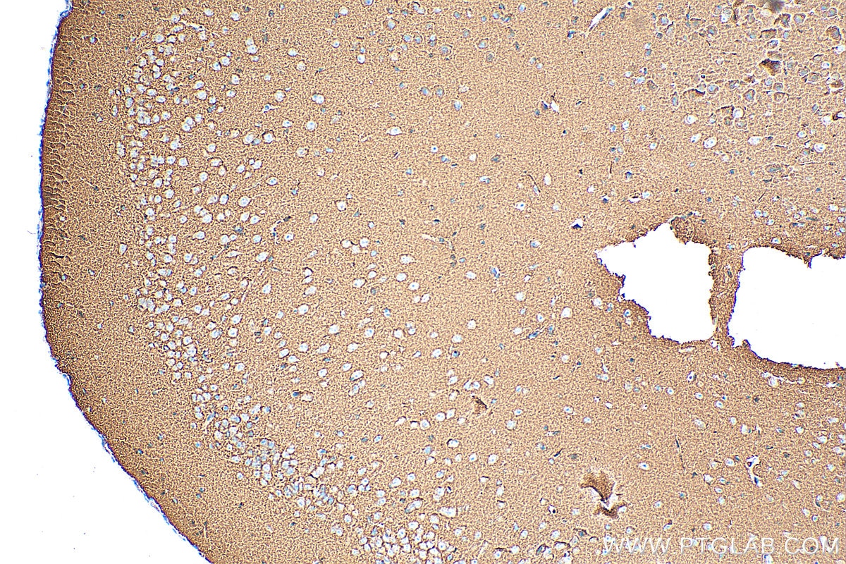
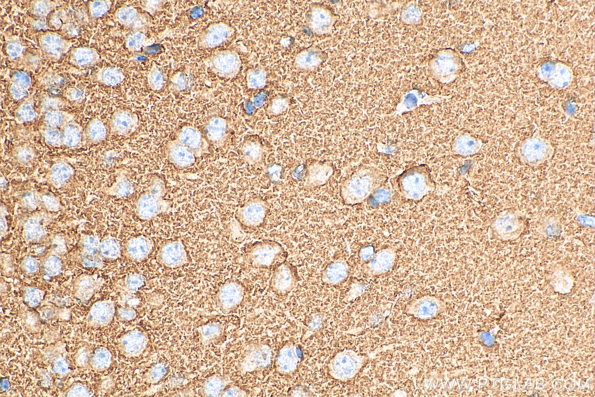
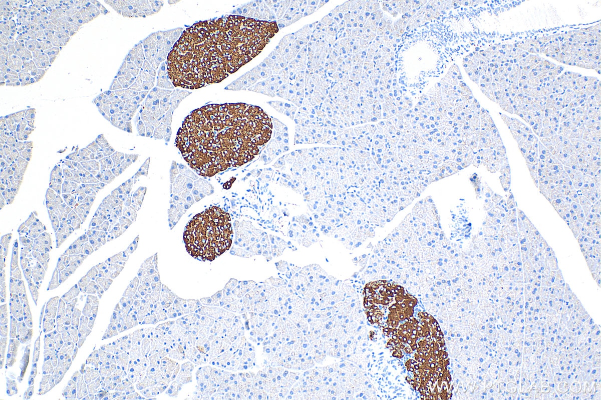
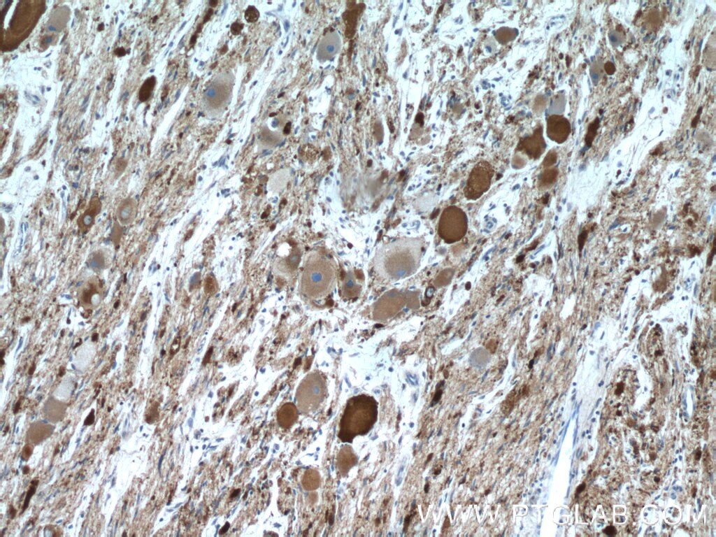
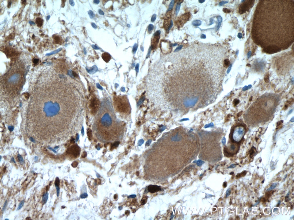
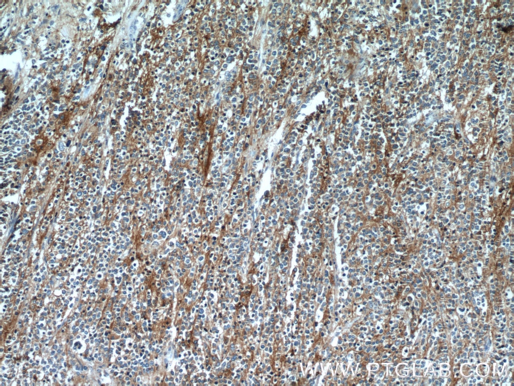
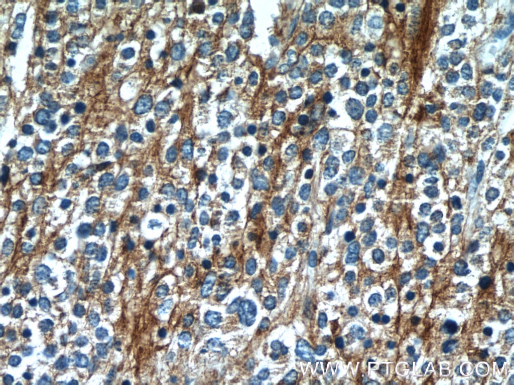
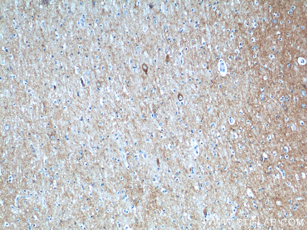
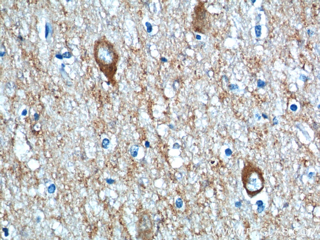
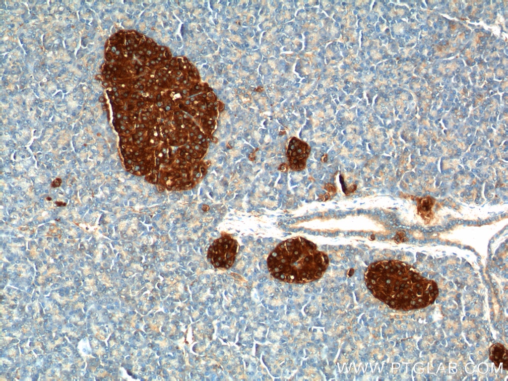
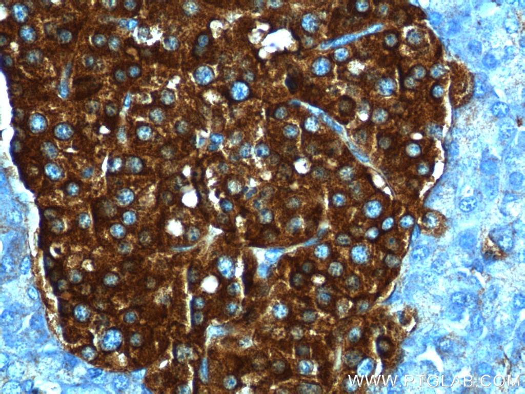
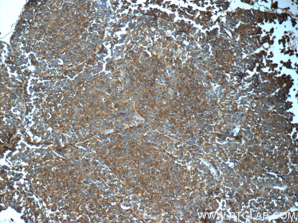
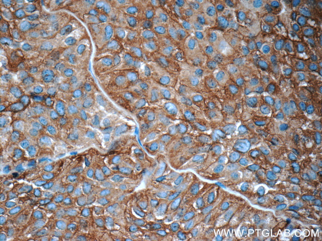
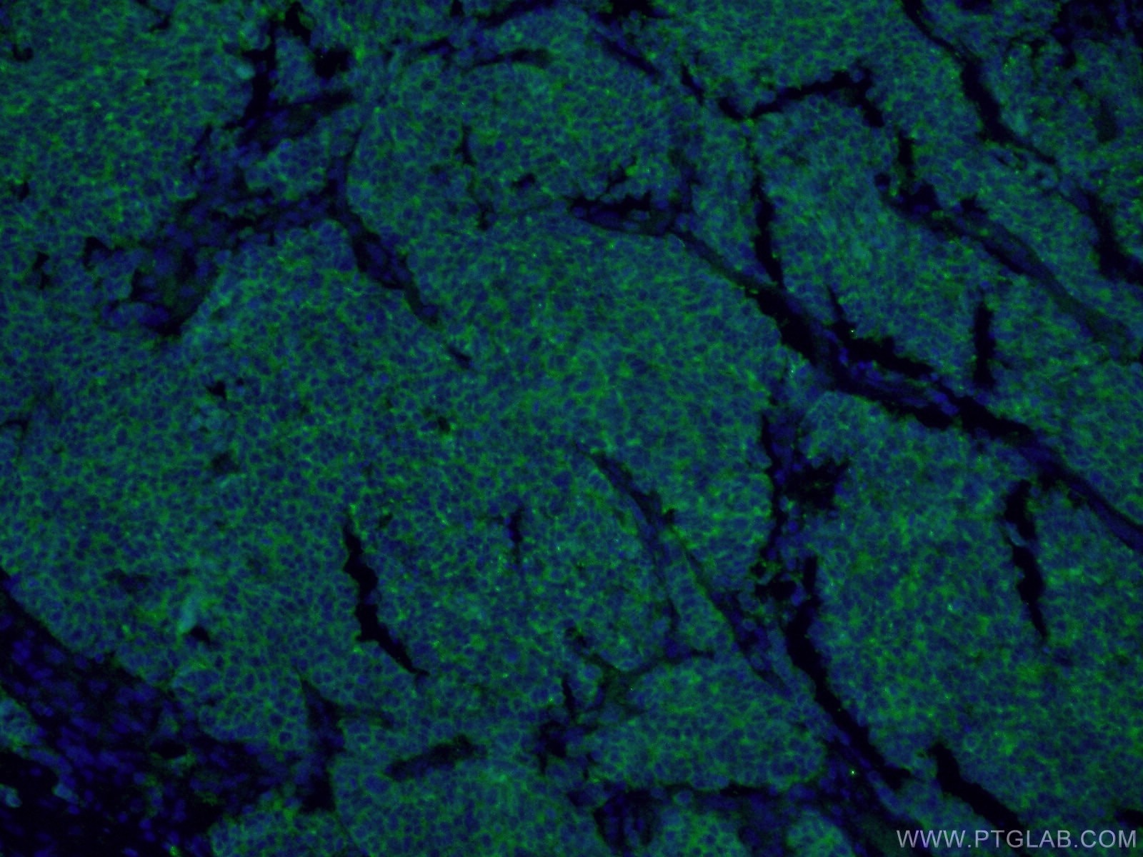
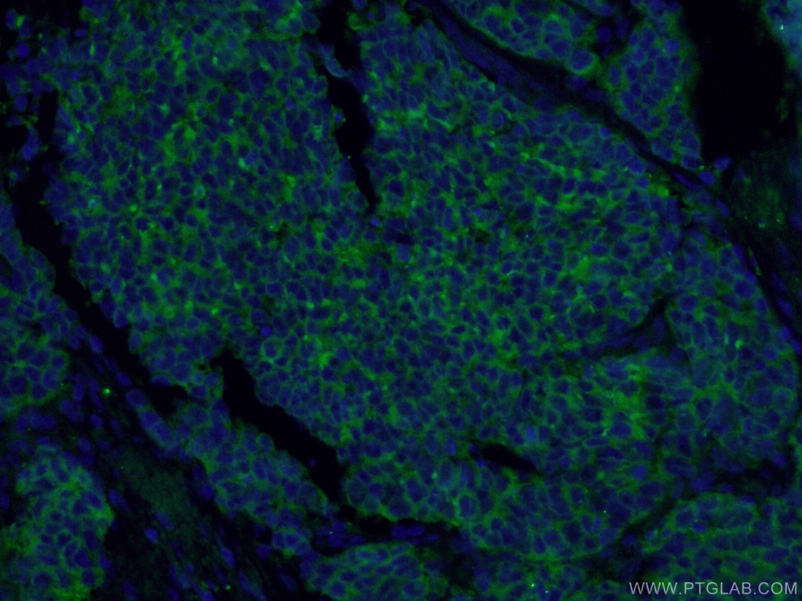
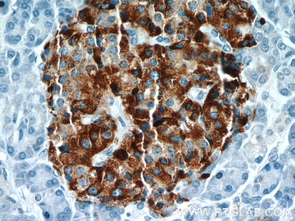
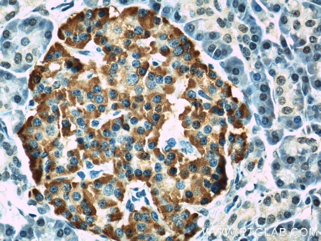
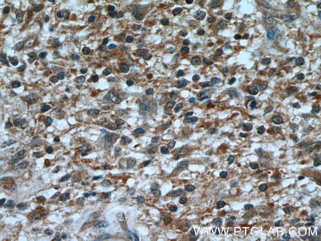
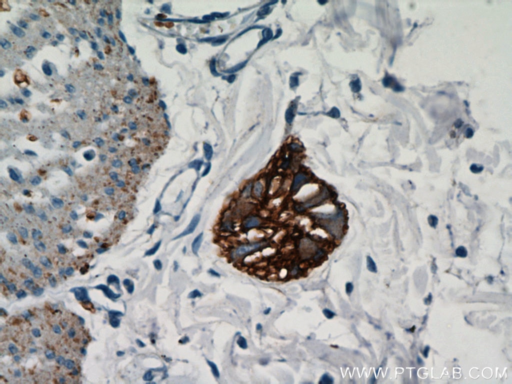
| Résultats positifs en IHC | tissu pancréatique de souris, tissu cérébral de souris, tissu cérébral humain, tissu d'adénome hypophysaire humain, tissu de gangliome, tissu de neuroblastome, tissu pancréatique humain il est suggéré de démasquer l'antigène avec un tampon de TE buffer pH 9.0; (*) À défaut, 'le démasquage de l'antigène peut être 'effectué avec un tampon citrate pH 6,0. |
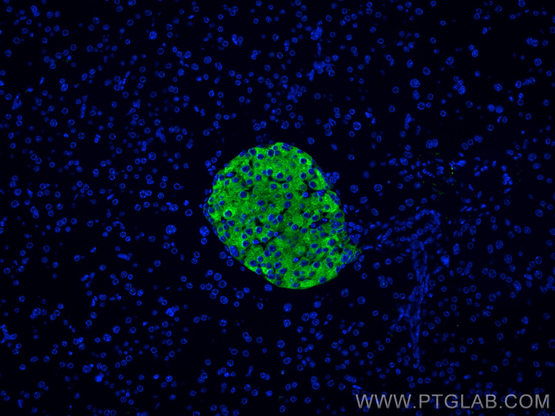
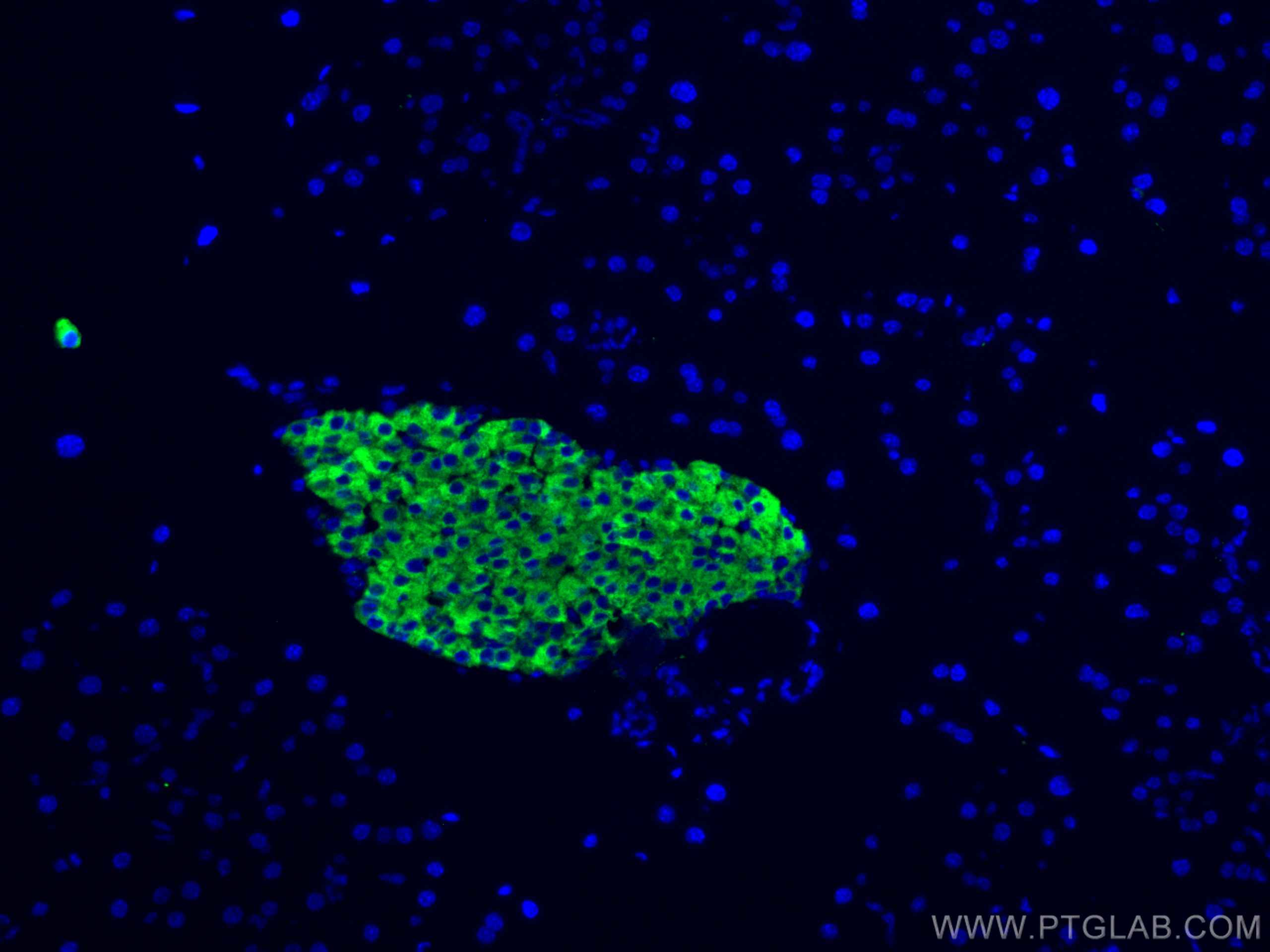
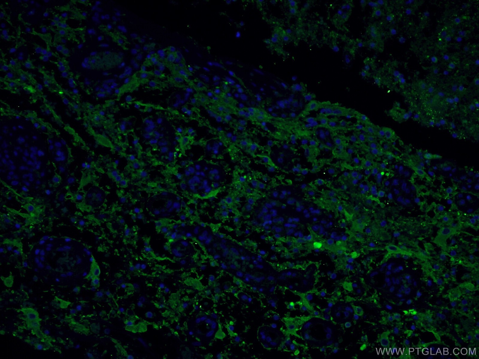
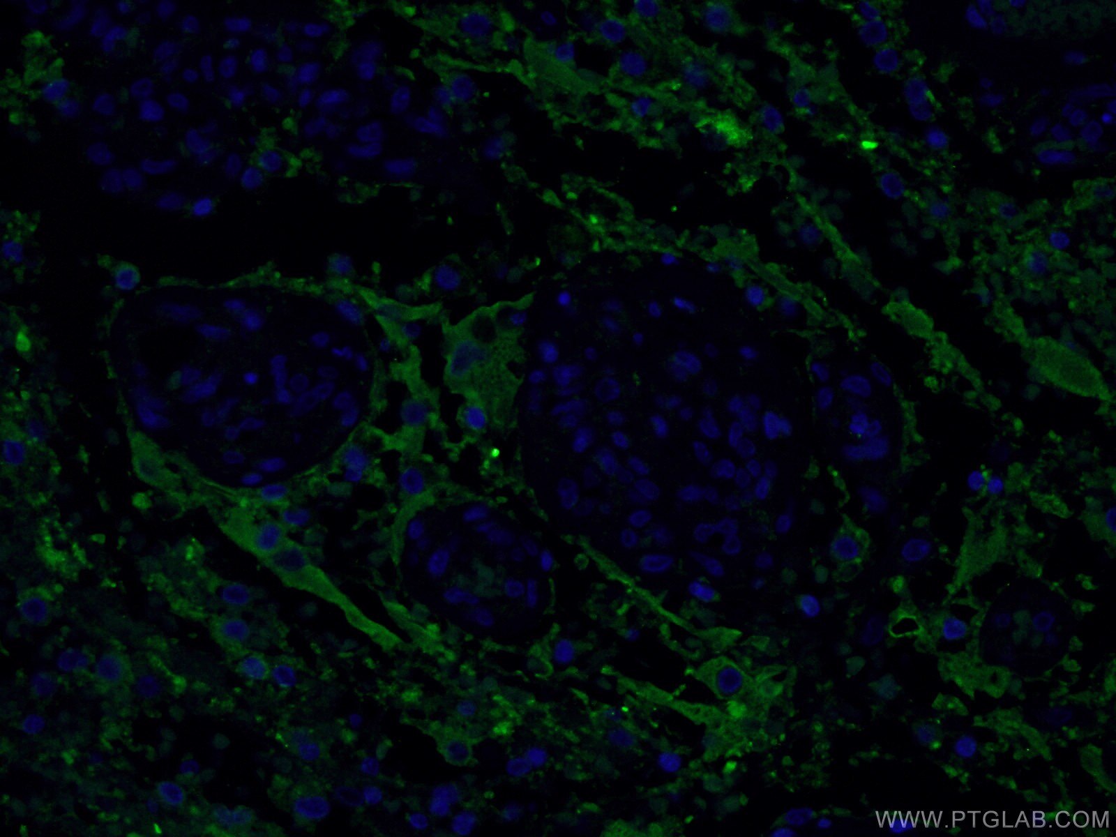
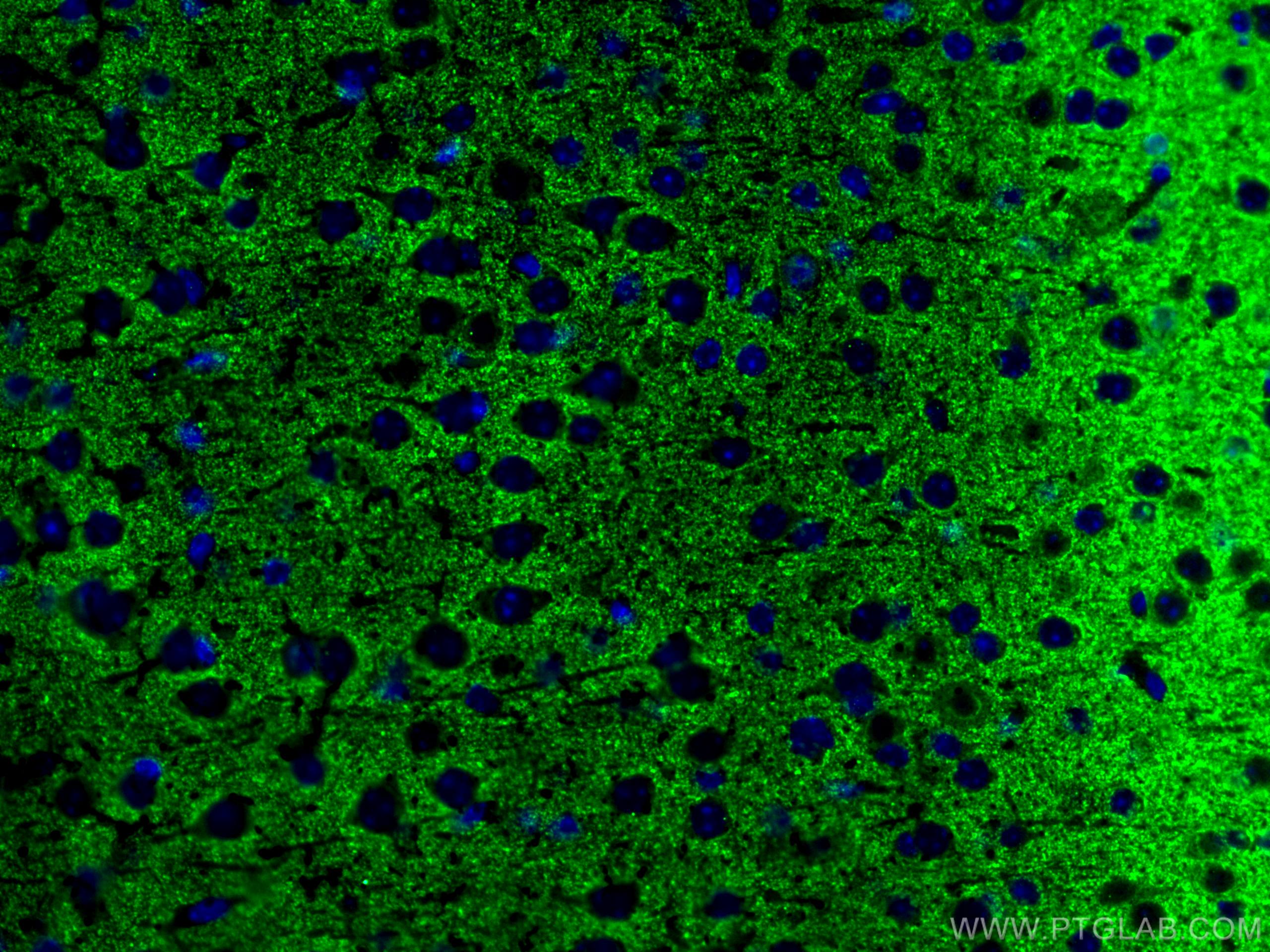
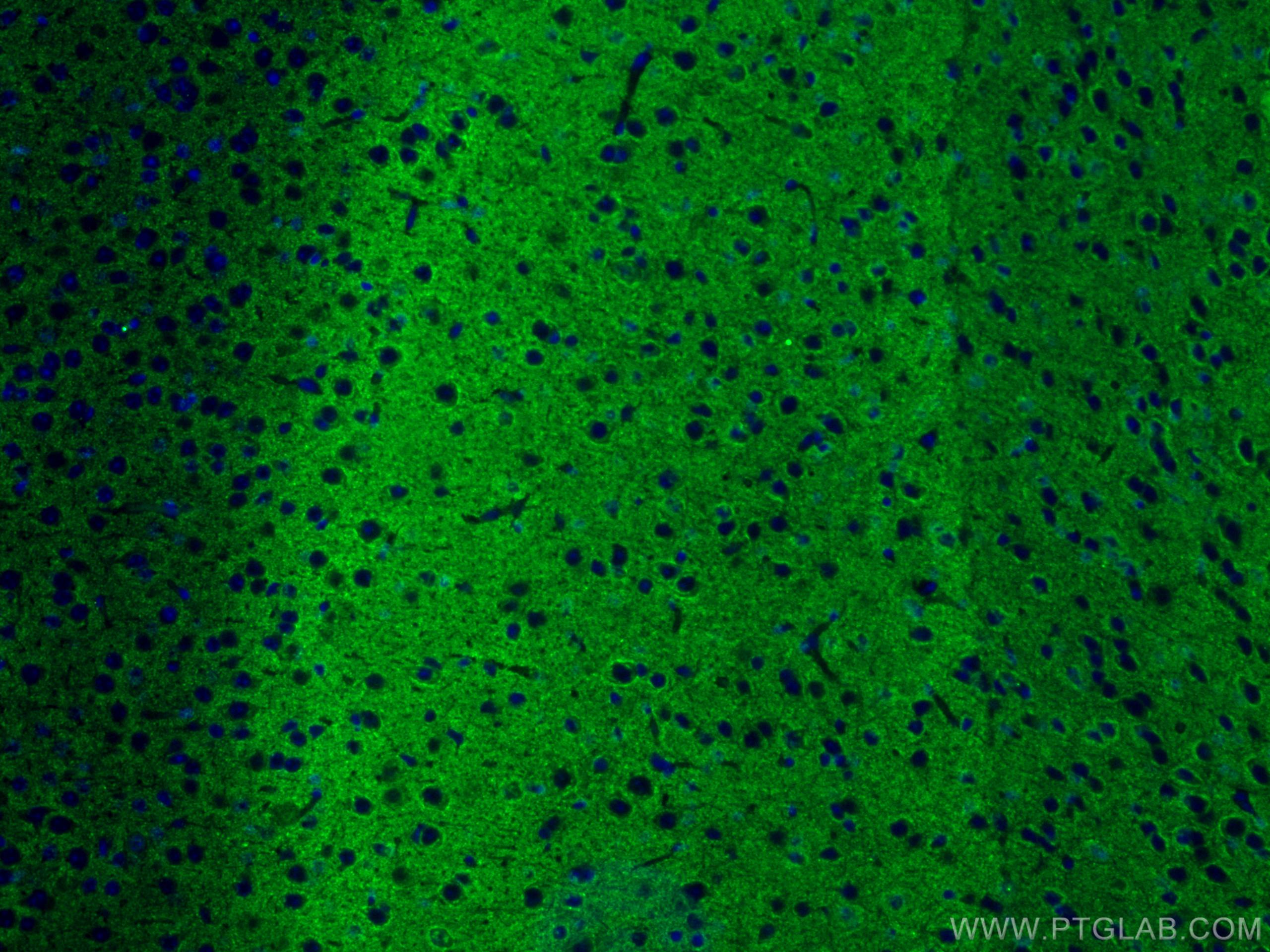
| Résultats positifs en IF-P | tissu pancréatique de souris, tissu cérébral de souris, tissu de cancer du poumon humain, tissu de gliome humain |
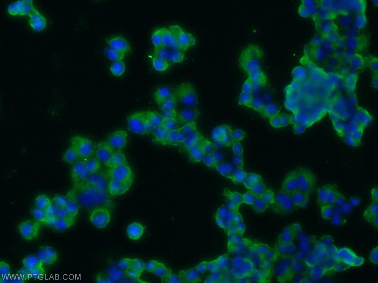
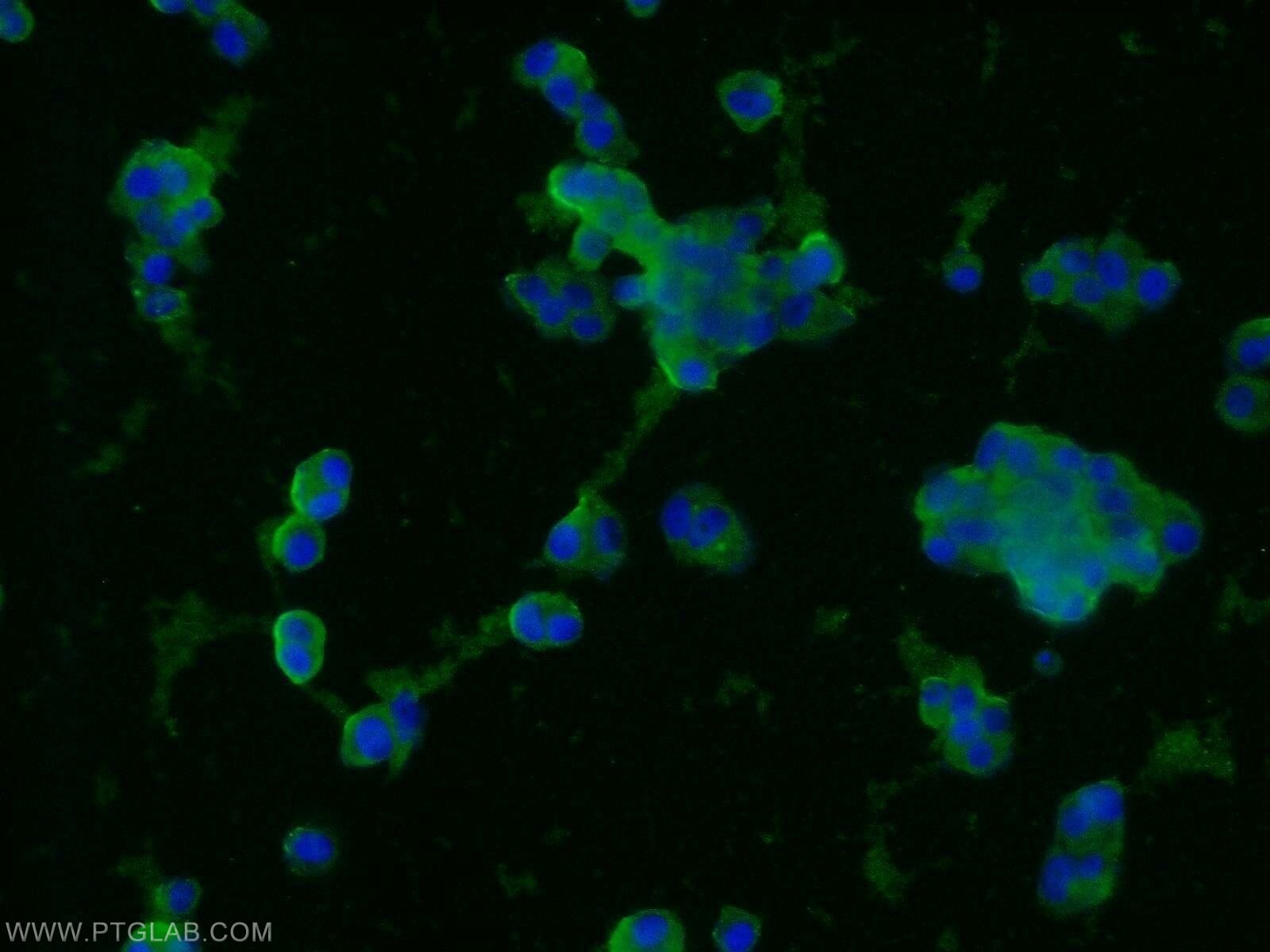
| Résultats positifs en IF/ICC | cellules PC-12, |
Dilution recommandée
| Application | Dilution |
|---|---|
| Western Blot (WB) | WB : 1:5000-1:50000 |
| Immunoprécipitation (IP) | IP : 0.5-4.0 ug for 1.0-3.0 mg of total protein lysate |
| Immunohistochimie (IHC) | IHC : 1:1000-1:4000 |
| Immunofluorescence (IF)-P | IF-P : 1:50-1:500 |
| Immunofluorescence (IF)/ICC | IF/ICC : 1:10-1:100 |
| It is recommended that this reagent should be titrated in each testing system to obtain optimal results. | |
| Sample-dependent, check data in validation data gallery | |
Applications publiées
| WB | See 135 publications below |
| IHC | See 16 publications below |
| IF | See 47 publications below |
Informations sur le produit
17785-1-AP cible Synaptophysin dans les applications de WB, IHC, IF/ICC, IF-P, IP, ELISA et montre une réactivité avec des échantillons Humain, rat, souris
| Réactivité | Humain, rat, souris |
| Réactivité citée | rat, canin, Humain, souris, Hamster |
| Hôte / Isotype | Lapin / IgG |
| Clonalité | Polyclonal |
| Type | Anticorps |
| Immunogène | Synaptophysin Protéine recombinante Ag11781 |
| Nom complet | synaptophysin |
| Masse moléculaire calculée | 313 aa, 34 kDa |
| Poids moléculaire observé | 38-40 kDa |
| Numéro d’acquisition GenBank | BC064550 |
| Symbole du gène | Synaptophysin |
| Identification du gène (NCBI) | 6855 |
| Conjugaison | Non conjugué |
| Forme | Liquide |
| Méthode de purification | Purification par affinité contre l'antigène |
| Tampon de stockage | PBS avec azoture de sodium à 0,02 % et glycérol à 50 % pH 7,3 |
| Conditions de stockage | Stocker à -20°C. Stable pendant un an après l'expédition. L'aliquotage n'est pas nécessaire pour le stockage à -20oC Les 20ul contiennent 0,1% de BSA. |
Informations générales
Synaptophysin (SYP, also known as major synaptic vesicle protein p38) is a 38-kDa integral membrane glycoprotein that regulates synaptic vesicle endocytosis. It is the most abundant synaptic vesicle protein by mass. Synaptophysin is present in neuroendocrine cells and neurons that participate in synaptic transmission. Synaptophysin is a useful marker for identification of neuroendocrine cells and neoplasms. (PMID: 3010302; 21658579)
Protocole
| Product Specific Protocols | |
|---|---|
| WB protocol for Synaptophysin antibody 17785-1-AP | Download protocol |
| IHC protocol for Synaptophysin antibody 17785-1-AP | Download protocol |
| IF protocol for Synaptophysin antibody 17785-1-AP | Download protocol |
| IP protocol for Synaptophysin antibody 17785-1-AP | Download protocol |
| Standard Protocols | |
|---|---|
| Click here to view our Standard Protocols |
Publications
| Species | Application | Title |
|---|---|---|
Nat Biotechnol Magnify is a universal molecular anchoring strategy for expansion microscopy | ||
Nat Neurosci Aerobic glycolysis is the predominant means of glucose metabolism in neuronal somata, which protects against oxidative damage | ||
Mol Cell Mixed Lineage Kinase Domain-like Protein MLKL Breaks Down Myelin following Nerve Injury. | ||
Neuron Astrocytic ApoE reprograms neuronal cholesterol metabolism and histone-acetylation-mediated memory. | ||
Neuron Loss of PYCR2 Causes Neurodegeneration by Increasing Cerebral Glycine Levels via SHMT2. |
Avis
The reviews below have been submitted by verified Proteintech customers who received an incentive forproviding their feedback.
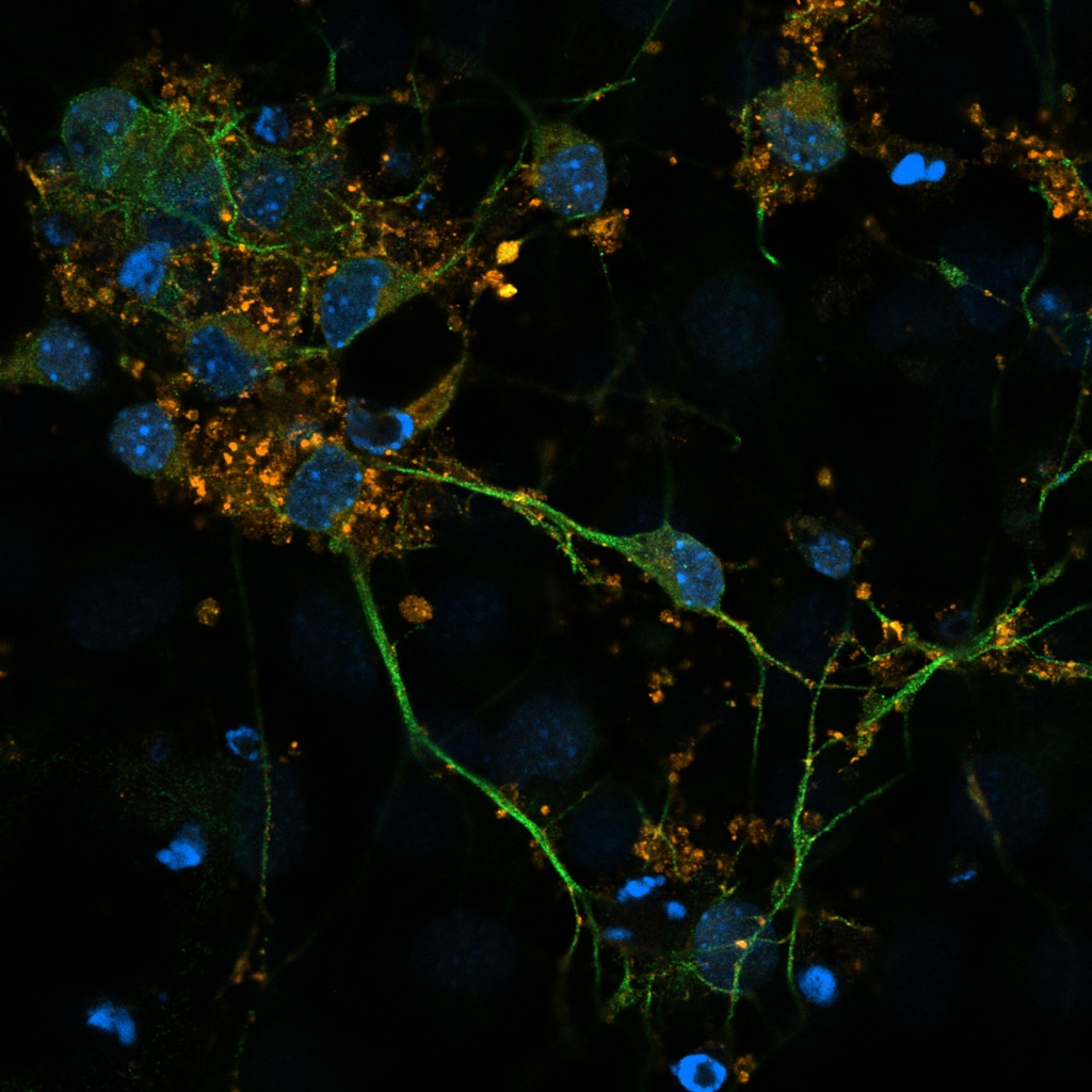
FH Alexandra (Verified Customer) (06-11-2023) | Hoechst-blue Green-Tubulin III (neuronal marker) Orange (Synaptophysin)
 |
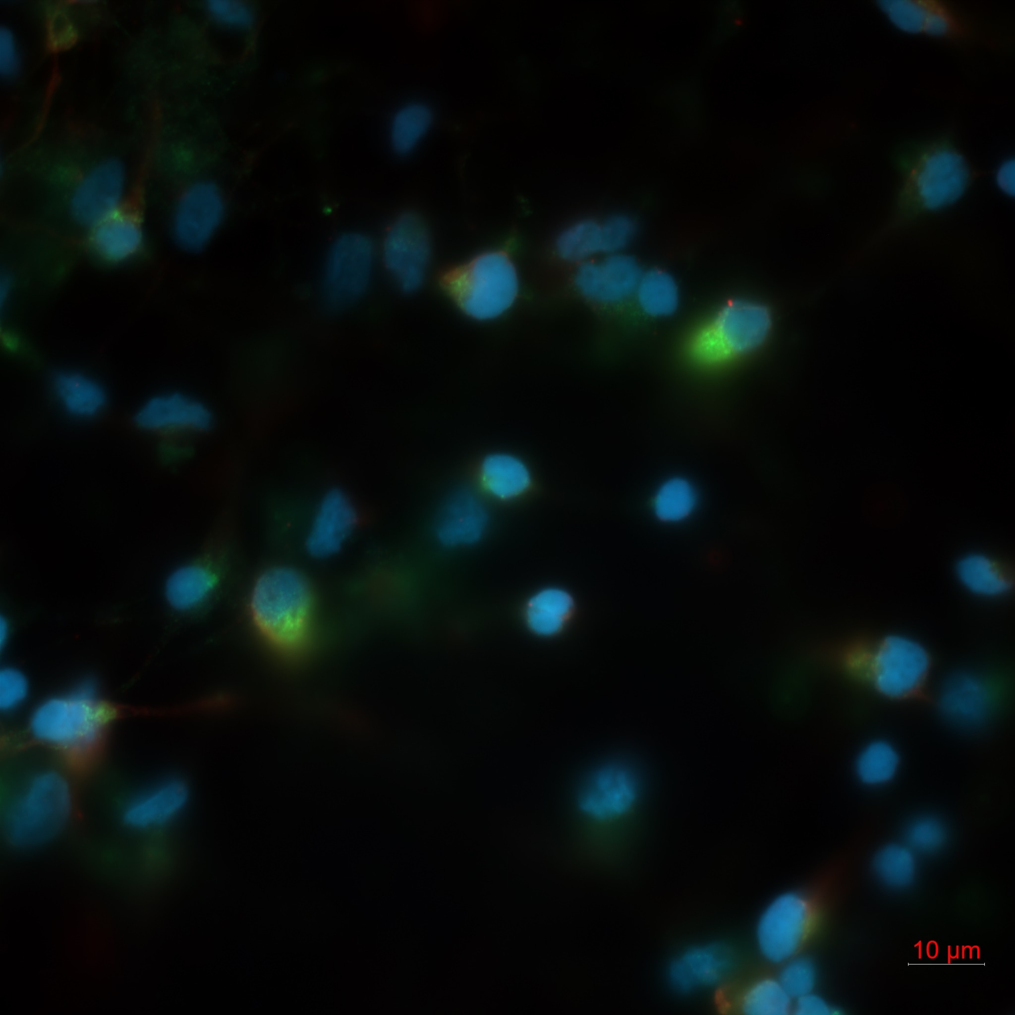
FH Azita (Verified Customer) (05-31-2021) | Human primary cortical neurons was labelled with 1/1000 of the Synaptophysin Polyclonal antibody. The labelling was strong and very good. Blue (Bisbenzimide) Green (Synaptophysin)
 |
