- Phare
- Validé par KD/KO
Anticorps Polyclonal de lapin anti-NRF2, NFE2L2
NRF2, NFE2L2 Polyclonal Antibody for WB, IP, IF, IHC, ELISA
Hôte / Isotype
Lapin / IgG
Réactivité testée
Humain et plus (6)
Applications
WB, IHC, IF/ICC, IP, CoIP, ChIP, ELISA
Conjugaison
Non conjugué
1568
N° de cat : 16396-1-AP
Synonymes
Galerie de données de validation
Applications testées
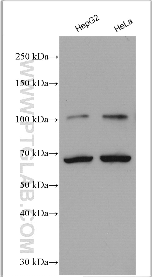
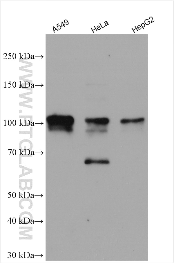
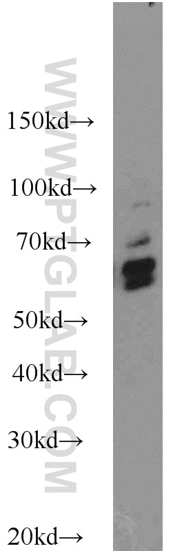
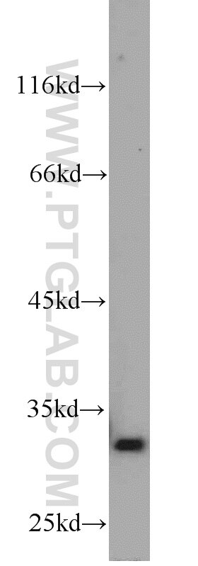
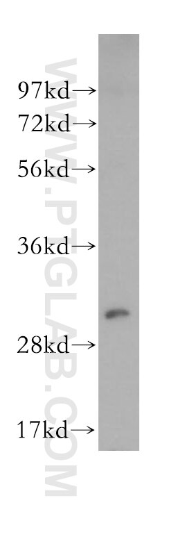
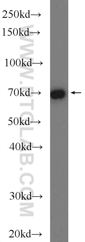
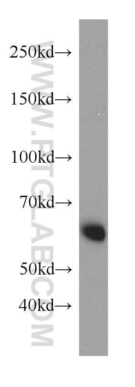
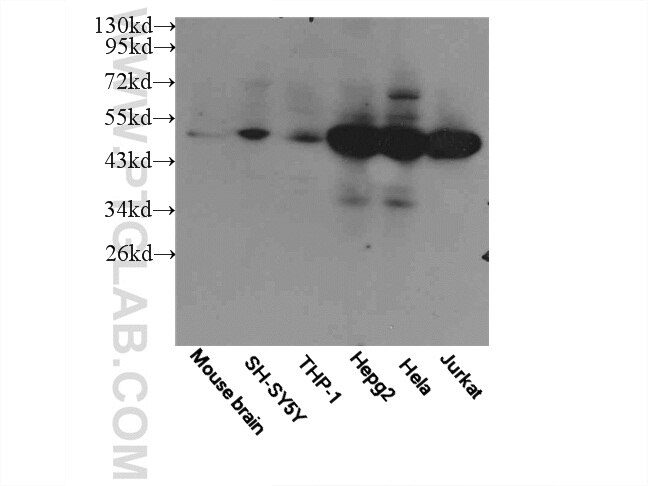
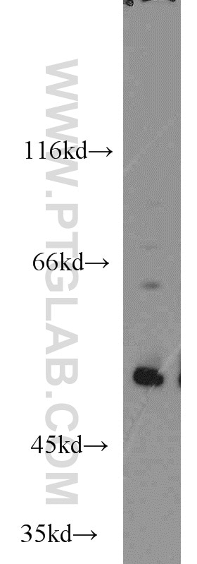
| Résultats positifs en WB | cellules HepG2, cellules A549, cellules HeLa traitées au DMSO |
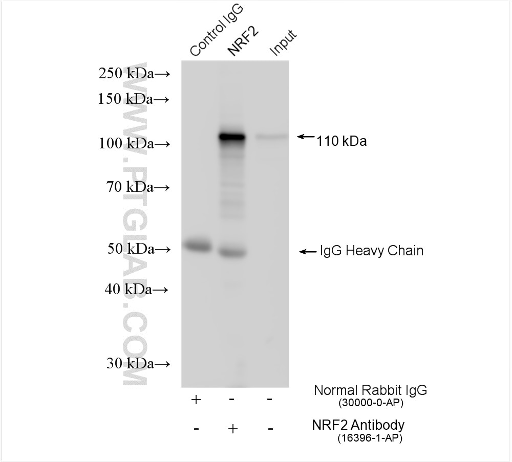
| Résultats positifs en IP | cellules HeLa, |
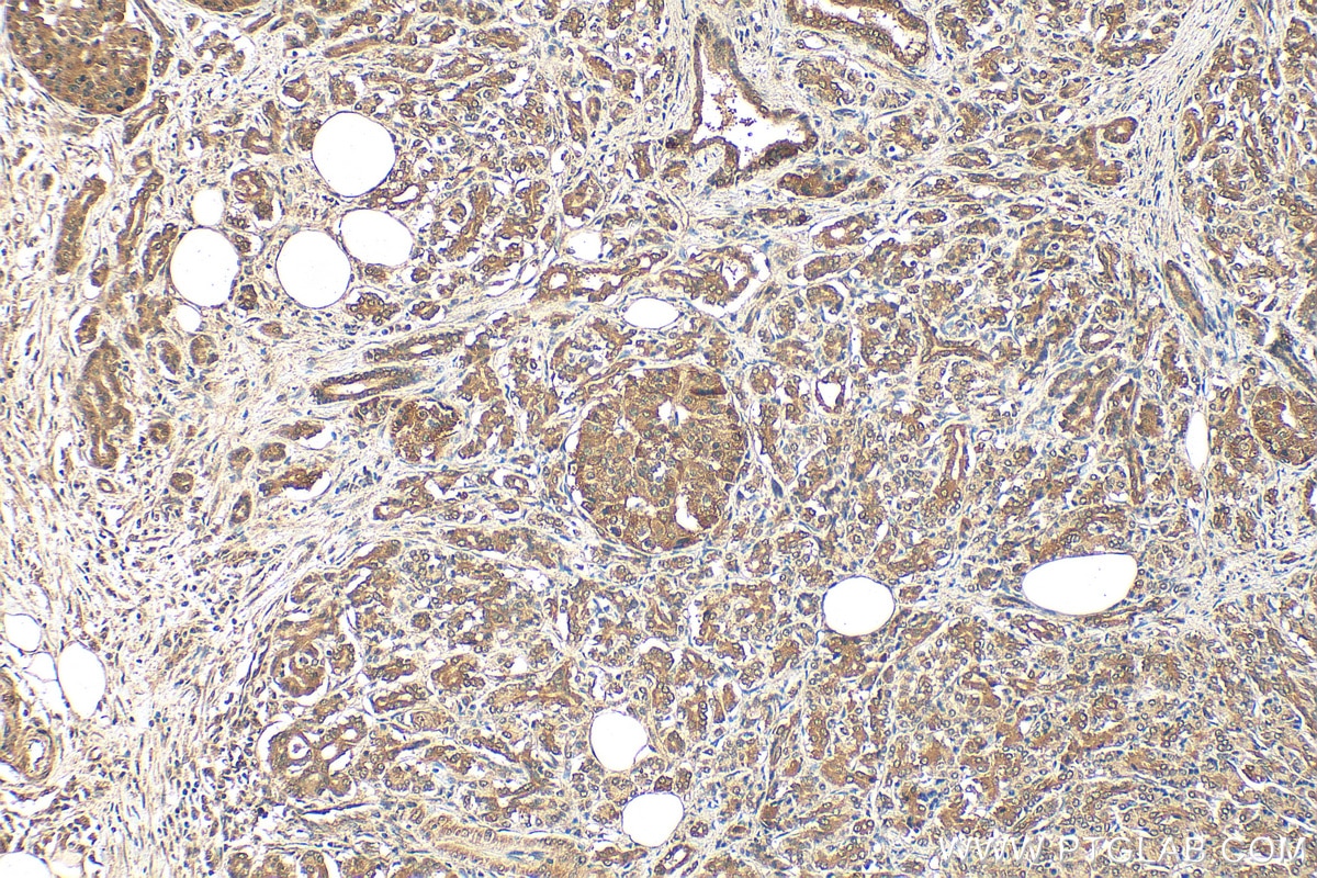
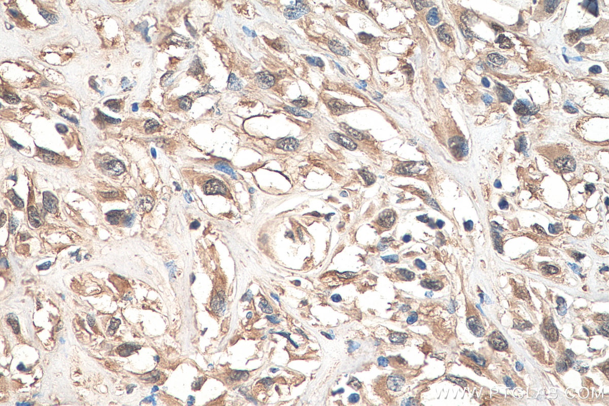
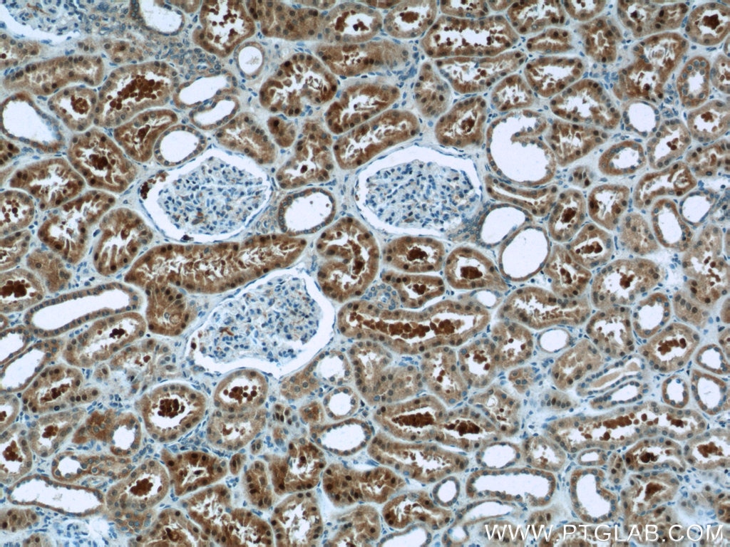
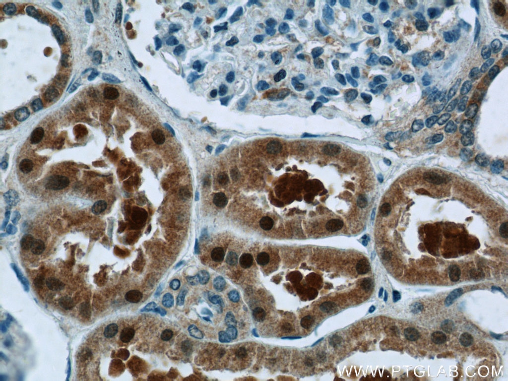
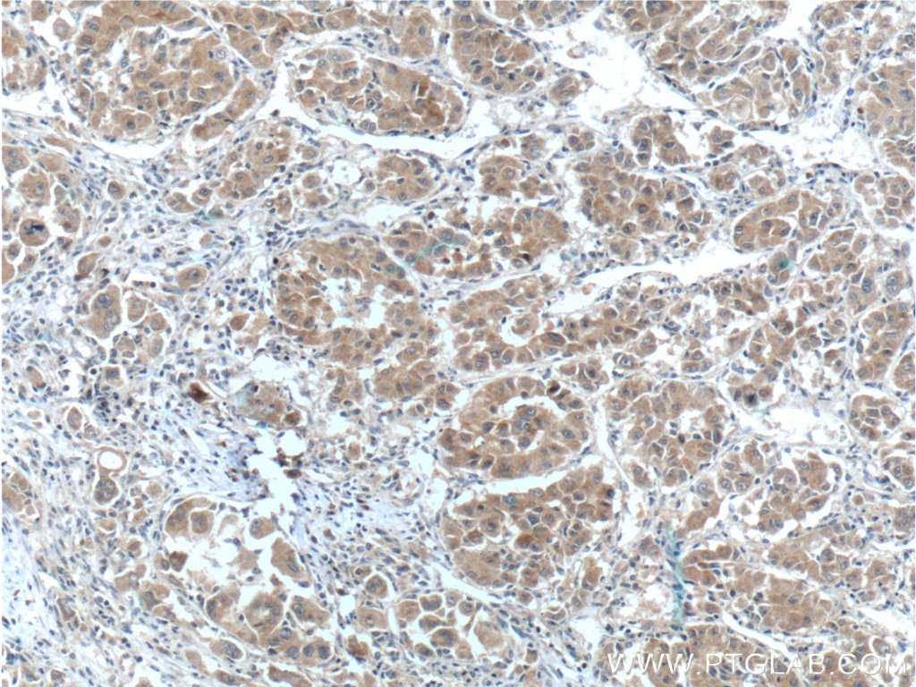
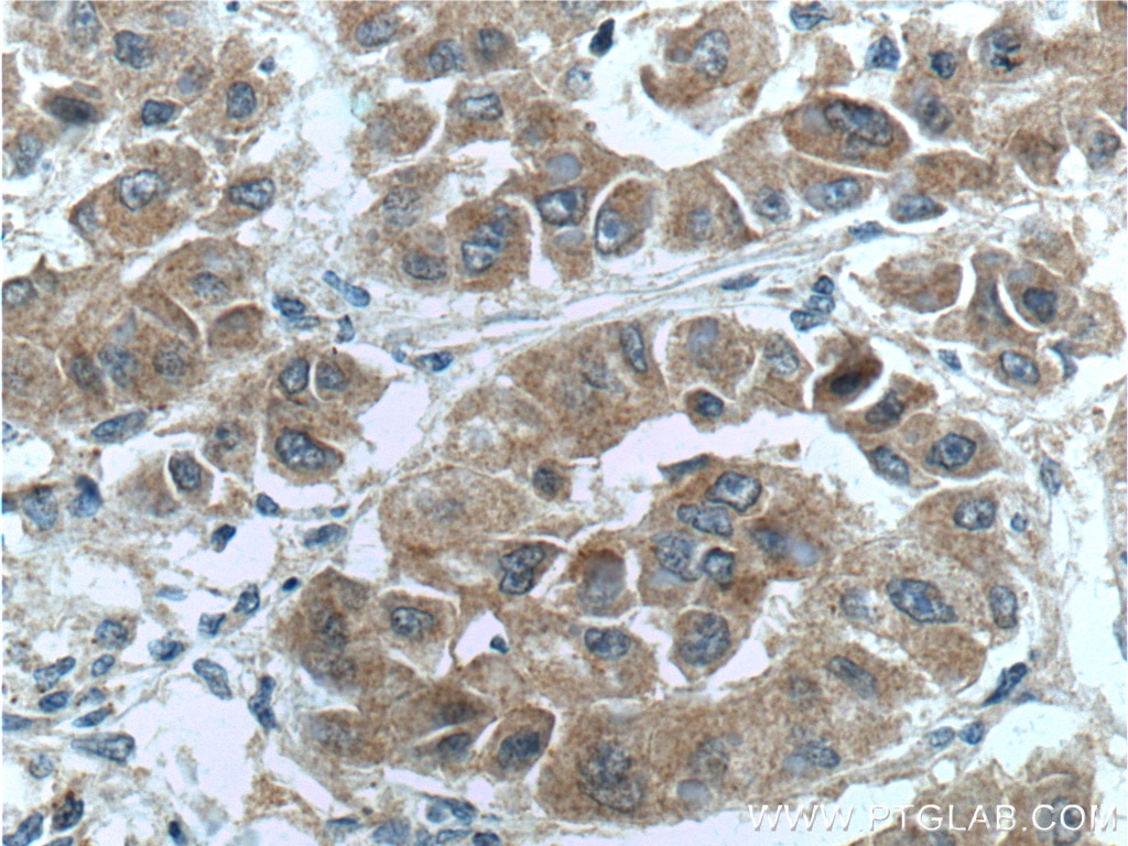
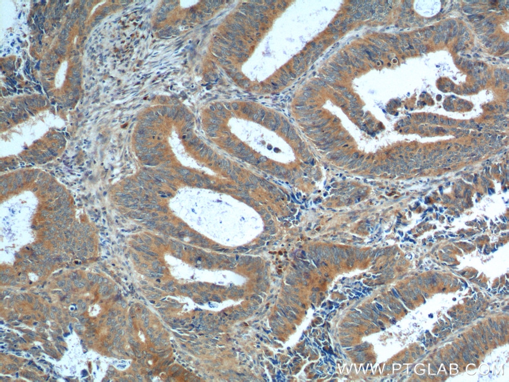
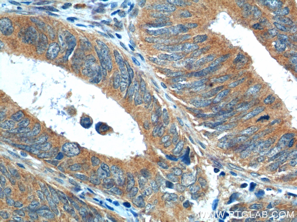
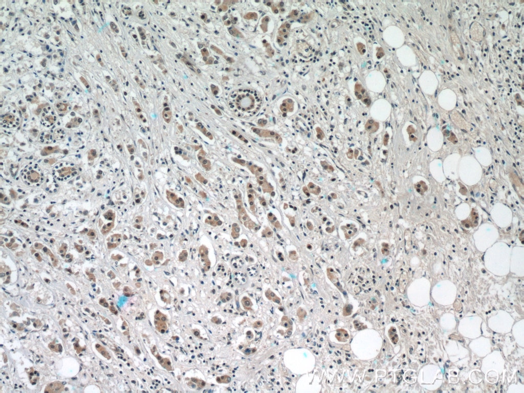
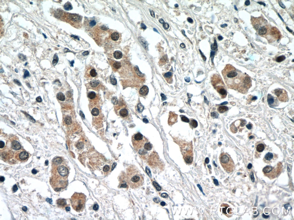
| Résultats positifs en IHC | tissu de cancer du foie humain, tissu de cancer du côlon humain, tissu de cancer du pancréas humain, tissu de cancer du sein humain, tissu de carcinome à cellules rénales humain, tissu rénal humain il est suggéré de démasquer l'antigène avec un tampon de TE buffer pH 9.0; (*) À défaut, 'le démasquage de l'antigène peut être 'effectué avec un tampon citrate pH 6,0. |
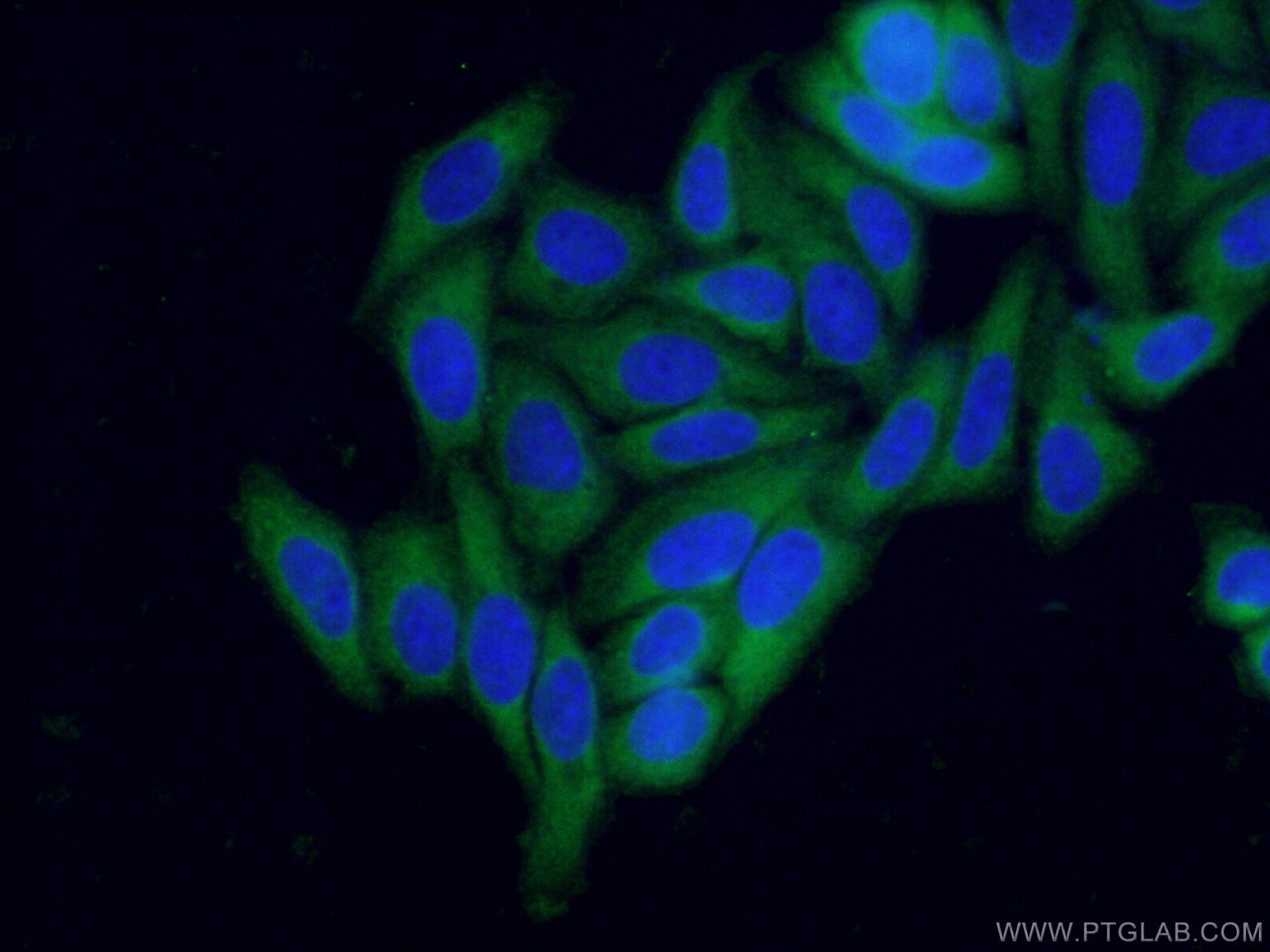
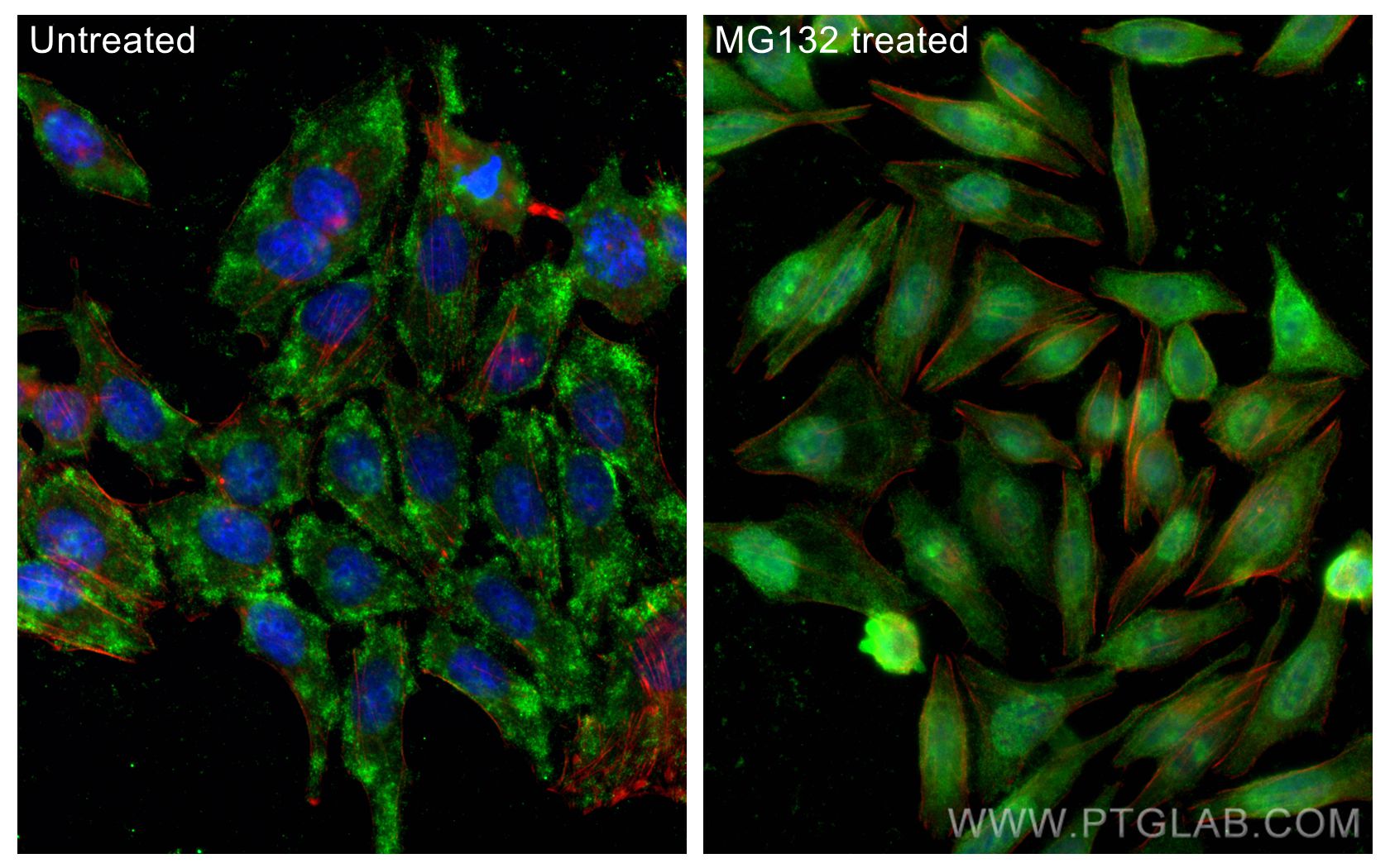
| Résultats positifs en IF/ICC | MG132 treated HepG2 cells, cellules HepG2 |
Dilution recommandée
| Application | Dilution |
|---|---|
| Western Blot (WB) | WB : 1:2000-1:12000 |
| Immunoprécipitation (IP) | IP : 0.5-4.0 ug for 1.0-3.0 mg of total protein lysate |
| Immunohistochimie (IHC) | IHC : 1:50-1:500 |
| Immunofluorescence (IF)/ICC | IF/ICC : 1:50-1:500 |
| It is recommended that this reagent should be titrated in each testing system to obtain optimal results. | |
| Sample-dependent, check data in validation data gallery | |
Informations sur le produit
16396-1-AP cible NRF2, NFE2L2 dans les applications de WB, IHC, IF/ICC, IP, CoIP, ChIP, ELISA et montre une réactivité avec des échantillons Humain
| Réactivité | Humain |
| Réactivité citée | rat, Chèvre, Humain, Lapin, poulet, singe, souris |
| Hôte / Isotype | Lapin / IgG |
| Clonalité | Polyclonal |
| Type | Anticorps |
| Immunogène | NRF2, NFE2L2 Protéine recombinante Ag9489 |
| Nom complet | nuclear factor (erythroid-derived 2)-like 2 |
| Masse moléculaire calculée | 605 aa, 68 kDa |
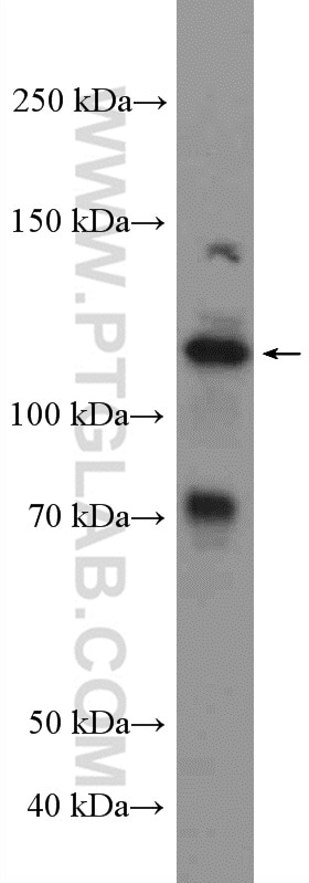
| Poids moléculaire observé | 110 kDa, 68 kDa |
| Numéro d’acquisition GenBank | BC011558 |
| Symbole du gène | NRF2 |
| Identification du gène (NCBI) | 4780 |
| Conjugaison | Non conjugué |
| Forme | Liquide |
| Méthode de purification | Purification par affinité contre l'antigène |
| Tampon de stockage | PBS avec azoture de sodium à 0,02 % et glycérol à 50 % pH 7,3 |
| Conditions de stockage | Stocker à -20°C. Stable pendant un an après l'expédition. L'aliquotage n'est pas nécessaire pour le stockage à -20oC Les 20ul contiennent 0,1% de BSA. |
Informations générales
NRF2, also named as NFE2L2, belongs to the bZIP family and CNC subfamily. It is a transcription activator that binds to antioxidant response (ARE) elements in the promoter regions of target genes. NRF2 is important for the coordinated up-regulation of genes in response to oxidative stress. It may be involved in the transcriptional activation of genes of the beta-globin cluster by mediating enhancer activity of hypersensitive site 2 of the beta-globin locus control region. Nrf2 is a key player in the regulation of genes encoding for many antioxidative response enzymes.The expression of NRF2 may be induced under oxidative stress (PMID:14567983).In lung cancer, Nrf2 activation in malignant cells has been associated with tumor progression and chemotherapy resistance(PMID:20534738). Identifying patients with abnormal NRF2 expression may be important for selection for chemotherapy in NSCLC. As new investigators break into the emerging field of Nrf2 research, confusion regarding the correct migratory pattern of Nrf2 is causing doubts about the accuracy and reproducibility of published results. This letter provides solid evidence that the actually observed molecular weight of Nrf2 is about 70kDa and 95-110 kDa. (PMID: 22703241).
Protocole
| Product Specific Protocols | |
|---|---|
| WB protocol for NRF2, NFE2L2 antibody 16396-1-AP | Download protocol |
| IHC protocol for NRF2, NFE2L2 antibody 16396-1-AP | Download protocol |
| IF protocol for NRF2, NFE2L2 antibody 16396-1-AP | Download protocol |
| IP protocol for NRF2, NFE2L2 antibody 16396-1-AP | Download protocol |
| Standard Protocols | |
|---|---|
| Click here to view our Standard Protocols |
Publications
- Journal Impact Factor
- Most recent
| Species | Application | Title |
|---|---|---|
Gastroenterology Nucleolar HEATR1 upregulated by mTORC1 signaling promotes hepatocellular carcinoma growth by dominating ribosome biogenesis and proteome homeostasis | ||
Cancer Cell iASPP Is an Antioxidative Factor and Drives Cancer Growth and Drug Resistance by Competing with Nrf2 for Keap1 Binding.
| ||
Cell Metab Transsulfuration Activity Can Support Cell Growth upon Extracellular Cysteine Limitation. | ||
Avis
The reviews below have been submitted by verified Proteintech customers who received an incentive forproviding their feedback.
FH SD (Verified Customer) (07-05-2024) | Excellent
 |
FH Guillermo (Verified Customer) (09-03-2022) | Very good product, great spots.
|
FH balawant (Verified Customer) (08-06-2022) | for westerblotting I have used this Antibody. this antibody is excellent and I will recommend if you are working with this protein
|
FH Georgina (Verified Customer) (07-04-2022) | This is a good antibody - it gives some background signal but the band of interest is clear. Sometimes need to use a higher concentration than recommended for cells with lower expression of NRF2.
|
FH Iram (Verified Customer) (03-11-2022) | Gave good bands around 100 kda in colon cancer cell lysates
|
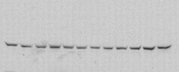
FH Giovanni (Verified Customer) (01-11-2022) | the antiboby worked very well at dilution 1:1000 in WB using Htr8/SVneo cell line lysates. It gave a specific band at around 100 kDa with very low background.
 |
FH Xiaoping (Verified Customer) (10-14-2021) | Gave good bands around 100 kda in renal cell lysates
|
FH Razia Sultana (Verified Customer) (08-20-2020) | The antibody gives good band at around 100KD(100 KD in HK2 cells and 90KD in kidney tissue lysate) along with some non specific bands at around 40-50KD. The trial size gave a band at 130KD which is unusual and lot of non specific bands,but the full size antibody is good. We are satisfied with the purchase
|
FH JIM (Verified Customer) (03-26-2019) | Strong Nrf2 signal can be observed in macrophages treated with LPS and D3T by western blots.
|
