- Phare
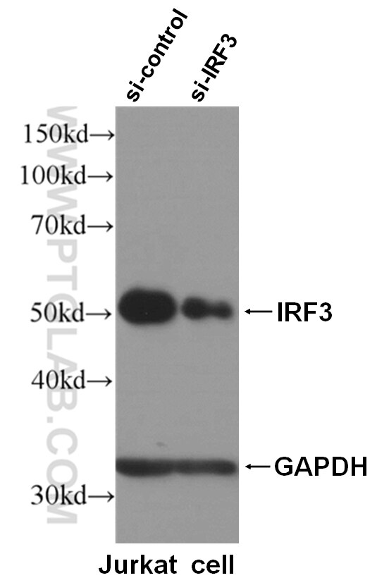
- Validé par KD/KO
Anticorps Polyclonal de lapin anti-MAVS; VISA
MAVS; VISA Polyclonal Antibody for WB, IP, IF, IHC, ELISA
Hôte / Isotype
Lapin / IgG
Réactivité testée
Humain et plus (2)
Applications
WB, IHC, IF/ICC, IP, ELISA
Conjugaison
Non conjugué
N° de cat : 14341-1-AP
Synonymes
Galerie de données de validation
Applications testées
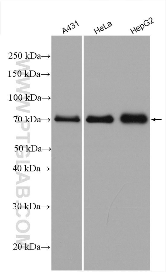
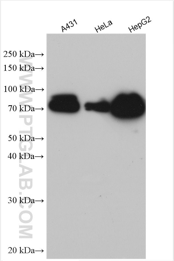
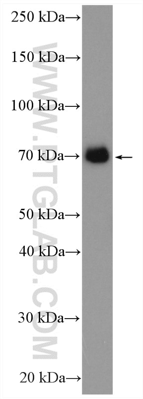
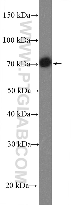
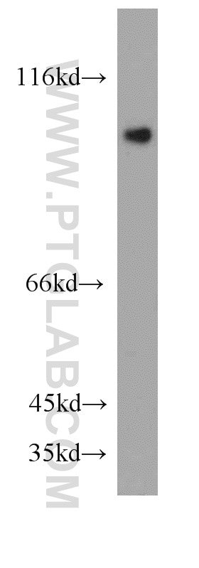
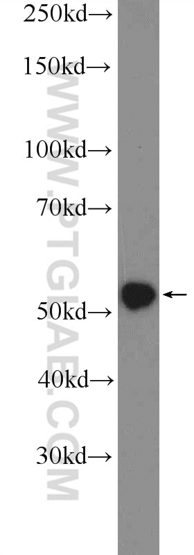
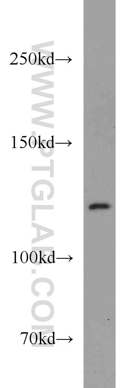
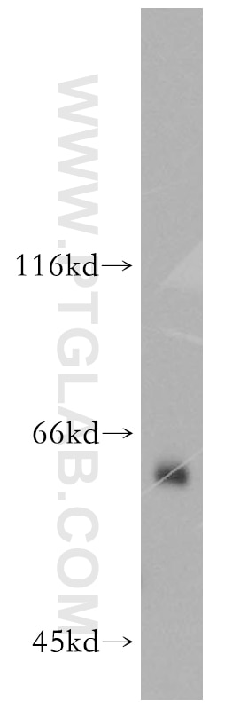
| Résultats positifs en WB | cellules A431, cellules HeLa, cellules HepG2, cellules HuH-7, cellules Jurkat |
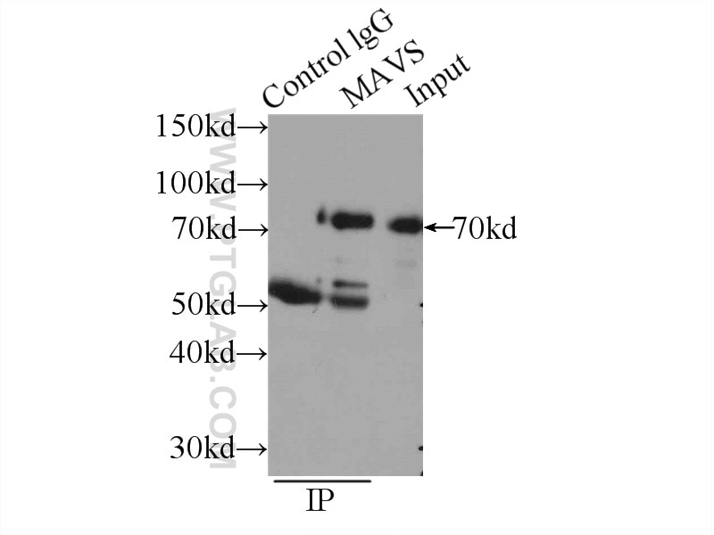
| Résultats positifs en IP | cellules HEK-293 |
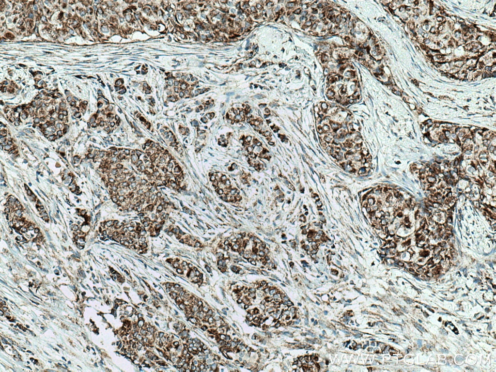
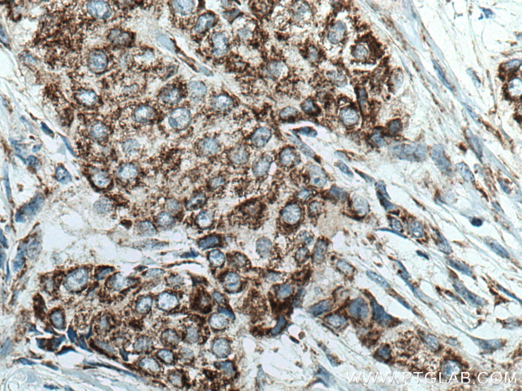
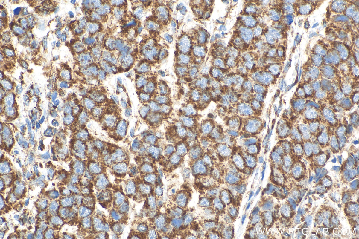
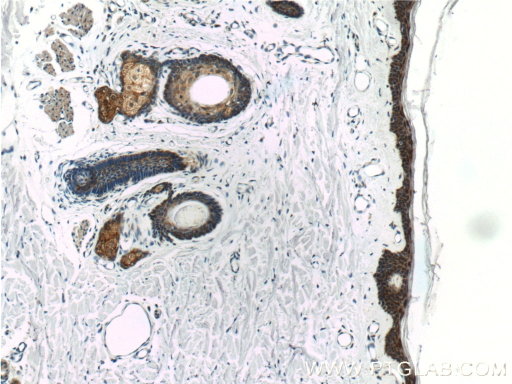
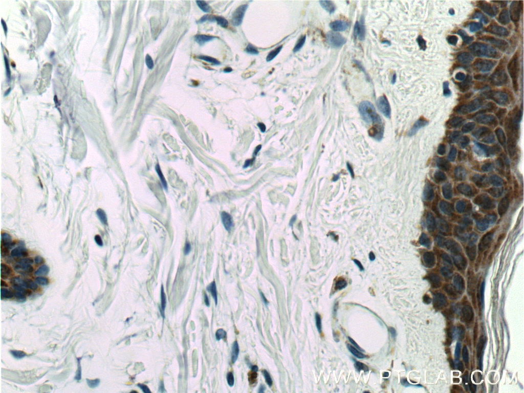
| Résultats positifs en IHC | tissu de cancer du sein humain, tissu cutané humain il est suggéré de démasquer l'antigène avec un tampon de TE buffer pH 9.0; (*) À défaut, 'le démasquage de l'antigène peut être 'effectué avec un tampon citrate pH 6,0. |
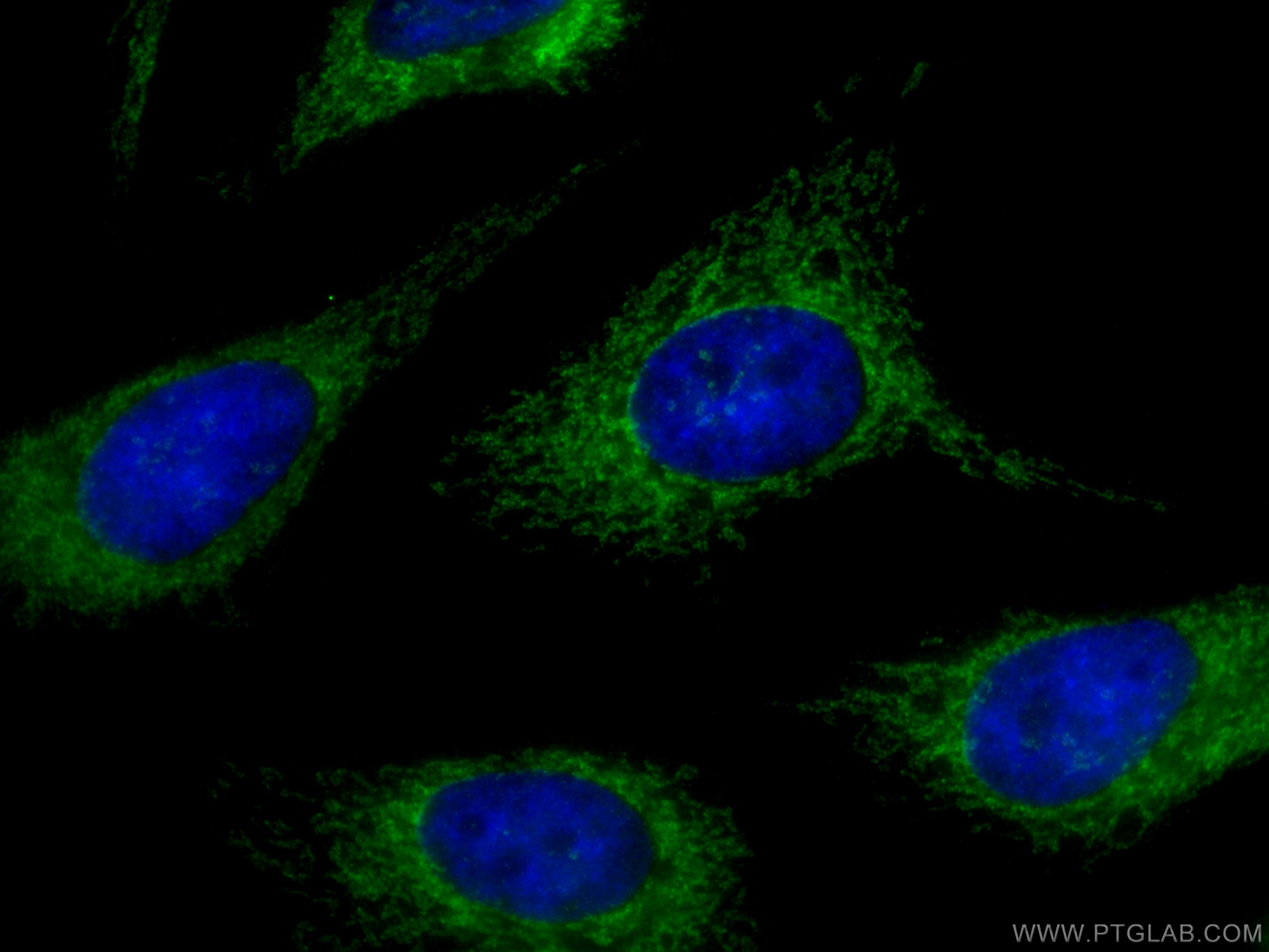
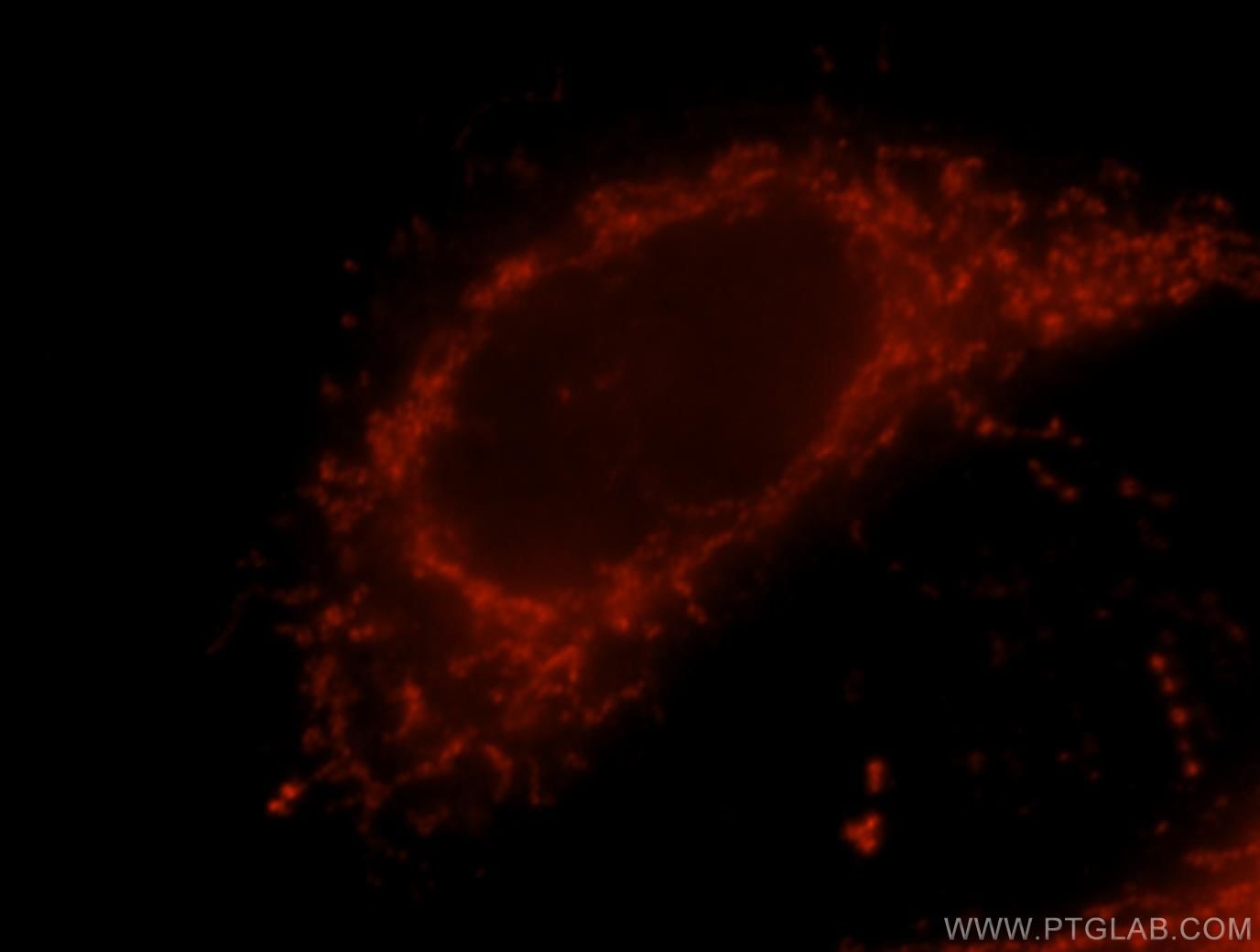
| Résultats positifs en IF/ICC | cellules HeLa, |
Dilution recommandée
| Application | Dilution |
|---|---|
| Western Blot (WB) | WB : 1:2000-1:16000 |
| Immunoprécipitation (IP) | IP : 0.5-4.0 ug for 1.0-3.0 mg of total protein lysate |
| Immunohistochimie (IHC) | IHC : 1:250-1:1000 |
| Immunofluorescence (IF)/ICC | IF/ICC : 1:50-1:500 |
| It is recommended that this reagent should be titrated in each testing system to obtain optimal results. | |
| Sample-dependent, check data in validation data gallery | |
Informations sur le produit
14341-1-AP cible MAVS; VISA dans les applications de WB, IHC, IF/ICC, IP, ELISA et montre une réactivité avec des échantillons Humain
| Réactivité | Humain |
| Réactivité citée | Humain, porc, singe |
| Hôte / Isotype | Lapin / IgG |
| Clonalité | Polyclonal |
| Type | Anticorps |
| Immunogène | MAVS; VISA Protéine recombinante Ag5655 |
| Nom complet | mitochondrial antiviral signaling protein |
| Masse moléculaire calculée | 57 kDa |
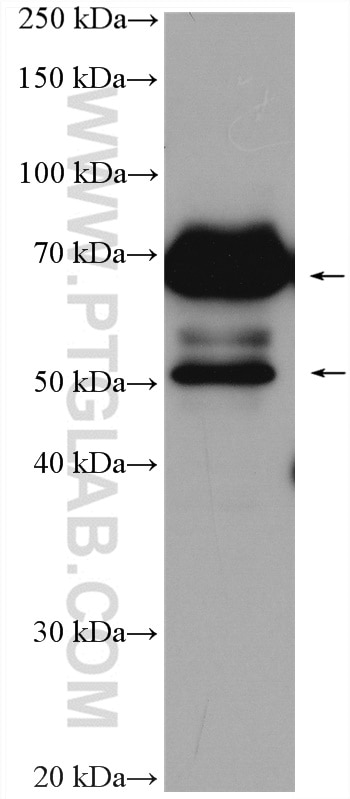
| Poids moléculaire observé | 50-55 kDa, 70-75 kDa |
| Numéro d’acquisition GenBank | BC044952 |
| Symbole du gène | MAVS |
| Identification du gène (NCBI) | 57506 |
| Conjugaison | Non conjugué |
| Forme | Liquide |
| Méthode de purification | Purification par affinité contre l'antigène |
| Tampon de stockage | PBS avec azoture de sodium à 0,02 % et glycérol à 50 % pH 7,3 |
| Conditions de stockage | Stocker à -20°C. Stable pendant un an après l'expédition. L'aliquotage n'est pas nécessaire pour le stockage à -20oC Les 20ul contiennent 0,1% de BSA. |
Informations générales
Mitochondrial antiviral-signaling protein (MAVS) is also known as virus-induced-signaling adapter (VISA) or IFN-β promoter stimulator protein 1 (IPS-1), it is widely involved and required for innate immune defense against viruses. MAVS, present in T cells, monocytes, epithelial cells and hepatocytes, contains CARD and transmembrane domains which are essential for antiviral functions. MAVS is able to interact with various cellular proteins including DDX58/RIG-I, IFIH1/MDA5, TRAF2, TRAF6, TMEM173/MITA, IFIT3 and etc. It can undergoe phosphorylation on multiple sites and ubiquitination, which may together cause the molecular weight migrate to about 70 kDa despite the predicated 57 kDa.
Protocole
| Product Specific Protocols | |
|---|---|
| WB protocol for MAVS; VISA antibody 14341-1-AP | Download protocol |
| IHC protocol for MAVS; VISA antibody 14341-1-AP | Download protocol |
| IF protocol for MAVS; VISA antibody 14341-1-AP | Download protocol |
| IP protocol for MAVS; VISA antibody 14341-1-AP | Download protocol |
| Standard Protocols | |
|---|---|
| Click here to view our Standard Protocols |
Publications
| Species | Application | Title |
|---|---|---|
Signal Transduct Target Ther TRAF3 activates STING-mediated suppression of EV-A71 and target of viral evasion | ||
Immunity Decreased Expression of the Host Long-Noncoding RNA-GM Facilitates Viral Escape by Inhibiting the Kinase activity TBK1 via S-glutathionylation. | ||
Mol Cell An Epstein-Barr virus protein interaction map reveals NLRP3 inflammasome evasion via MAVS UFMylation | ||
Nat Commun MLL5 suppresses antiviral innate immune response by facilitating STUB1-mediated RIG-I degradation. | ||
EMBO J CircPVT1 promotes ER-positive breast tumorigenesis and drug resistance by targeting ESR1 and MAVS
| ||
Avis
The reviews below have been submitted by verified Proteintech customers who received an incentive forproviding their feedback.
FH Damien (Verified Customer) (09-03-2019) | No convince by this anti-MAVS antibody.In fact, in human cells MAVS protein is expressed in two differents forms : short-MAVS (55kDa) and full-length MAVS (72 kDa). So I have to observe two bands by WB. However, after using different conditions with this antibody according to the manufacturer's protocol I always observe a smear of protein, and one band at 72 kDa. The expected on around 55 kDa never appear. Note that my cells lysates were not degraded since the others proteins that I want to reveal were great.
|
