- Phare
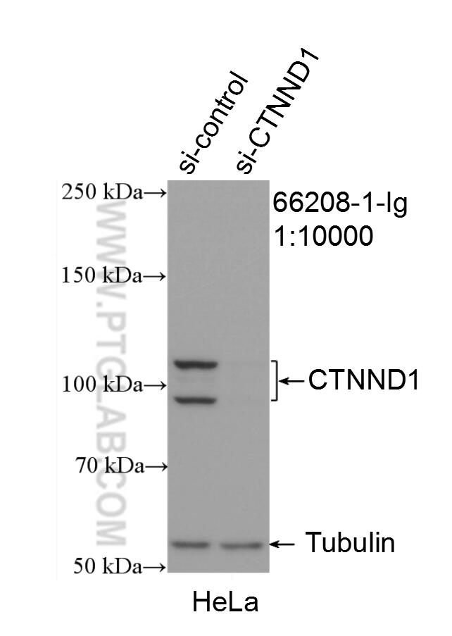
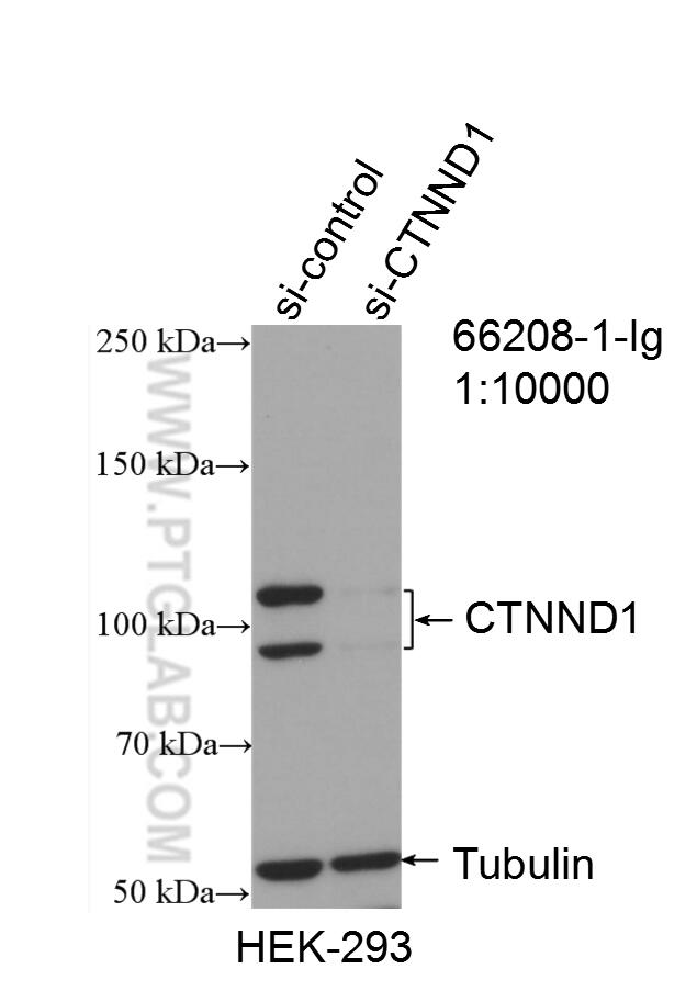
- Validé par KD/KO
Anticorps Monoclonal anti-p120 Catenin
p120 Catenin Monoclonal Antibody for IF, IHC, WB,ELISA
Hôte / Isotype
Mouse / IgG2b
Réactivité testée
Humain, rat, souris
Applications
WB, IP, IHC, IF, ELISA
Conjugaison
Non conjugué
CloneNo.
2F7H8
N° de cat : 66208-1-Ig
Synonymes
Galerie de données de validation
Applications testées
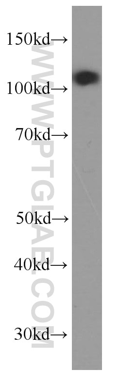
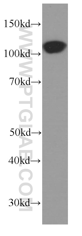
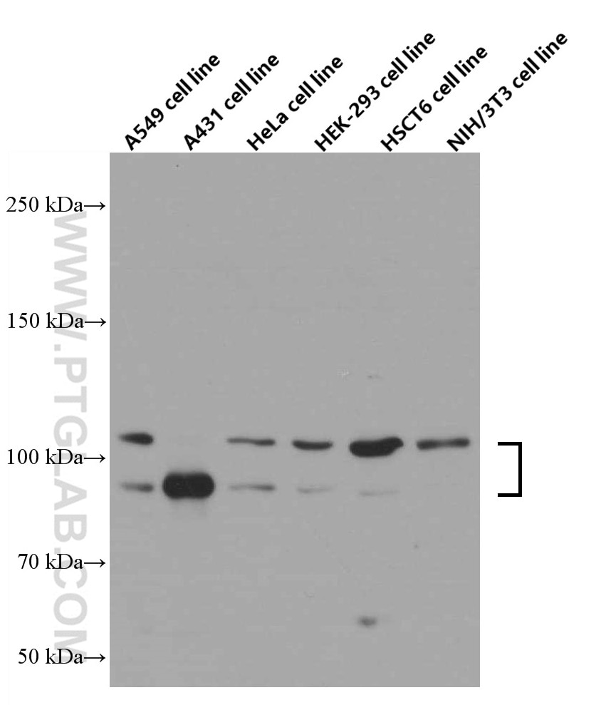
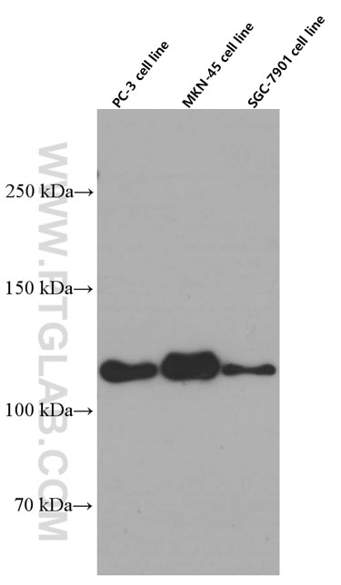
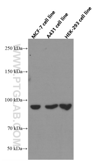
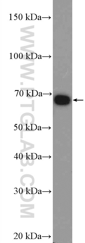
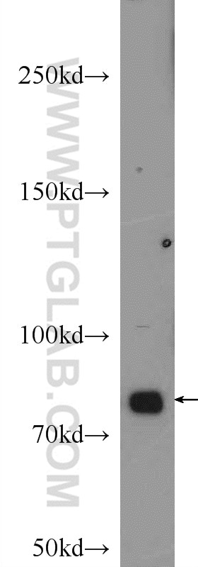
| Résultats positifs en WB | tissu cérébral humain fœtal, cellules A431, cellules A549, cellules HEK-293, cellules HeLa, cellules HSC-T6, cellules NIH/3T3, tissu cérébral de souris |
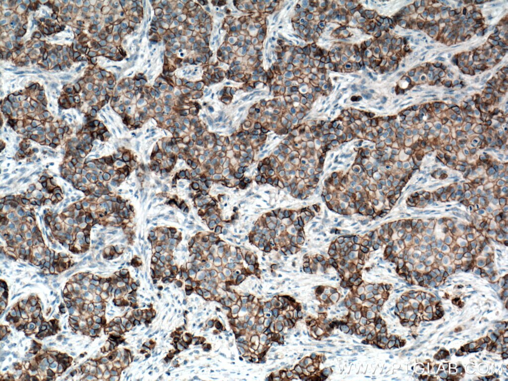
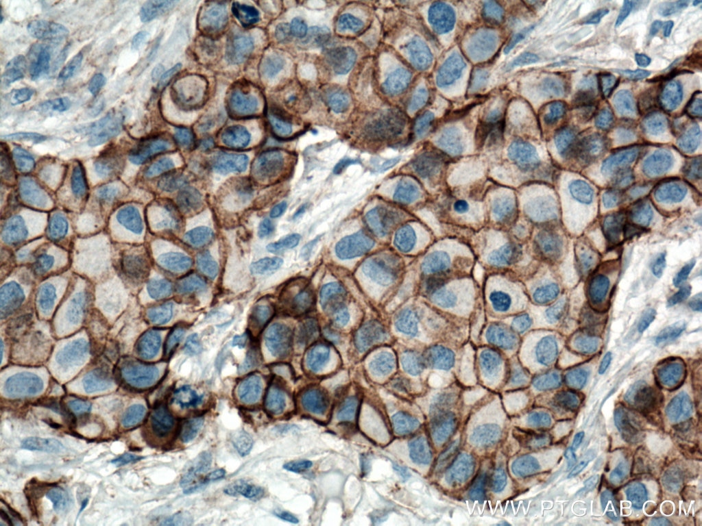
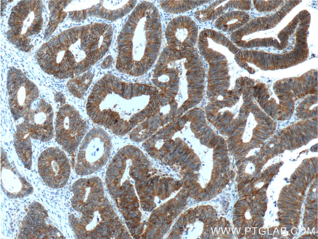
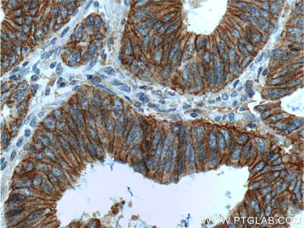
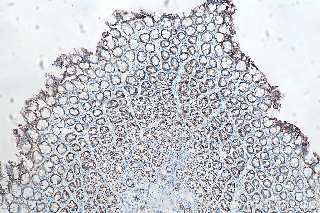
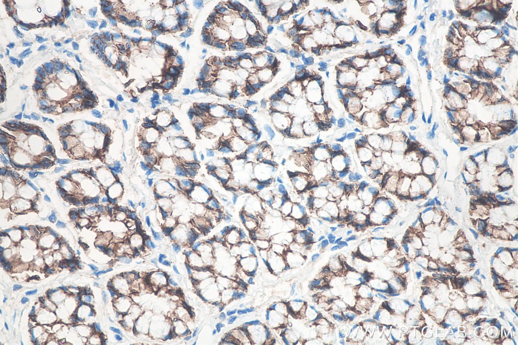
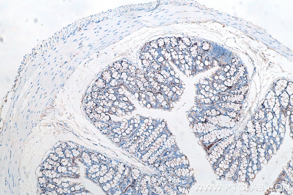
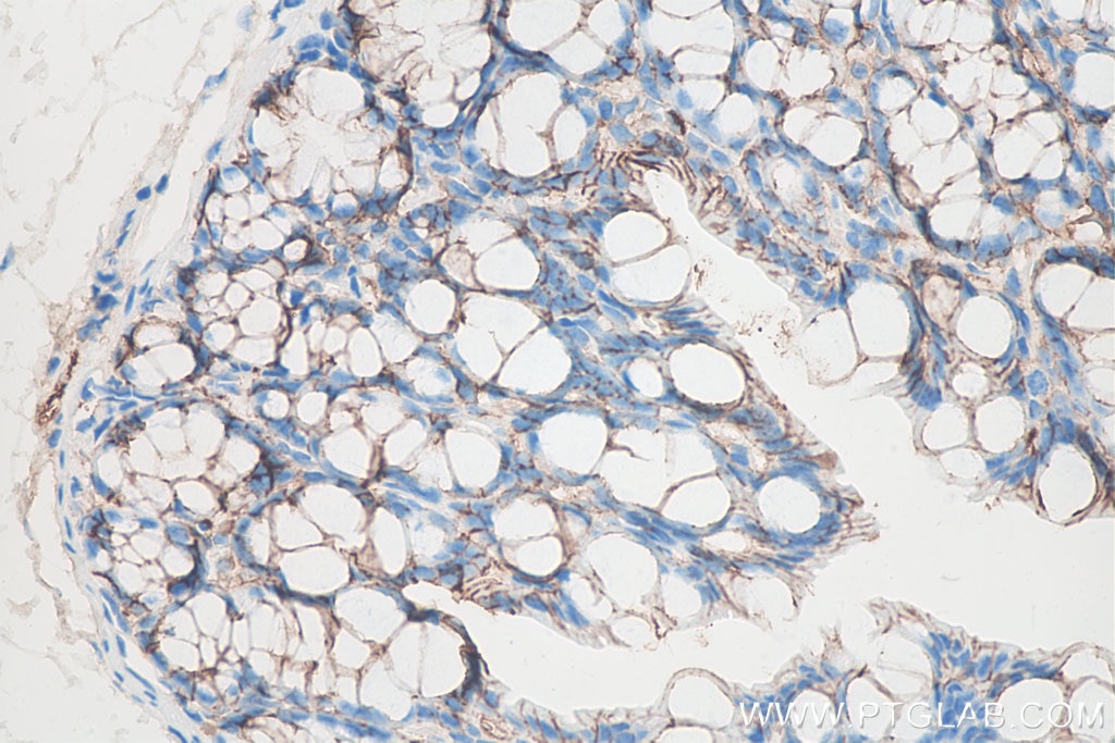
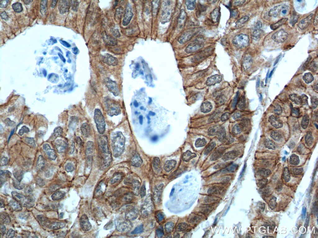
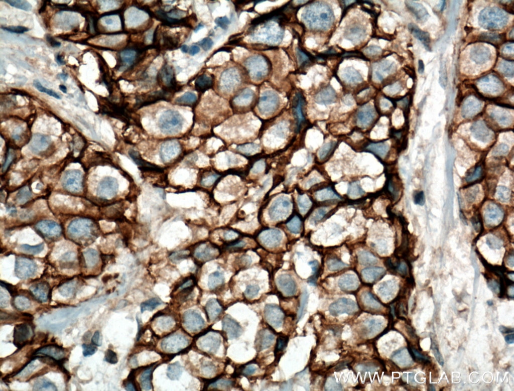
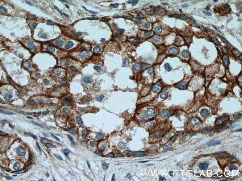
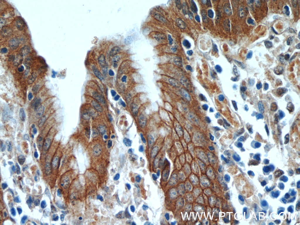
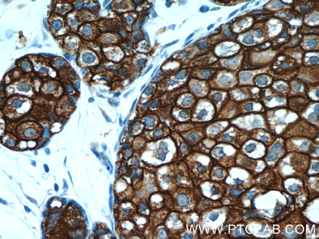
| Résultats positifs en IHC | tissu de cancer du sein humain, tissu de cancer du côlon humain, tissu de côlon de rat, tissu de côlon de souris il est suggéré de démasquer l'antigène avec un tampon de TE buffer pH 9.0; (*) À défaut, 'le démasquage de l'antigène peut être 'effectué avec un tampon citrate pH 6,0. |
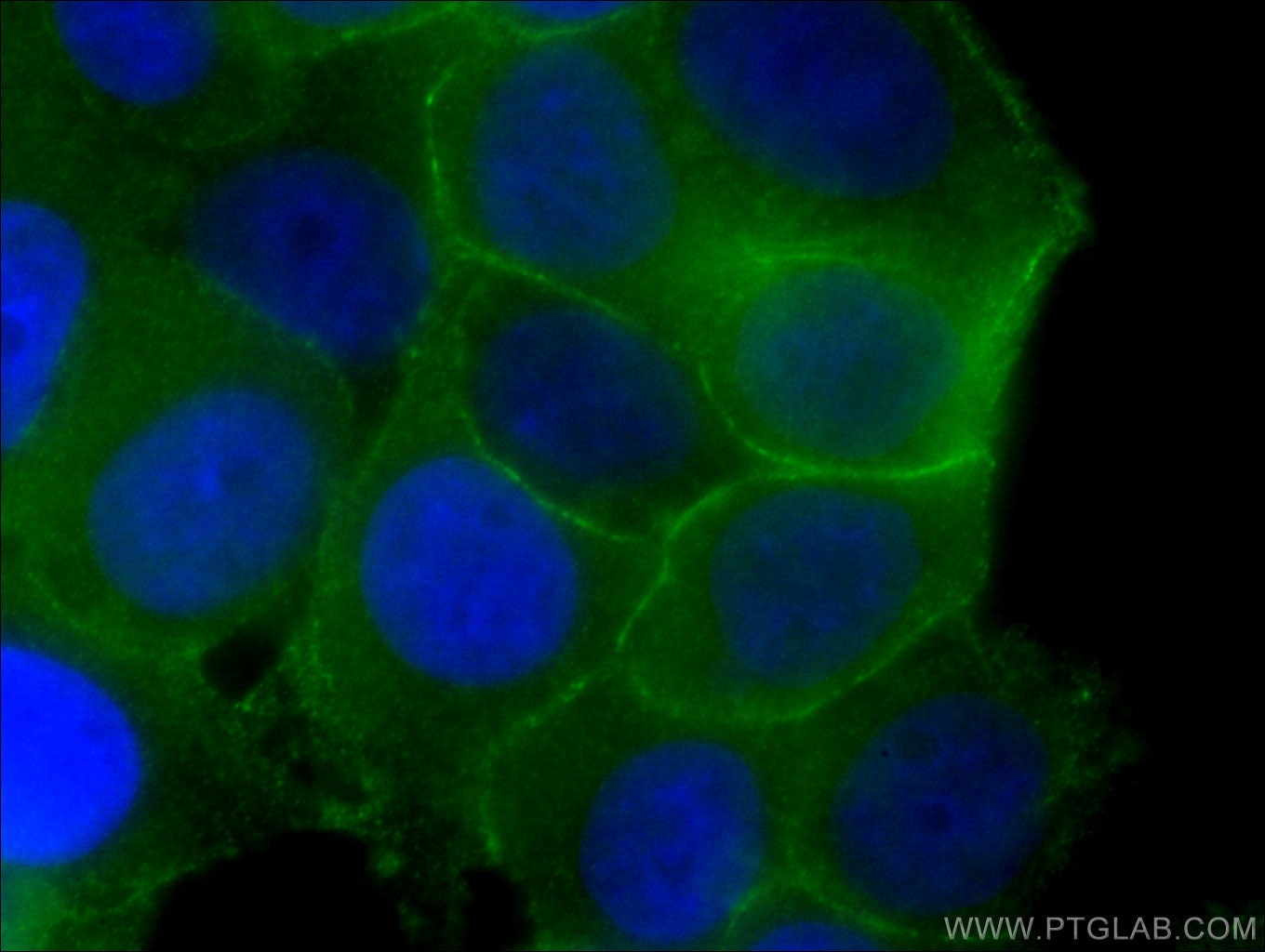
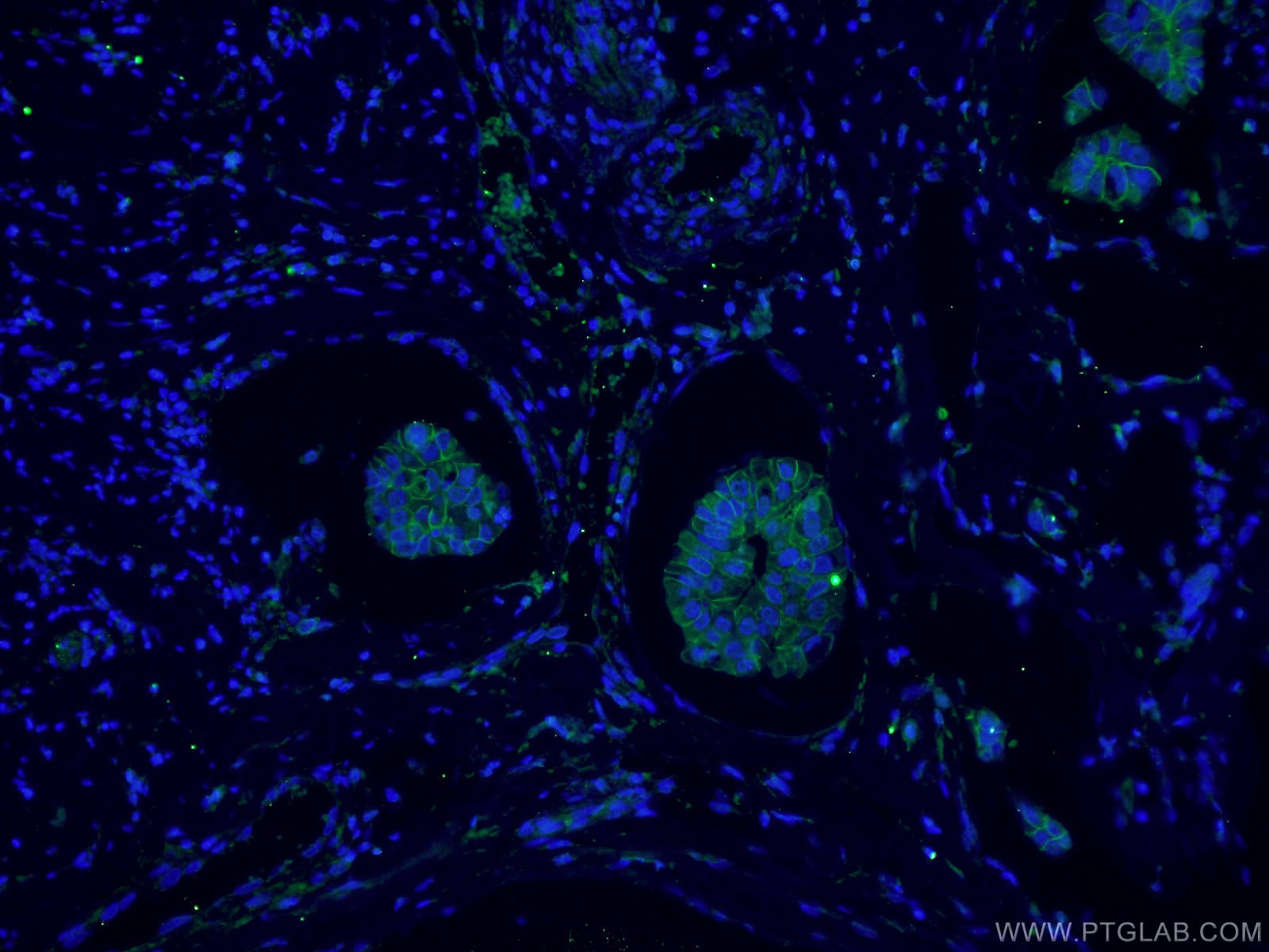
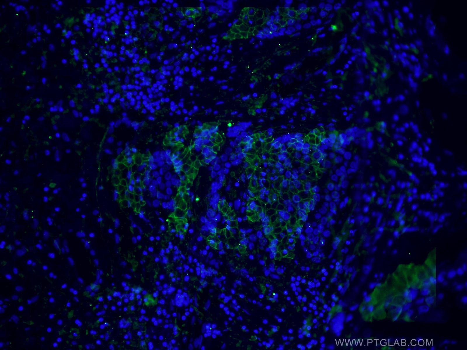
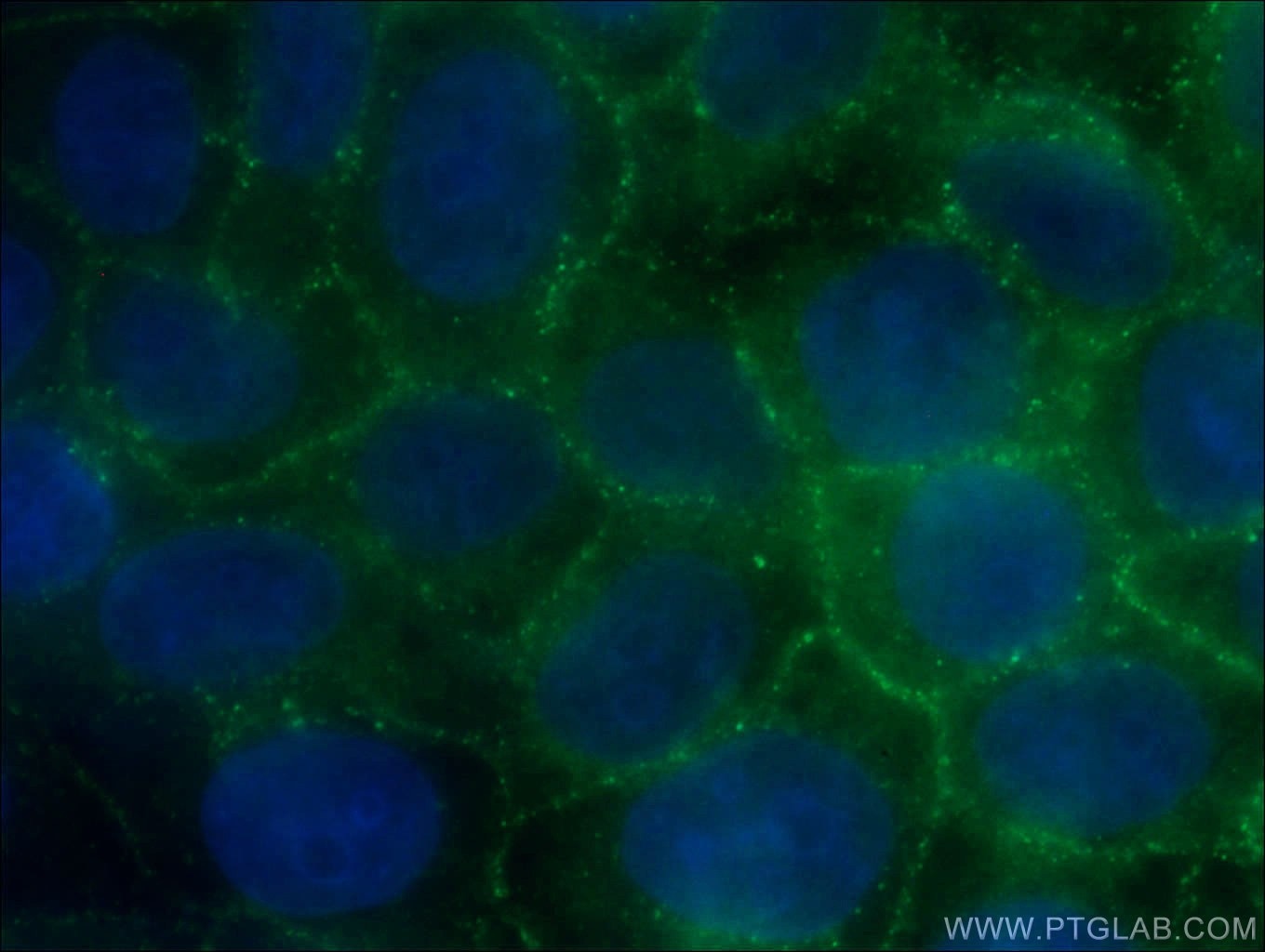
| Résultats positifs en IF | cellules MCF-7, cellules HeLa, tissu de cancer du sein humain |
Dilution recommandée
| Application | Dilution |
|---|---|
| Western Blot (WB) | WB : 1:500-1:2000 |
| Immunohistochimie (IHC) | IHC : 1:250-1:1000 |
| Immunofluorescence (IF) | IF : 1:50-1:500 |
| It is recommended that this reagent should be titrated in each testing system to obtain optimal results. | |
| Sample-dependent, check data in validation data gallery | |
Applications publiées
| WB | See 2 publications below |
| IF | See 3 publications below |
| IP | See 2 publications below |
Informations sur le produit
66208-1-Ig cible p120 Catenin dans les applications de WB, IP, IHC, IF, ELISA et montre une réactivité avec des échantillons Humain, rat, souris
| Réactivité | Humain, rat, souris |
| Réactivité citée | rat, Humain, souris |
| Hôte / Isotype | Mouse / IgG2b |
| Clonalité | Monoclonal |
| Type | Anticorps |
| Immunogène | p120 Catenin Protéine recombinante Ag2824 |
| Nom complet | catenin (cadherin-associated protein), delta 1 |
| Masse moléculaire calculée | 948 aa, 105 kDa |
| Poids moléculaire observé | 90-120 kDa |
| Numéro d’acquisition GenBank | BC010501 |
| Symbole du gène | CTNND1 |
| Identification du gène (NCBI) | 1500 |
| Conjugaison | Non conjugué |
| Forme | Liquide |
| Méthode de purification | Purification par protéine A |
| Tampon de stockage | PBS avec azoture de sodium à 0,02 % et glycérol à 50 % pH 7,3 |
| Conditions de stockage | Stocker à -20°C. Stable pendant un an après l'expédition. L'aliquotage n'est pas nécessaire pour le stockage à -20oC Les 20ul contiennent 0,1% de BSA. |
Informations générales
Catenins were discovered as proteins that are linked to the cytoplasmic domain of transmembrane cadherins (PMID: 9653641). p120 catenin, also called p120 ctn or catenin delta-1, regulates cell-cell adhesion through its interaction with the cytoplasmic tail of classical and type II cadherins. p120 catenin is a tyrosine kinase substrate implicated in cell transformation by SRC, as well as in ligand-induced receptor signaling through the EGF receptor, the PDGF receptor, and the CSF1 receptor. Different expression patterns of p120 catenin in lobular and ductal carcinomas of breast have been reported: membrane stain for ductal carcinoma and cytoplasmic stain for lobular carcinoma (PMID: 24966968). Different isoforms of p120 catenin are variably expressed in different tissues as a result of alternative splicing and the use of multiple translation initiation codons (PMID: 19150613).
Protocole
| Product Specific Protocols | |
|---|---|
| WB protocol for p120 Catenin antibody 66208-1-Ig | Download protocol |
| IHC protocol for p120 Catenin antibody 66208-1-Ig | Download protocol |
| IF protocol for p120 Catenin antibody 66208-1-Ig | Download protocol |
| Standard Protocols | |
|---|---|
| Click here to view our Standard Protocols |
Publications
| Species | Application | Title |
|---|---|---|
Mol Oncol Kaiso phosphorylation at threonine 606 leads to its accumulation in the cytoplasm, reducing its transcriptional repression of the tumour suppressor CDH1. | ||
Development Ontogenesis of the tear drainage system requires Prickle1-driven polarized basement membrane deposition. | ||
Proc Natl Acad Sci U S A SENCR stabilizes vascular endothelial cell adherens junctions through interaction with CKAP4. | ||
Biomed Pharmacother Inhibition of autophagy and RIP1/RIP3/MLKL-mediated necroptosis by edaravone attenuates blood spinal cord barrier disruption following spinal cord injury |
