- Featured Product
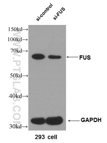
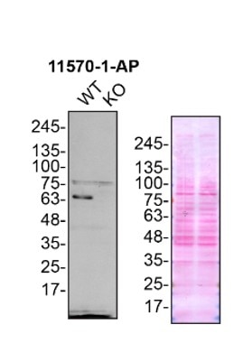
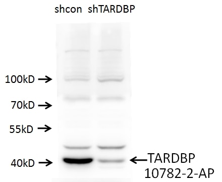
- KD/KO Validated
FUS/TLS Polyklonaler Antikörper
FUS/TLS Polyklonal Antikörper für WB, IHC, IF/ICC, IF-P, IP, ELISA
Wirt / Isotyp
Kaninchen / IgG
Getestete Reaktivität
human, Maus, Ratte und mehr (1)
Anwendung
WB, IHC, IF/ICC, IF-P, IP, CoIP, ChIP, RIP, ELISA
Konjugation
Unkonjugiert
Kat-Nr. : 11570-1-AP
Synonyme
Galerie der Validierungsdaten
Geprüfte Anwendungen
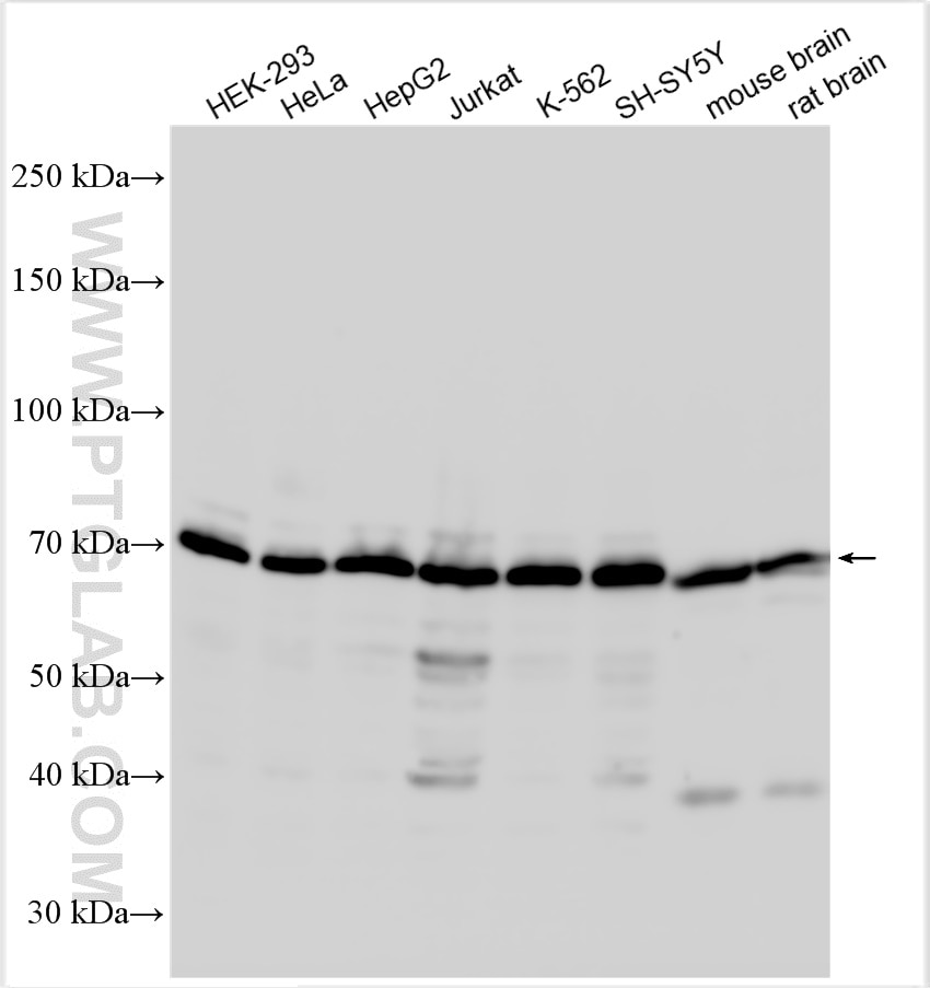
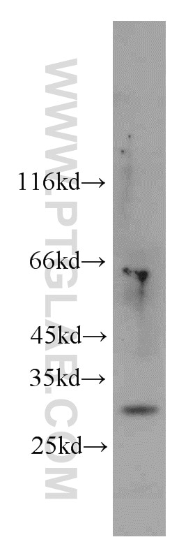
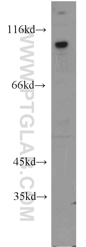
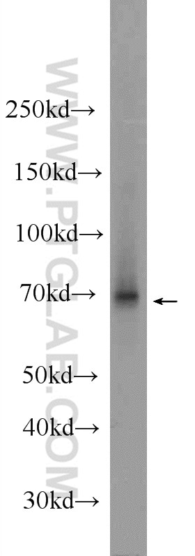
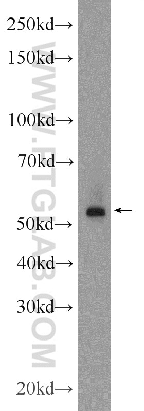
| Erfolgreiche Detektion in WB | HEK-293-Zellen, HeLa-Zellen, HepG2-Zellen, Jurkat-Zellen, K-562-Zellen, Maushirngewebe, Rattenhirngewebe, SH-SY5Y-Zellen |
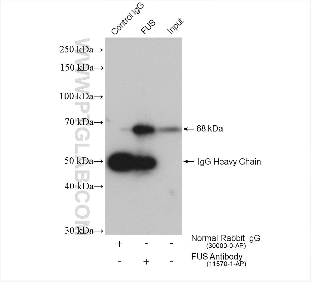
| Erfolgreiche IP | K-562-Zellen |
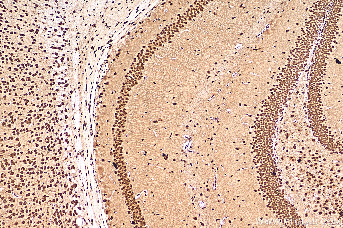
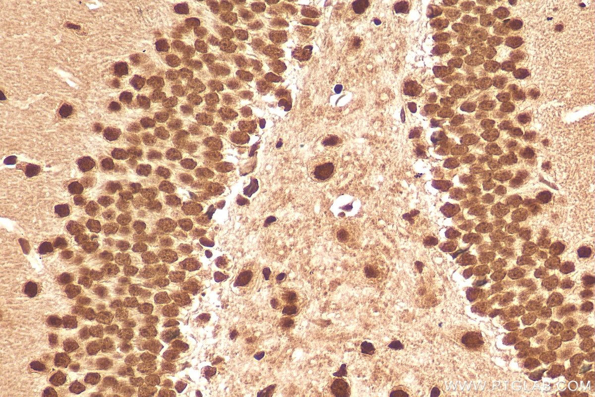
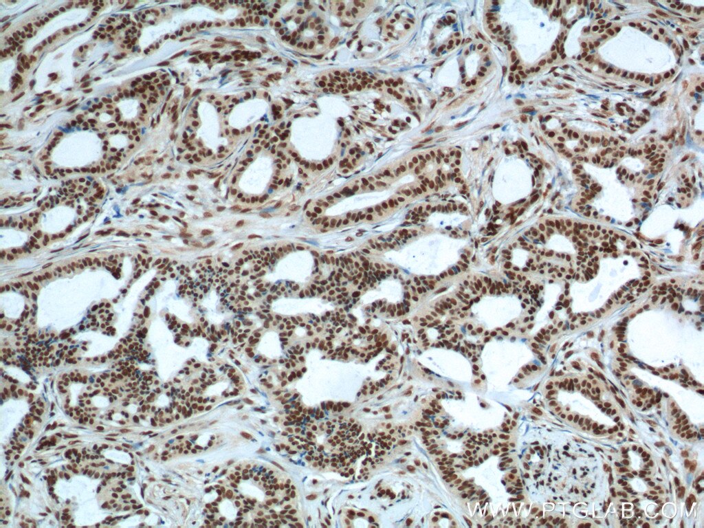
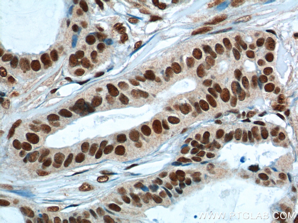
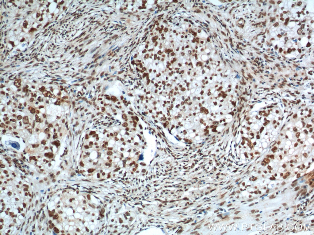
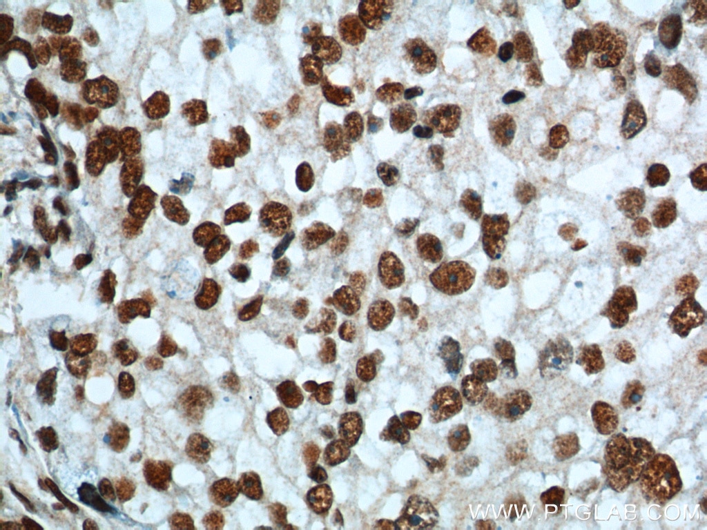
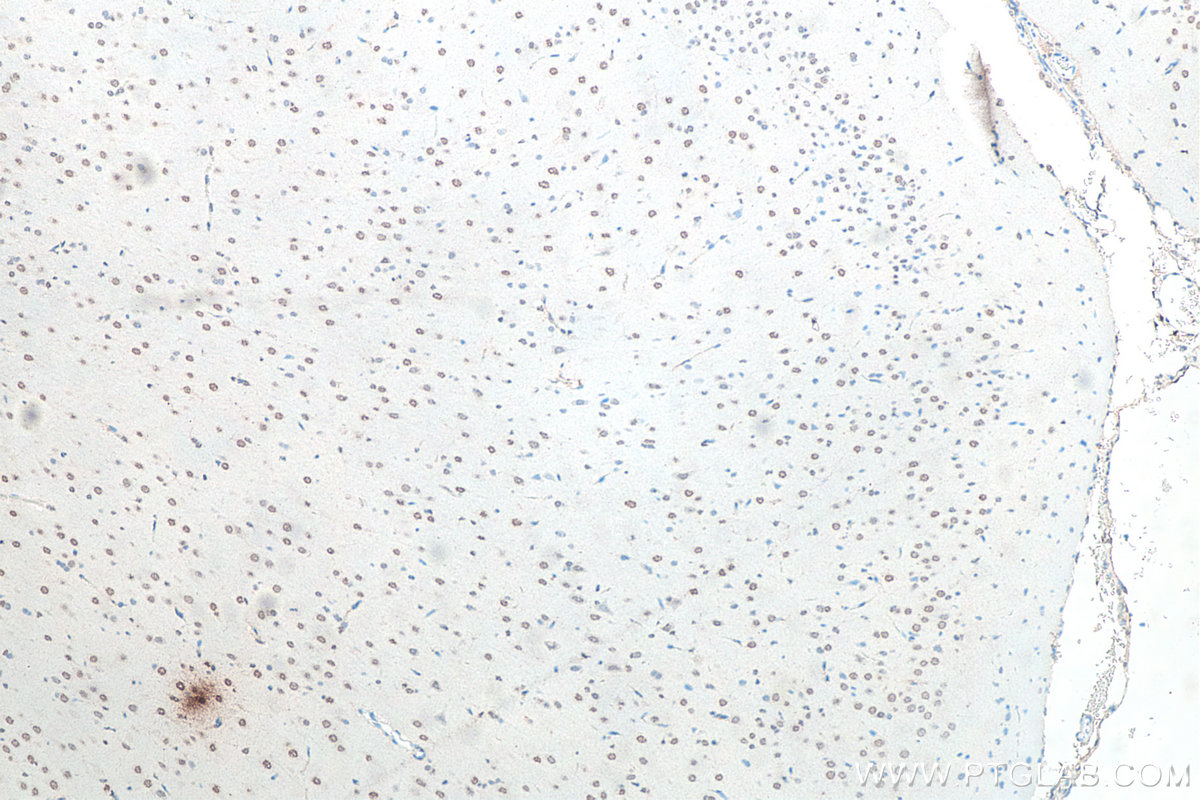
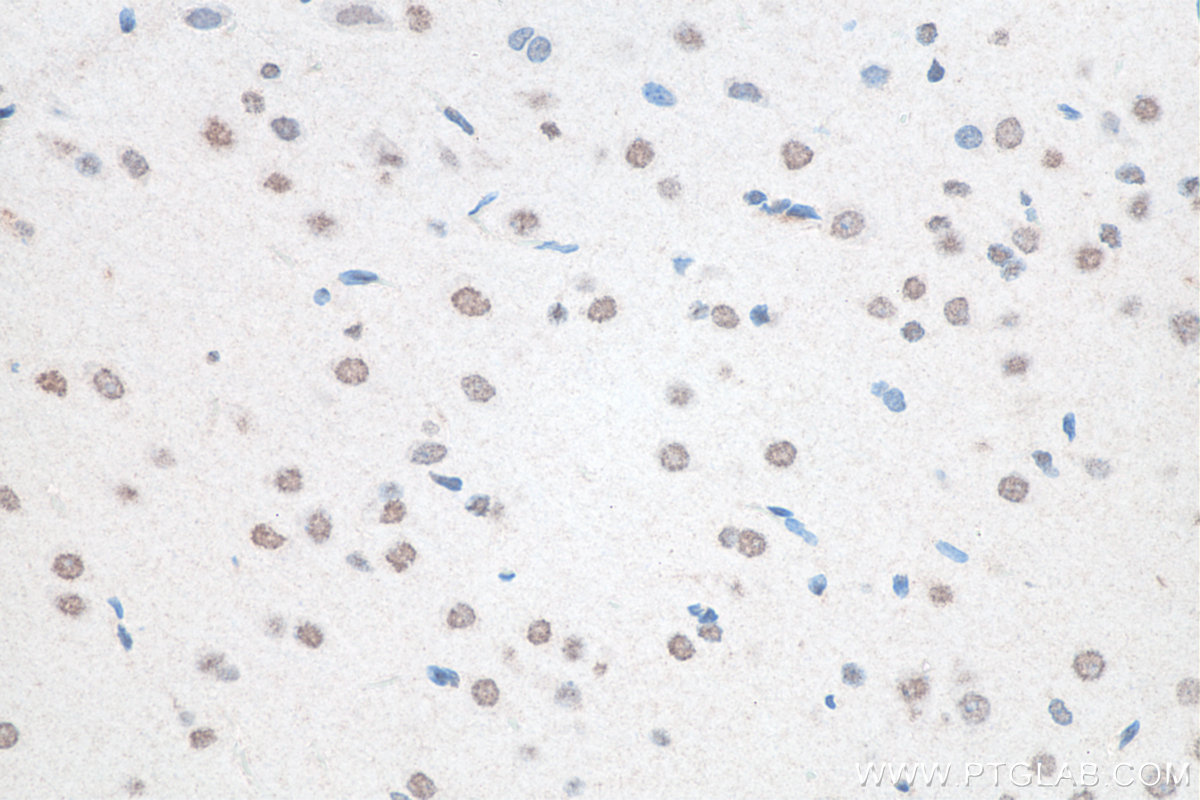
| Erfolgreiche Detektion in IHC | Maushirngewebe, humanes Mammakarzinomgewebe, humanes Ovarialkarzinomgewebe, Rattenhirngewebe Hinweis: Antigendemaskierung mit TE-Puffer pH 9,0 empfohlen. (*) Wahlweise kann die Antigendemaskierung auch mit Citratpuffer pH 6,0 erfolgen. |
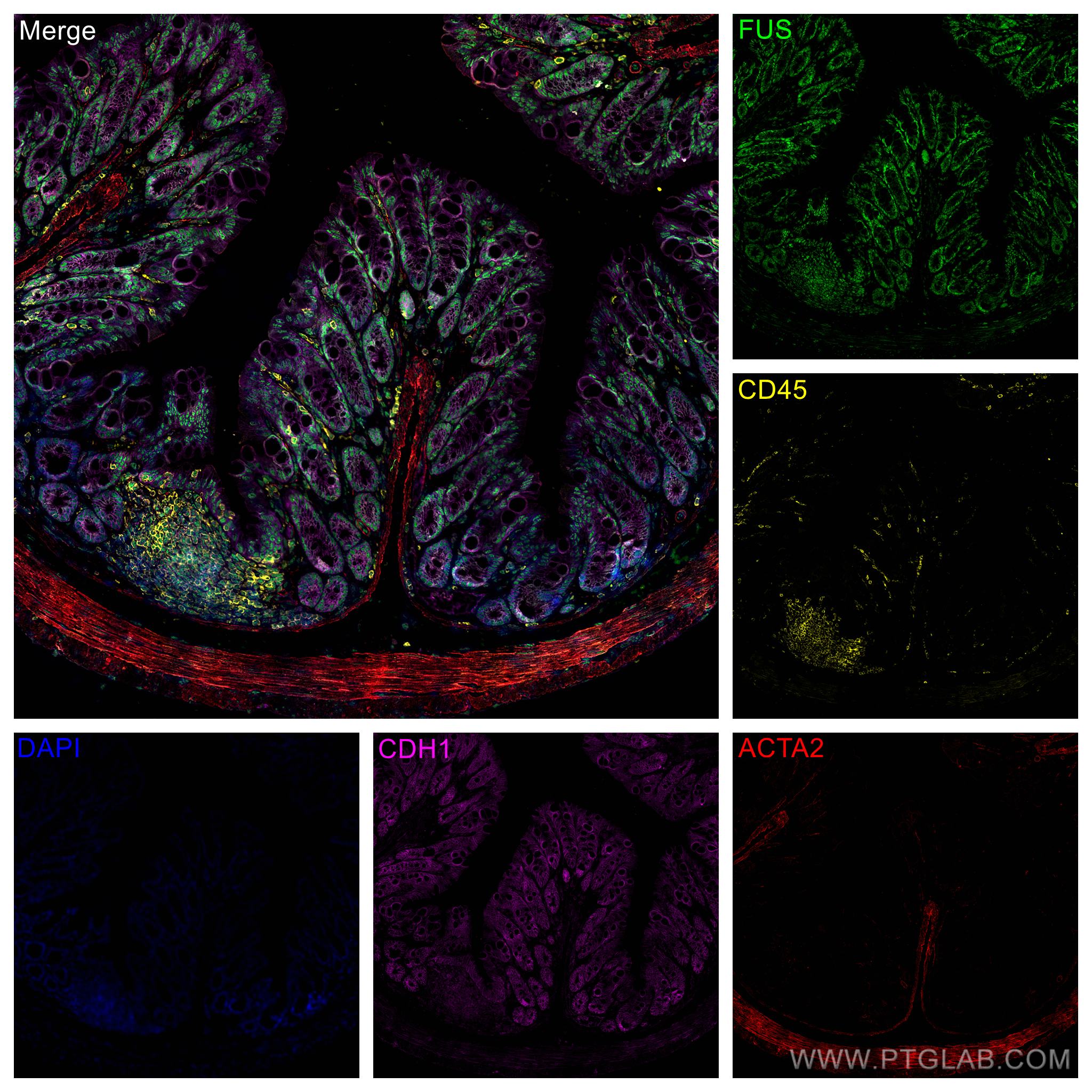
| Erfolgreiche Detektion in IF-P | Maus-Kolongewebe, HeLa-Zellen, HepG2-Zellen |
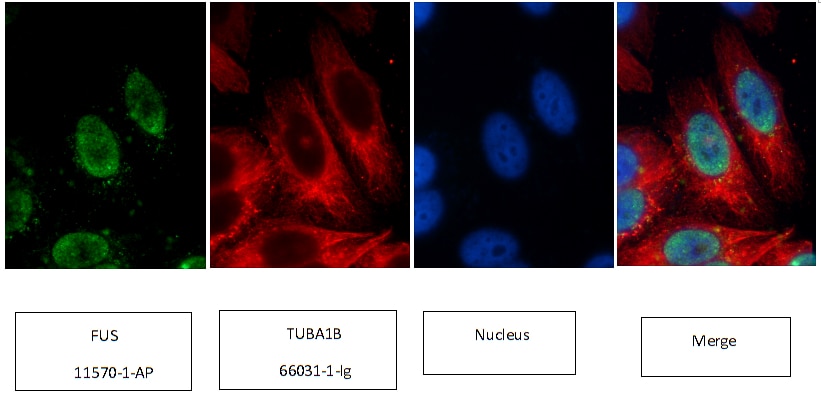
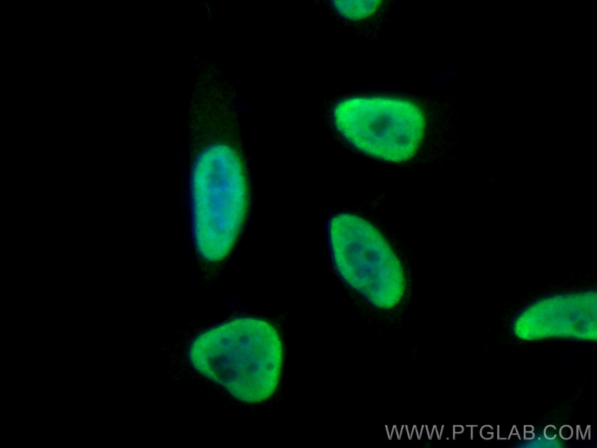
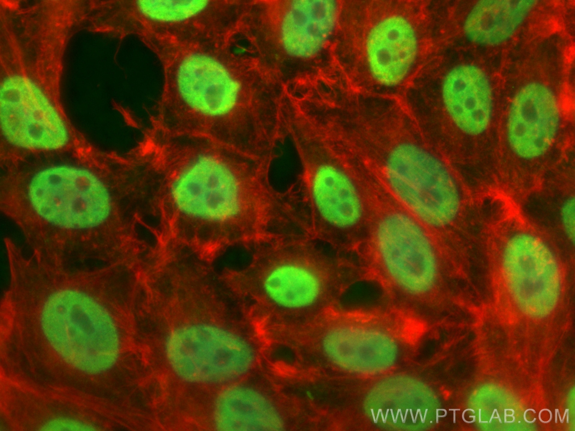
| Erfolgreiche Detektion in IF/ICC | HepG2-Zellen, HeLa-Zellen |
Empfohlene Verdünnung
| Anwendung | Verdünnung |
|---|---|
| Western Blot (WB) | WB : 1:5000-1:50000 |
| Immunpräzipitation (IP) | IP : 0.5-4.0 ug for 1.0-3.0 mg of total protein lysate |
| Immunhistochemie (IHC) | IHC : 1:50-1:500 |
| Immunfluoreszenz (IF)-P | IF-P : 1:50-1:500 |
| Immunfluoreszenz (IF)/ICC | IF/ICC : 1:50-1:500 |
| It is recommended that this reagent should be titrated in each testing system to obtain optimal results. | |
| Sample-dependent, check data in validation data gallery | |
Veröffentlichte Anwendungen
Produktinformation
11570-1-AP bindet in WB, IHC, IF/ICC, IF-P, IP, CoIP, ChIP, RIP, ELISA FUS/TLS und zeigt Reaktivität mit human, Maus, Ratten
| Getestete Reaktivität | human, Maus, Ratte |
| In Publikationen genannte Reaktivität | human, Huhn, Maus, Ratte |
| Wirt / Isotyp | Kaninchen / IgG |
| Klonalität | Polyklonal |
| Typ | Antikörper |
| Immunogen | FUS/TLS fusion protein Ag2150 |
| Vollständiger Name | fusion (involved in t(12;16) in malignant liposarcoma) |
| Berechnetes Molekulargewicht | 75 kDa |
| Beobachtetes Molekulargewicht | 68-75 kDa |
| GenBank-Zugangsnummer | BC026062 |
| Gene symbol | FUS/TLS |
| Gene ID (NCBI) | 2521 |
| Konjugation | Unkonjugiert |
| Form | Liquid |
| Reinigungsmethode | Antigen-Affinitätsreinigung |
| Lagerungspuffer | PBS mit 0.02% Natriumazid und 50% Glycerin pH 7.3. |
| Lagerungsbedingungen | Bei -20°C lagern. Nach dem Versand ein Jahr lang stabil Aliquotieren ist bei -20oC Lagerung nicht notwendig. 20ul Größen enthalten 0,1% BSA. |
Hintergrundinformationen
FUS (also named TLS and POMp75) belongs to the RRM TET family. FUS may play a role in the maintenance of genomic integrity; it binds both single-stranded and double-stranded DNA and promotes ATP-independent annealing of complementary single-stranded DNAs and D-loop formation in superhelical double-stranded DNA. FUS is also an RNA-binding protein, and its links to neurodegenerative disease proffer the intriguing possibility that altered RNA metabolism or RNA processing may underlie or contribute to neuron degeneration[PMID: 22640227]. FUS may be a cause of angiomatoid fibrous histiocytoma (AFH) and is implicated in certain forms of amyotrophic lateral sclerosis (ALS) and frontotemporal dementias (FTDs) such as frontotemporal lobar dementia with ubiquitin inclusions (FTLD-U)[PMID: 22640227]. This antibody is a rabbit polyclonal antibody raised against an internal region of human FUS. FUS was detected double bands of 68-74 kDa (PMID:31519807).
Protokolle
| Produktspezifische Protokolle | |
|---|---|
| WB protocol for FUS/TLS antibody 11570-1-AP | Protokoll herunterladen |
| IHC protocol for FUS/TLS antibody 11570-1-AP | Protokoll herunterladen |
| IF protocol for FUS/TLS antibody 11570-1-AP | Protokoll herunterladen |
| IP protocol for FUS/TLS antibody 11570-1-AP | Protokoll herunterladen |
| Standard-Protokolle | |
|---|---|
| Klicken Sie hier, um unsere Standardprotokolle anzuzeigen |
Publikationen
- Journal Impact Factor
- Most recent
| Species | Application | Title |
|---|---|---|
Nature Mutations in UBQLN2 cause dominant X-linked juvenile and adult-onset ALS and ALS/dementia. | ||
Nat Med Antisense oligonucleotide silencing of FUS expression as a therapeutic approach in amyotrophic lateral sclerosis. | ||
Cell Nuclear-Import Receptors Reverse Aberrant Phase Transitions of RNA-Binding Proteins with Prion-like Domains. | ||
Cell Metab NEAT1 is essential for metabolic changes that promote breast cancer growth and metastasis. | ||
Nat Neurosci FUS-mediated regulation of acetylcholine receptor transcription at neuromuscular junctions is compromised in amyotrophic lateral sclerosis.
|
Rezensionen
The reviews below have been submitted by verified Proteintech customers who received an incentive for providing their feedback.
FH Xiaochen (Verified Customer) (07-08-2024) | Sensitivie for IF and show image with good quelity.
 |
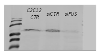
FH Xhuljana (Verified Customer) (03-01-2024) | Used in siRNA transfected C2C12 cells
 |
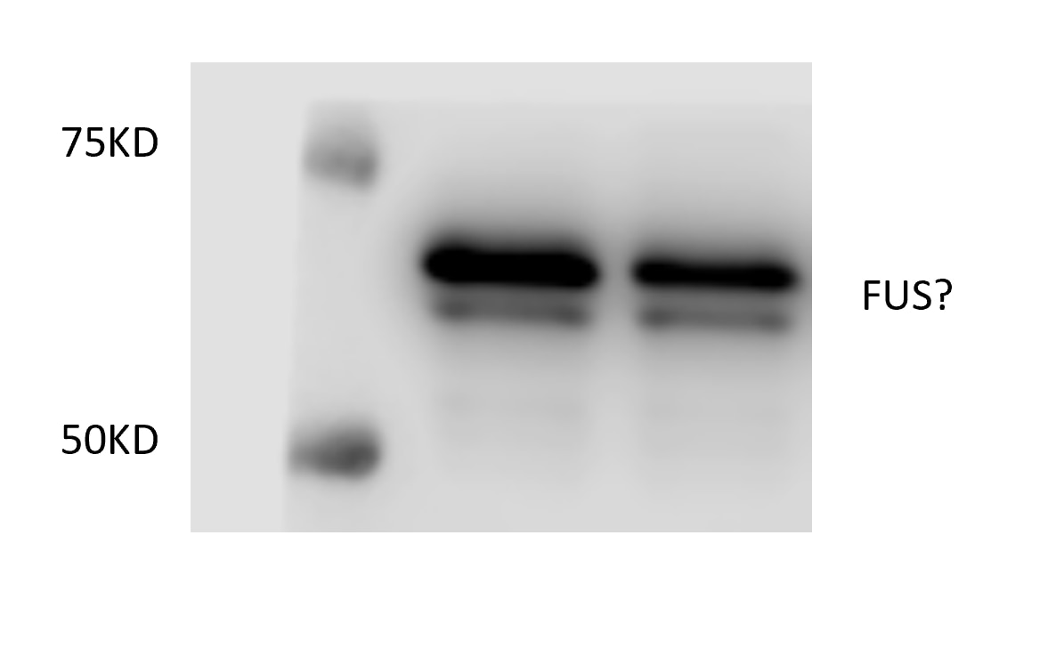
FH Zhongwen (Verified Customer) (09-25-2023) | I can find two bands in the target region. I am not sure which one is the target band.
 |
FH manohar (Verified Customer) (07-10-2023) | Nitrocellulose membrane is used with 5% milk as blocking and antibody diluted in 1% milk and incubated overnight.
|
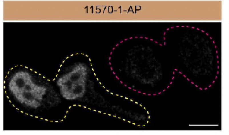
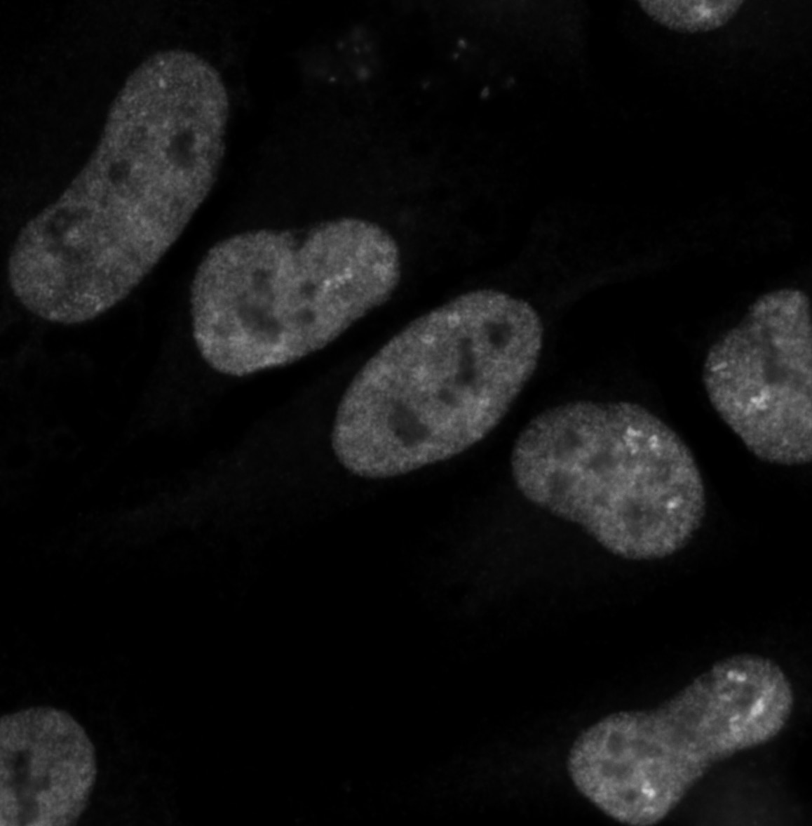
FH Tatyana (Verified Customer) (05-14-2023) | ICC using 5% goat serum/PBST buffer, 2 hours at RT. Good specific nuclear signal.
 |
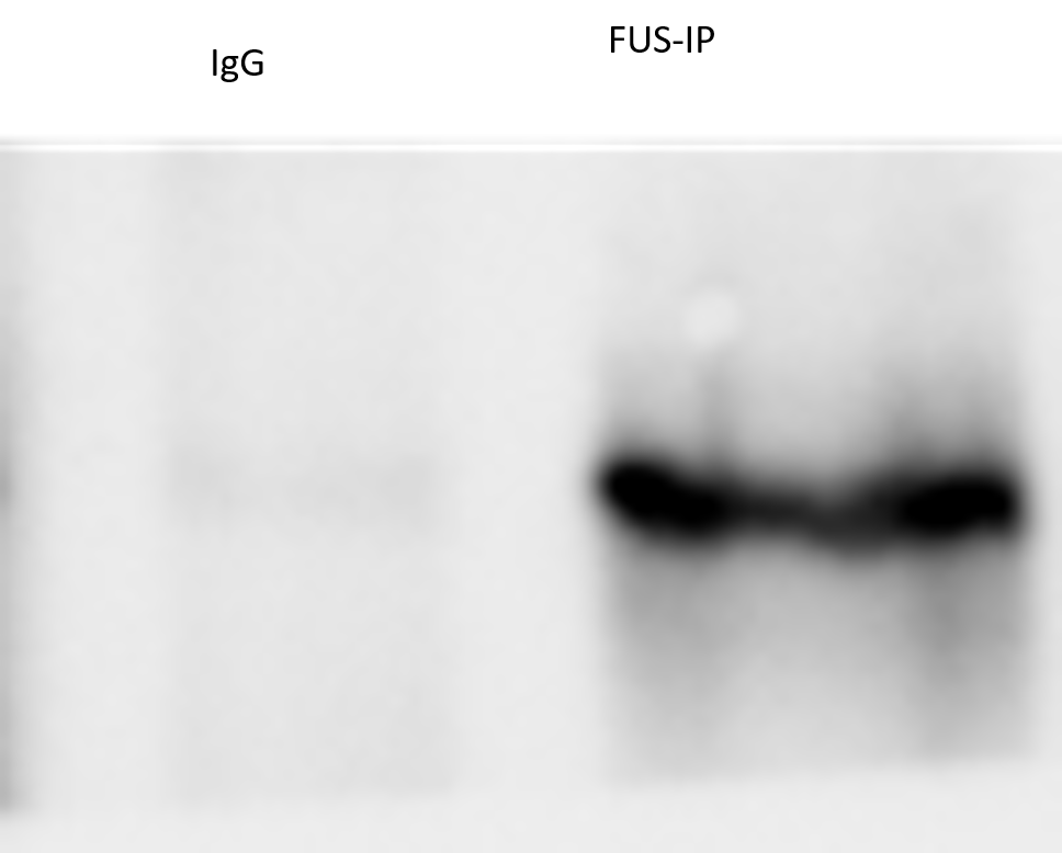
FH shashirekha (Verified Customer) (12-23-2020) | Used for immunopreciptation at 1:1000 dilution. Works very well
 |
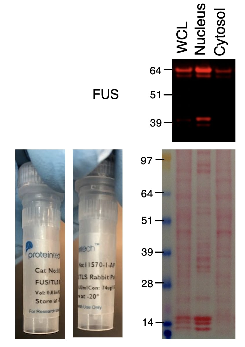
FH H (Verified Customer) (04-06-2020) | The antibody worked well for HCT116 cell line. Nuclear cytosolic fractionation clearly showed that FUS is dominantly in nucleus, and sub-fraction is present in cytosol.
 |
FH Karthik (Verified Customer) (04-24-2019) | Magenta- FUSBlue- MAP2FUS staining consistent obtained with this antibody is consistent with literature
|
FH Yen-Chen (Verified Customer) (12-03-2018) |
|
