- Featured Product
- KD/KO Validated
APAF1 Polyklonaler Antikörper
APAF1 Polyklonal Antikörper für IF, IHC, IP, WB, ELISA
Wirt / Isotyp
Kaninchen / IgG
Getestete Reaktivität
human, Ratte und mehr (1)
Anwendung
WB, IP, IHC, IF, ELISA
Konjugation
Unkonjugiert
Kat-Nr. : 21710-1-AP
Synonyme
Galerie der Validierungsdaten
Geprüfte Anwendungen
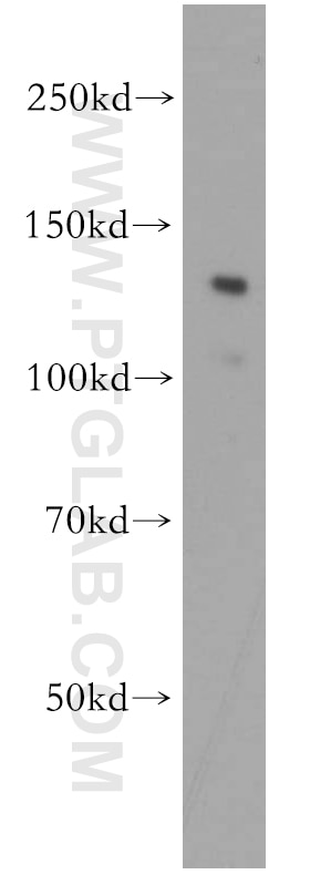
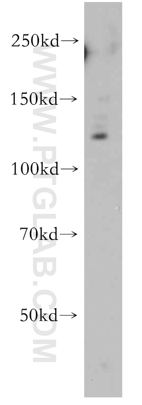
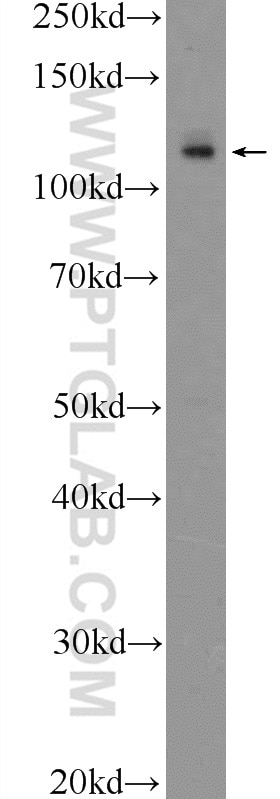
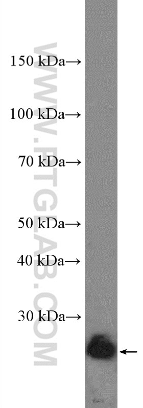
| Erfolgreiche Detektion in WB | HEK-293-Zellen, A549-Zellen, COLO 320-Zellen |
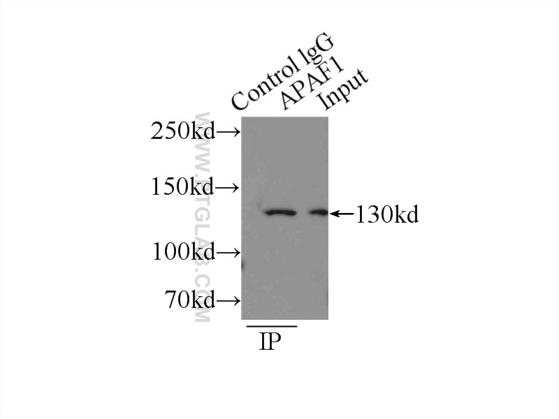
| Erfolgreiche IP | HEK-293-Zellen |
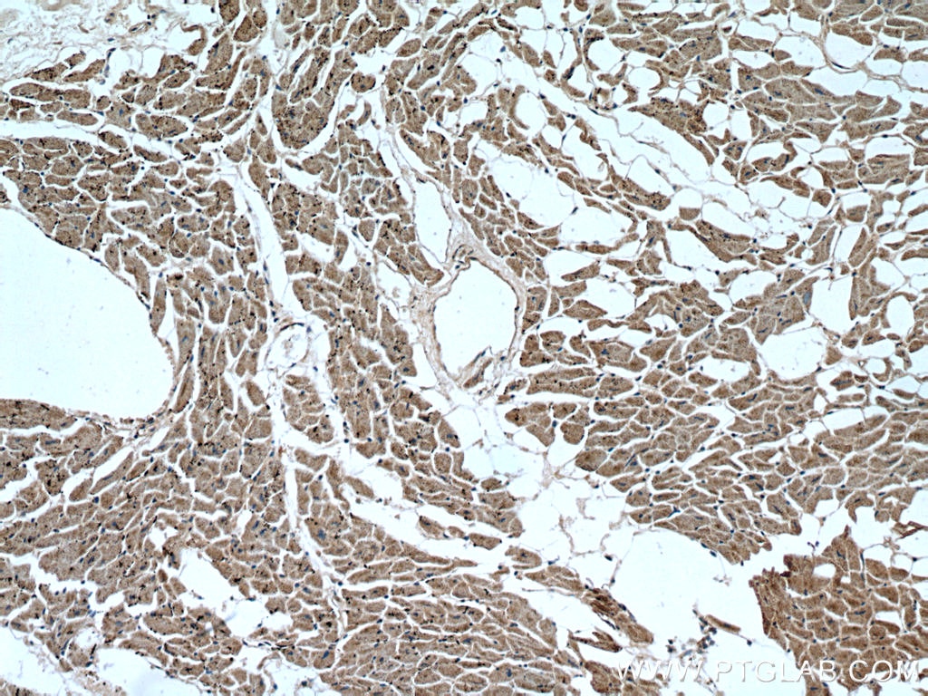
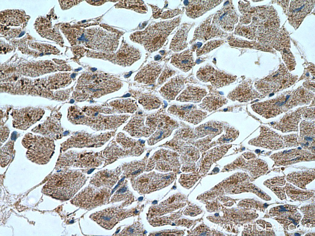
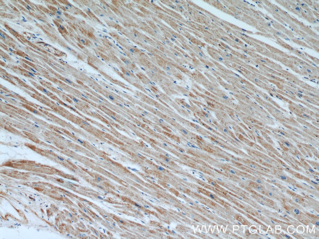
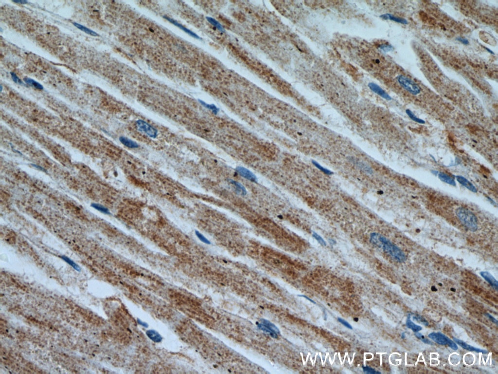
| Erfolgreiche Detektion in IHC | humanes Herzgewebe Hinweis: Antigendemaskierung mit TE-Puffer pH 9,0 empfohlen. (*) Wahlweise kann die Antigendemaskierung auch mit Citratpuffer pH 6,0 erfolgen. |
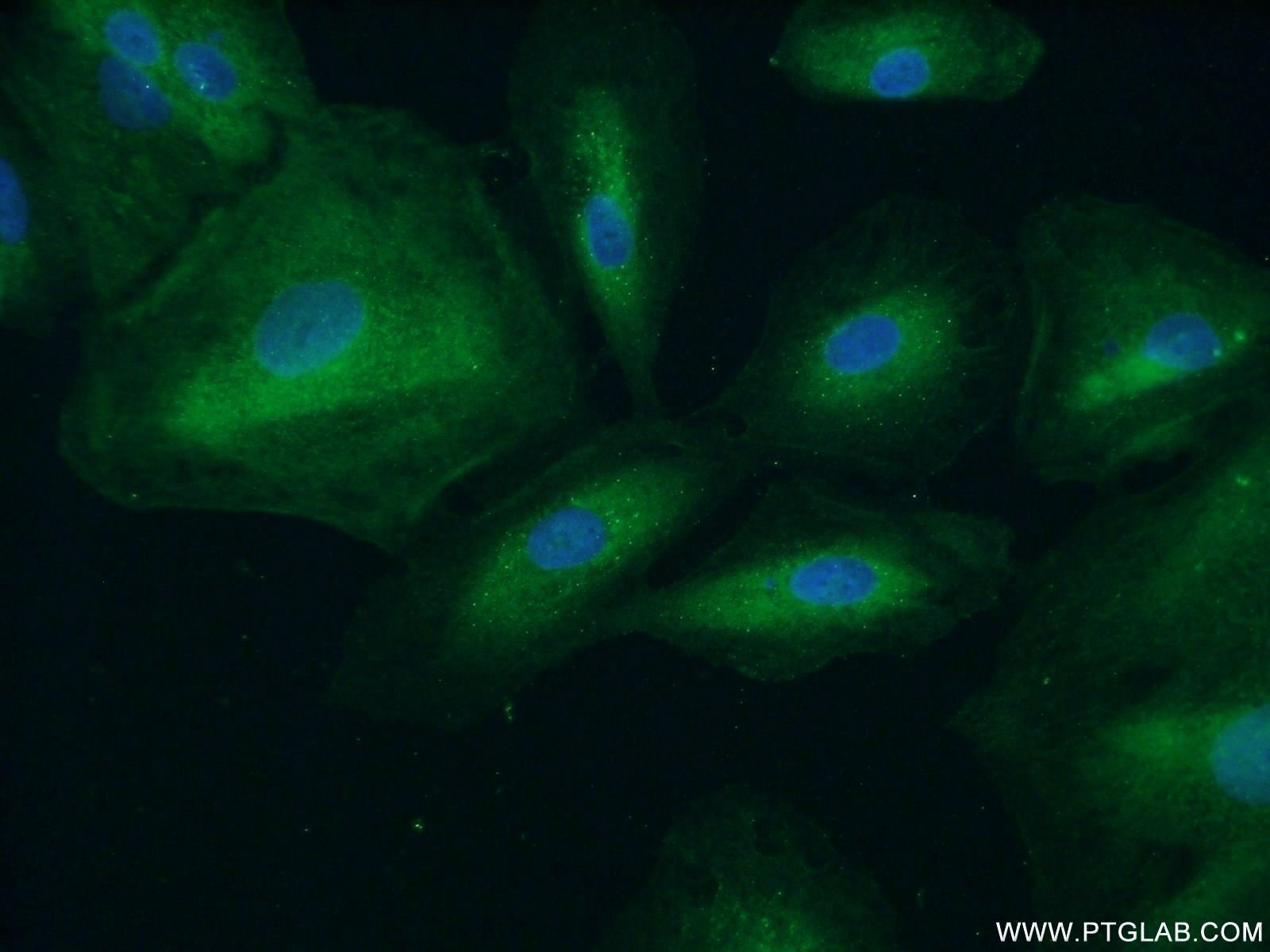
| Erfolgreiche Detektion in IF | A549-Zellen |
Empfohlene Verdünnung
| Anwendung | Verdünnung |
|---|---|
| Western Blot (WB) | WB : 1:500-1:2000 |
| Immunpräzipitation (IP) | IP : 0.5-4.0 ug for 1.0-3.0 mg of total protein lysate |
| Immunhistochemie (IHC) | IHC : 1:50-1:500 |
| Immunfluoreszenz (IF) | IF : 1:20-1:200 |
| It is recommended that this reagent should be titrated in each testing system to obtain optimal results. | |
| Sample-dependent, check data in validation data gallery | |
Veröffentlichte Anwendungen
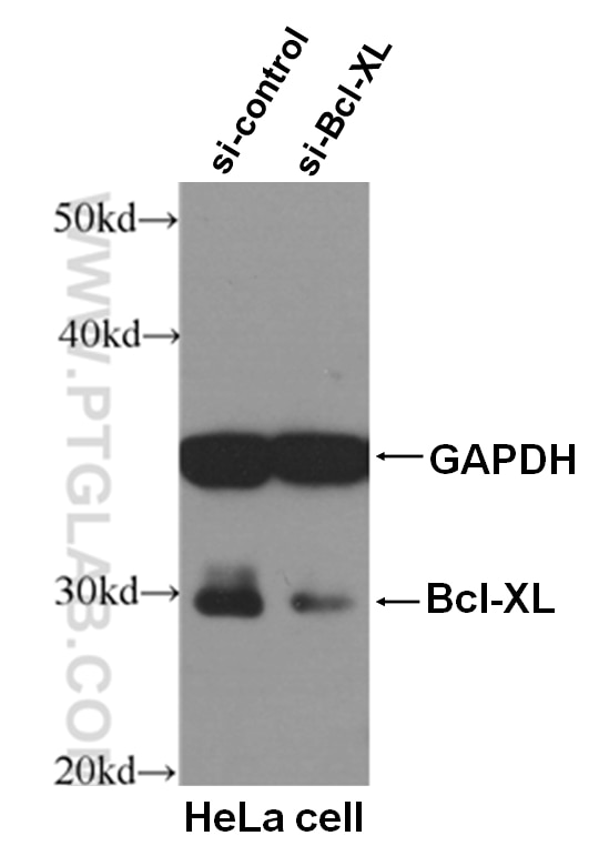
| KD/KO | See 1 publications below |
| WB | See 42 publications below |
| IHC | See 3 publications below |
| IF | See 2 publications below |
Produktinformation
21710-1-AP bindet in WB, IP, IHC, IF, ELISA APAF1 und zeigt Reaktivität mit human, Ratten
| Getestete Reaktivität | human, Ratte |
| In Publikationen genannte Reaktivität | human, Maus, Ratte |
| Wirt / Isotyp | Kaninchen / IgG |
| Klonalität | Polyklonal |
| Typ | Antikörper |
| Immunogen | APAF1 fusion protein Ag16336 |
| Vollständiger Name | apoptotic peptidase activating factor 1 |
| Berechnetes Molekulargewicht | 1248 aa, 142 kDa |
| Beobachtetes Molekulargewicht | 130-142 kDa |
| GenBank-Zugangsnummer | BC136532 |
| Gene symbol | APAF1 |
| Gene ID (NCBI) | 317 |
| Konjugation | Unkonjugiert |
| Form | Liquid |
| Reinigungsmethode | Antigen-Affinitätsreinigung |
| Lagerungspuffer | PBS mit 0.02% Natriumazid und 50% Glycerin pH 7.3. |
| Lagerungsbedingungen | Bei -20°C lagern. Nach dem Versand ein Jahr lang stabil Aliquotieren ist bei -20oC Lagerung nicht notwendig. 20ul Größen enthalten 0,1% BSA. |
Hintergrundinformationen
APAF1, also named as Apoptotic protease-activating factor 1 or KIAA0413, is a 1248 amino acid protein, which contains one CARD domain, contains one NB-ARC domain and Contains fifteen WD repeats. APAF1 localizes in the cytoplasm and is widely expressed in many tissues. Highest levels of expression is in adult spleen, peripheral blood leukocytes, in fetal brain, kidney and lung. Oligomeric APAF1 mediates the cytochrome c-dependent autocatalytic activation of pro-caspase-9 (Apaf-3), leading to the activation of caspase-3 and apoptosis.
Protokolle
| Produktspezifische Protokolle | |
|---|---|
| WB protocol for APAF1 antibody 21710-1-AP | Protokoll herunterladen |
| IHC protocol for APAF1 antibody 21710-1-AP | Protokoll herunterladen |
| IF protocol for APAF1 antibody 21710-1-AP | Protokoll herunterladen |
| IP protocol for APAF1 antibody 21710-1-AP | Protokoll herunterladen |
| Standard-Protokolle | |
|---|---|
| Klicken Sie hier, um unsere Standardprotokolle anzuzeigen |
Publikationen
| Species | Application | Title |
|---|---|---|
Theranostics Pro-Inflammatory CXCR3 Impairs Mitochondrial Function in Experimental Non-Alcoholic Steatohepatitis. | ||
Oxid Med Cell Longev Apoptotic Protease Activating Factor-1 Inhibitor Mitigates Myocardial Ischemia Injury via Disturbing Procaspase-9 Recruitment by Apaf-1. | ||
Mol Ther Nucleic Acids LNC473 Regulating APAF1 IRES-Dependent Translation via Competitive Sponging miR574 and miR15b: Implications in Colorectal Cancer. | ||
Cell Biol Toxicol Inhibition of ER stress attenuates kidney injury and apoptosis induced by 3-MCPD via regulating mitochondrial fission/fusion and Ca2+ homeostasis. | ||
Front Cell Dev Biol Antagonizing CDK8 Sensitizes Colorectal Cancer to Radiation Through Potentiating the Transcription of e2f1 Target Gene apaf1. | ||
Cells FUT2 Facilitates Autophagy and Suppresses Apoptosis via p53 and JNK Signaling in Lung Adenocarcinoma Cells |
