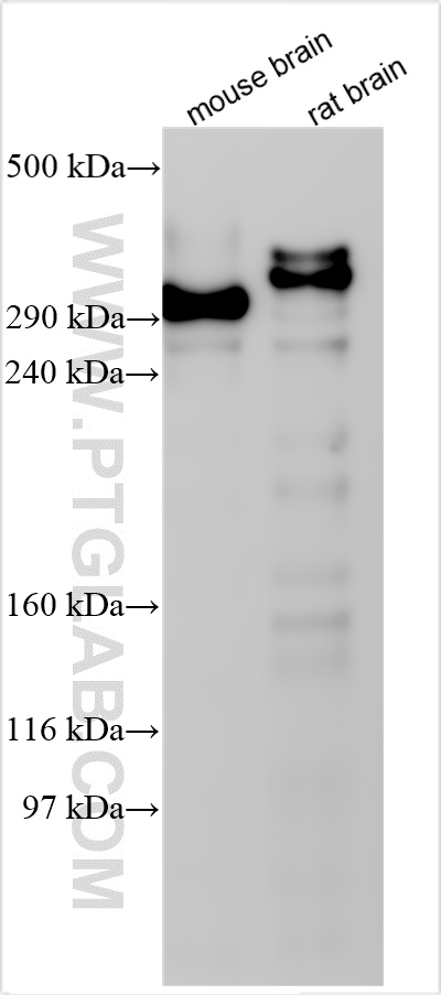
Various lysates were subjected to SDS PAGE followed by western blot with 84306-3-RR (MAP2 antibody) at dilution of 1:1000 incubated at room temperature for 1.5 hours.
Various lysates were subjected to SDS PAGE followed by western blot with 84306-3-RR (MAP2 antibody) at dilution of 1:1000 incubated at room temperature for 1.5 hours.
WB analysis using 84306-3-RR
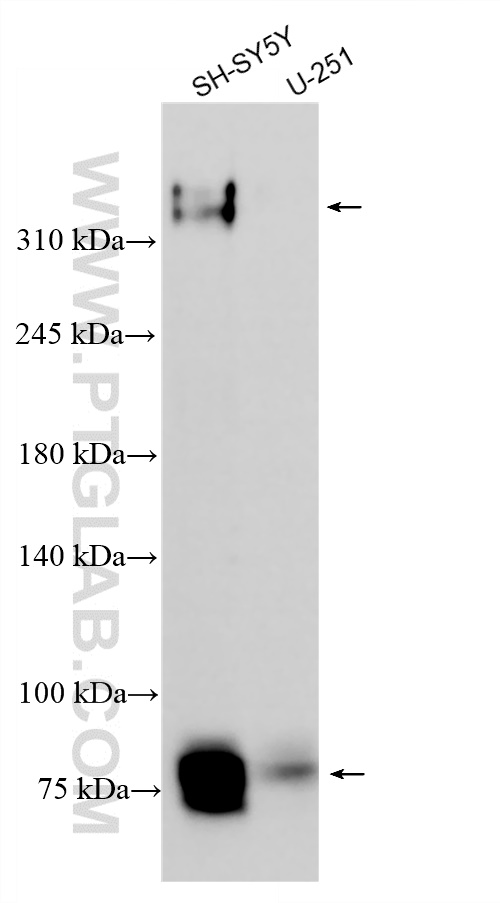
SH-SY5Y cells were subjected to SDS PAGE followed by western blot with 84306-3-RR (MAP2 antibody) at dilution of 1:2000 incubated at room temperature for 1.5 hours.
SH-SY5Y cells were subjected to SDS PAGE followed by western blot with 84306-3-RR (MAP2 antibody) at dilution of 1:2000 incubated at room temperature for 1.5 hours.
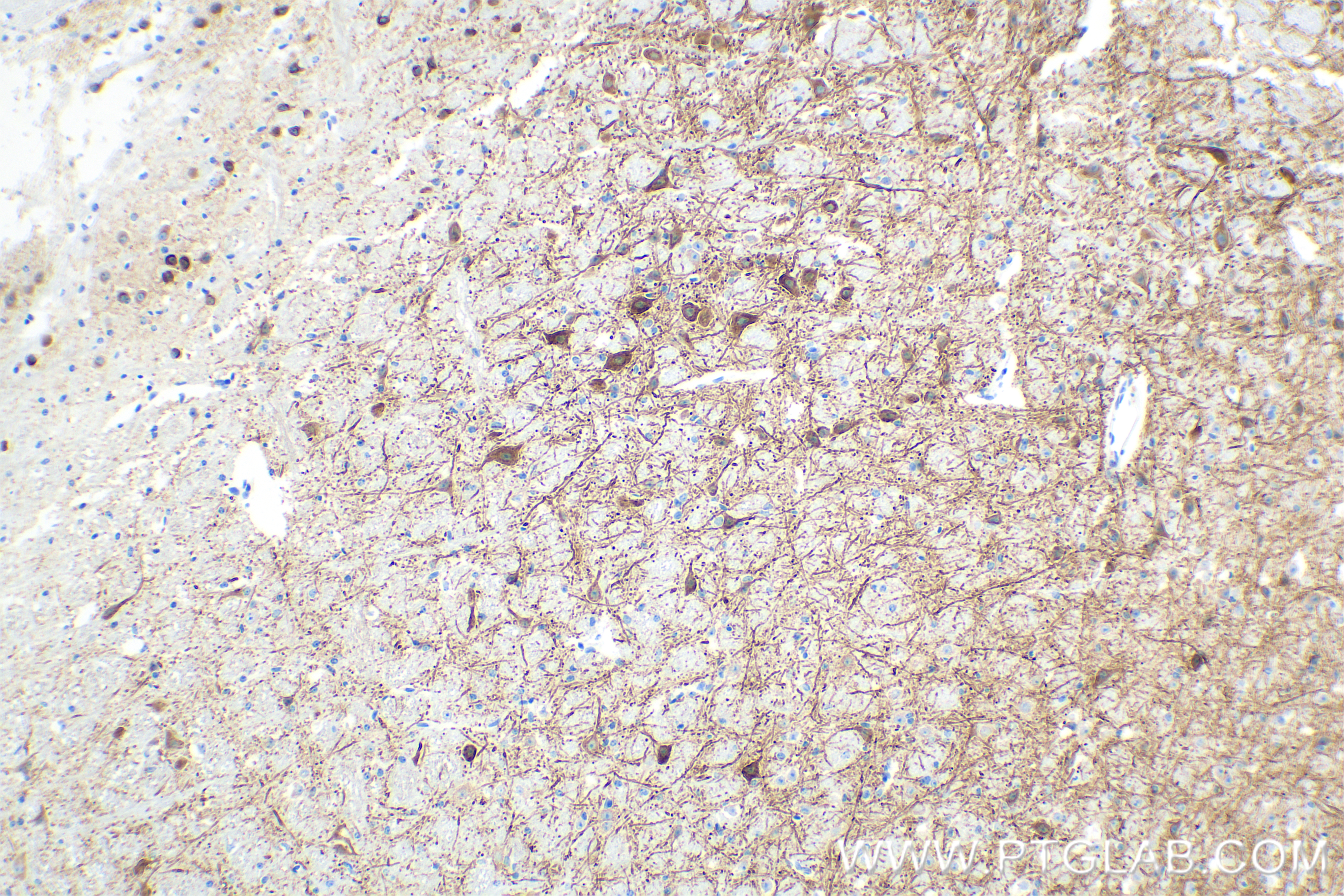
IHC staining of mouse cerebellum using 84306-3-RR
Immunohistochemical analysis of paraffin-embedded mouse cerebellum tissue slide using 84306-3-RR (MAP2 antibody) at dilution of 1:4000 (under 10x lens). Heat mediated antigen retrieval with Tris-EDTA buffer (pH 9.0).
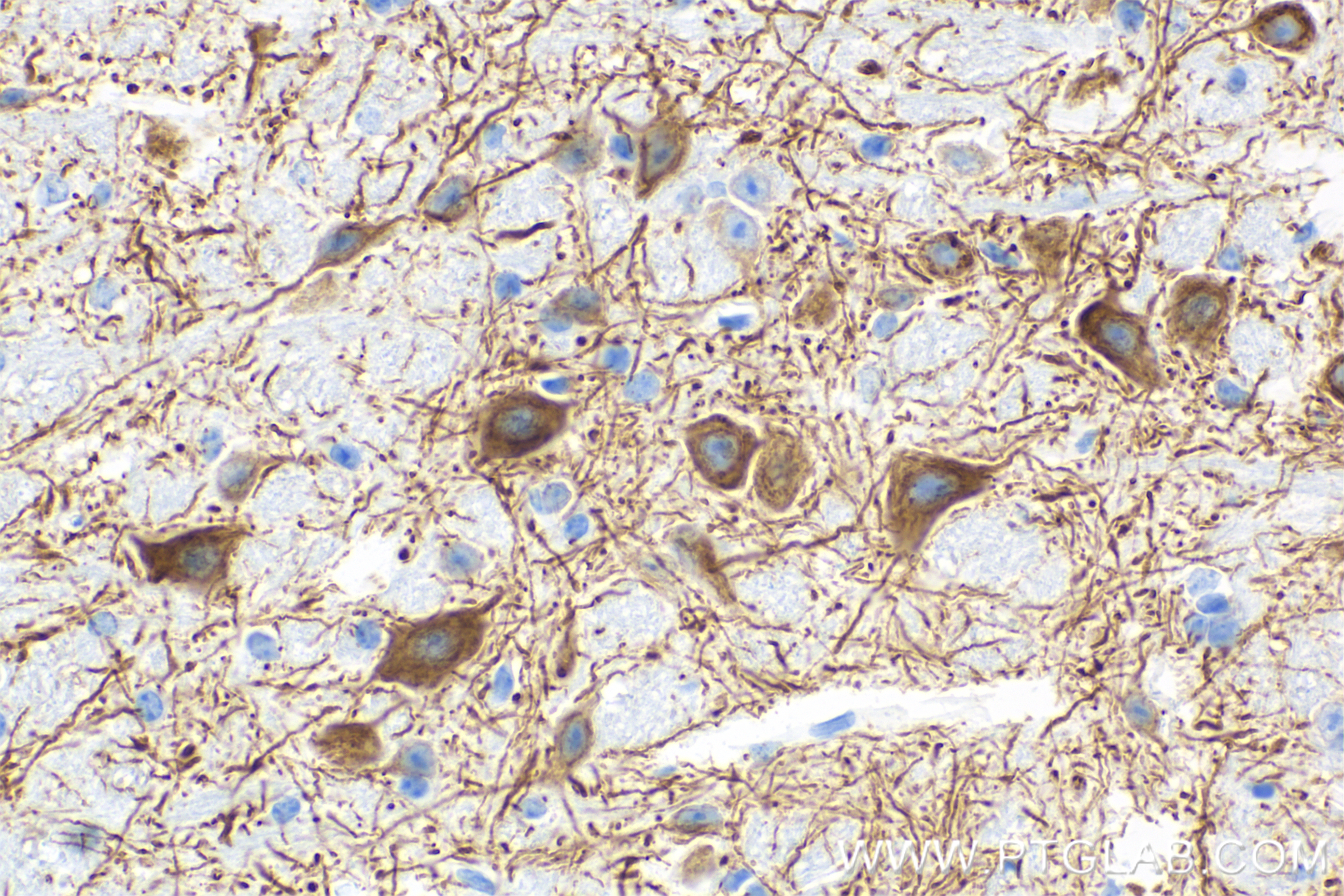
Immunohistochemical analysis of paraffin-embedded mouse cerebellum tissue slide using 84306-3-RR (MAP2 antibody) at dilution of 1:4000 (under 40x lens). Heat mediated antigen retrieval with Tris-EDTA buffer (pH 9.0).
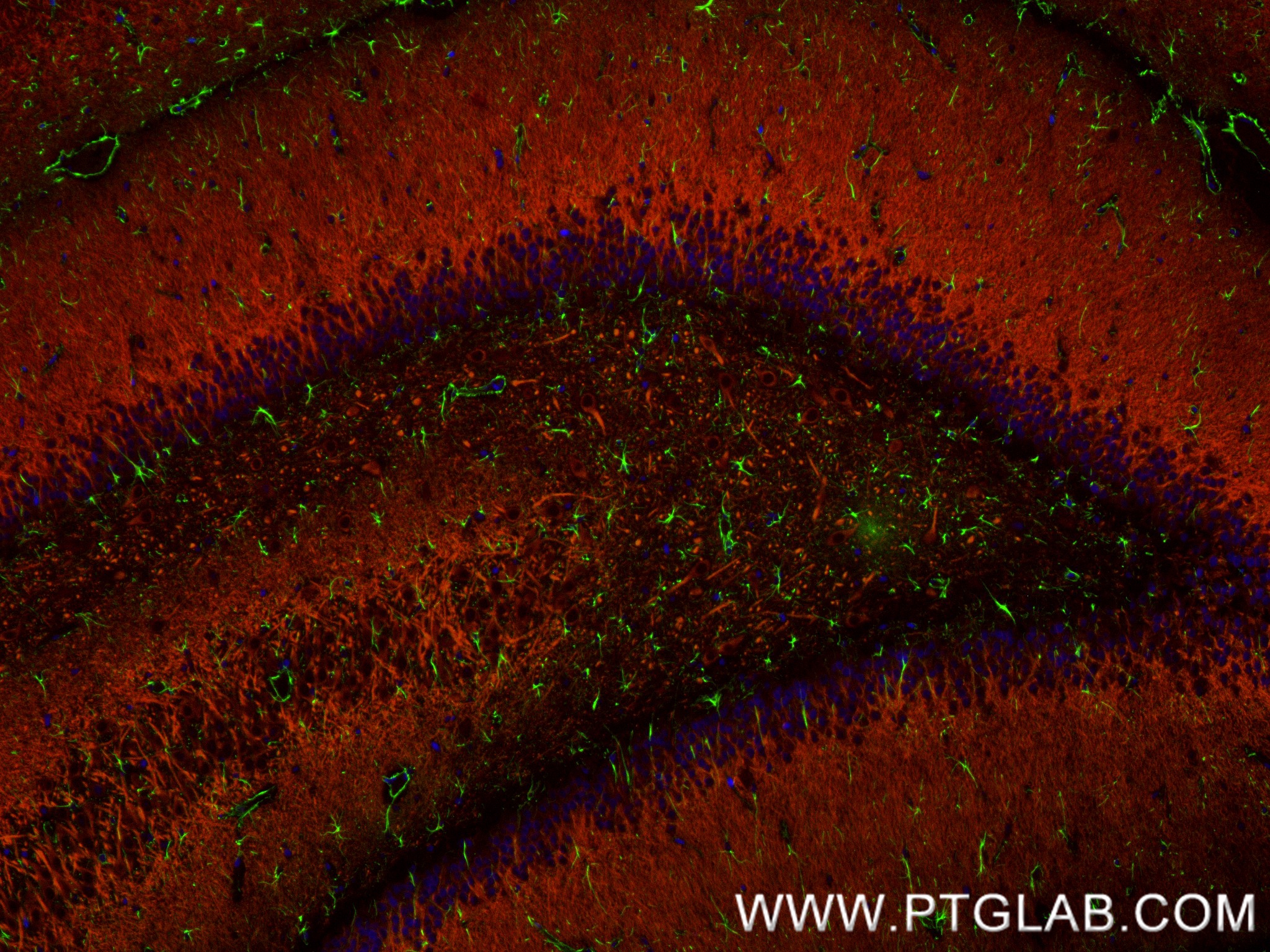
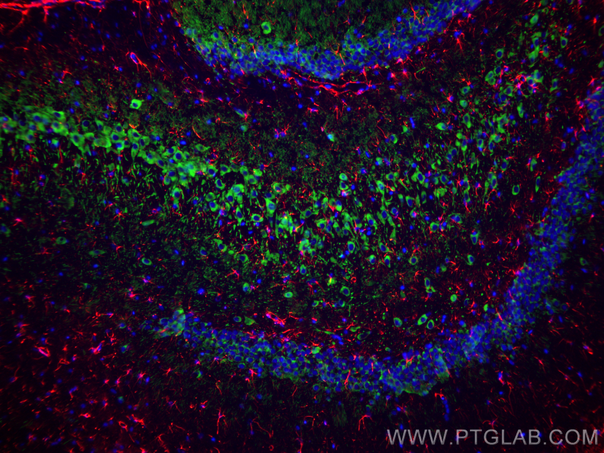
IF Staining of rat brain using 84306-3-RR
Immunofluorescent analysis of (4% PFA) fixed paraffin-embedded rat brain tissue using GFAP antibody (81063-1-RR, Clone: 4C6, green) at dilution of 1:250 and CoraLite®488-Conjugated Goat Anti-Rabbit IgG(H+L) (SA00013-2).MAP2 antibody (84306-3-RR, Clone: 241653E9,red ) at dilution of 1:210 and FlexAble CoraLite® Plus 594 Antibody Labeling Kit for Rabbit IgG(KFA009). Heat mediated antigen retrieval with Tris-EDTA buffer (pH 9.0).
Immunofluorescent analysis of (4% PFA) fixed paraffin-embedded rat brain tissue using GFAP antibody (81063-1-RR, Clone: 4C6, green) at dilution of 1:250 and CoraLite®488-Conjugated Goat Anti-Rabbit IgG(H+L) (SA00013-2).MAP2 antibody (84306-3-RR, Clone: 241653E9,red ) at dilution of 1:210 and FlexAble CoraLite® Plus 594 Antibody Labeling Kit for Rabbit IgG(KFA009). Heat mediated antigen retrieval with Tris-EDTA buffer (pH 9.0).
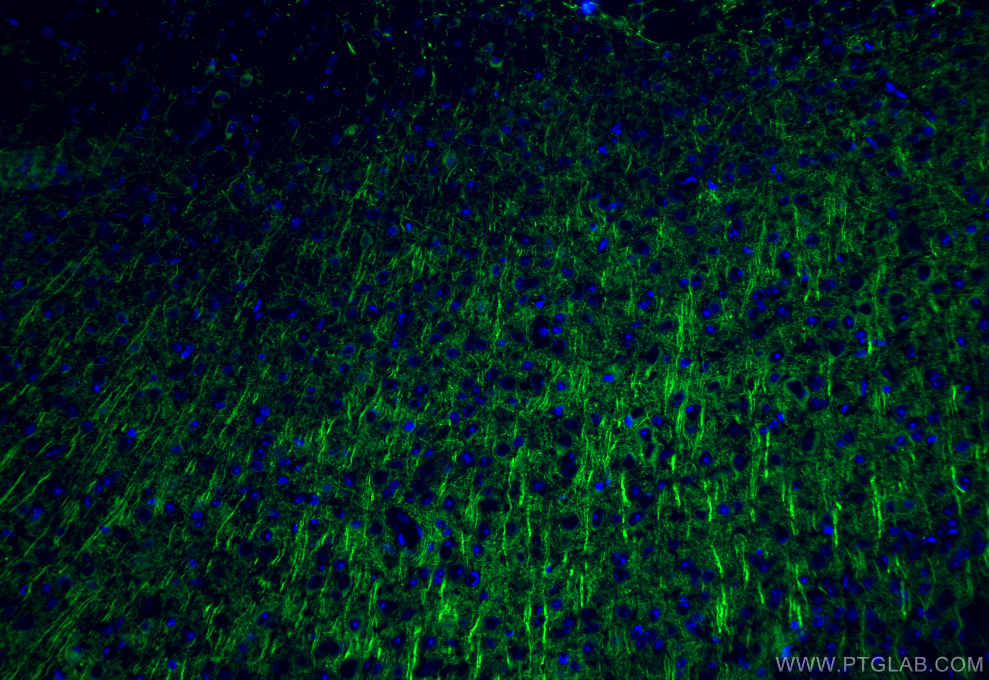
IF Staining of mouse brain using 84306-3-RR
Immunofluorescent analysis of (4% PFA) fixed frozen OCT-embedded mouse brain tissue using MAP2 antibody (84306-3-RR, Clone: 241653E9 ) at dilution of 1:200 and CoraLite®488-Conjugated Goat Anti-Rabbit IgG(H+L) (SA00013-2).
The Proteintech guarantee covers Proteintech antibodies in any species and any application, including those not listed on the datasheet. If the antibody doesn’t perform, you can receive a hassle-free refund or credit note.
SH-SY5Y cells, mouse brain tissue, rat brain tissue
Positive IHC detected in
mouse cerebellum tissue Note: suggested antigen retrieval with TE buffer pH 9.0; (*) Alternatively, antigen retrieval may be performed with citrate buffer pH 6.0
Positive IF-P detected in
rat brain tissue
Positive IF-Fro detected in
mouse brain tissue, rat brain tissue
Recommended dilution
Application
Dilution
Western Blot (WB)
WB : 1:1000-1:4000
Immunohistochemistry (IHC)
IHC : 1:2000-1:8000
Immunofluorescence (IF)-P
IF-P : 1:125-1:500
Immunofluorescence (IF)-FRO
IF-FRO : 1:50-1:500
It is recommended that this reagent should be titrated in each testing system to obtain optimal results.
Sample-dependent, Check data in validation data gallery.
Product Information
84306-3-RR targets MAP2 in WB, IHC, IF-P, IF-Fro, ELISA applications and shows reactivity with human, mouse, rat samples.
PBS with 0.02% sodium azide and 50% glycerol, pH 7.3.
Storage Conditions
Store at -20°C. Stable for one year after shipment. Aliquoting is unnecessary for -20oC storage. 20ul sizes contain 0.1% BSA.
Background Information
MAP2 (microtubule-associated protein 2) is a cytoskeleton protein abundant in the brain and has an important role in neuronal morphogenesis. Multiple high MW and low MW MAP2 isoforms are expressed within the proximal segment of axons, dendrites, and cell bodies. The expression of MAP2 is regulated in both a tissue- and developmentally-specific manner. The 280 kDa MAP2B is present throughout rat brain development, and the slightly larger MAP2A appears first during the end of the second week of postnatal life. MAP2C, composed of several bands of about 70 kDa, is present during early brain development and largely disappears from the mature brain except for the retina, olfactory bulb, and cerebellum. In addition, some isoforms with lower MW around 50-60 kDa also exist. MAP2 antibodies have been widely used to mark the neuron or dendrite formation.
Various lysates were subjected to SDS PAGE followed by western blot with 84306-3-RR (MAP2 antibody) at dilution of 1:1000 incubated at room temperature for 1.5 hours.
WB analysis using 84306-3-RR
SH-SY5Y cells were subjected to SDS PAGE followed by western blot with 84306-3-RR (MAP2 antibody) at dilution of 1:2000 incubated at room temperature for 1.5 hours.
IHC Figures
IHC staining of mouse cerebellum using 84306-3-RR
Immunohistochemical analysis of paraffin-embedded mouse cerebellum tissue slide using 84306-3-RR (MAP2 antibody) at dilution of 1:4000 (under 10x lens). Heat mediated antigen retrieval with Tris-EDTA buffer (pH 9.0).
IHC staining of mouse cerebellum using 84306-3-RR
Immunohistochemical analysis of paraffin-embedded mouse cerebellum tissue slide using 84306-3-RR (MAP2 antibody) at dilution of 1:4000 (under 40x lens). Heat mediated antigen retrieval with Tris-EDTA buffer (pH 9.0).
IF-P Figures
IF Staining of rat brain using 84306-3-RR
Immunofluorescent analysis of (4% PFA) fixed paraffin-embedded rat brain tissue using GFAP antibody (81063-1-RR, Clone: 4C6, green) at dilution of 1:250 and CoraLite®488-Conjugated Goat Anti-Rabbit IgG(H+L) (SA00013-2).MAP2 antibody (84306-3-RR, Clone: 241653E9,red ) at dilution of 1:210 and FlexAble CoraLite® Plus 594 Antibody Labeling Kit for Rabbit IgG(KFA009). Heat mediated antigen retrieval with Tris-EDTA buffer (pH 9.0).
IF-FRO Figures
IF Staining of mouse brain using 84306-3-RR
Immunofluorescent analysis of (4% PFA) fixed frozen OCT-embedded mouse brain tissue using MAP2 antibody (84306-3-RR, Clone: 241653E9 ) at dilution of 1:200 and CoraLite®488-Conjugated Goat Anti-Rabbit IgG(H+L) (SA00013-2).
IF Staining of rat brain using 84306-3-RR
Immunofluorescent analysis of (4% PFA) fixed frozen OCT-embedded rat brain tissue using MAP2 antibody (84306-3-RR, Clone: 241653E9 ) at dilution of 1:200 and CoraLite®488-Conjugated Goat Anti-Rabbit IgG(H+L) (SA00013-2), CoraLite®594 GFAP antibody (CL594-16825, red).
The species listed in Tested Reactivity are in-house verified and applicable species. For unlisted species, please refer to the homology analysis of the immunogen sequence and related species. For rabbit polyclonal antibodies, homology >70% is recommended. For mouse monoclonal antibodies and rabbit recombinant antibodies, homology >90% is recommended. Generally, the higher the homology, the greater the applicability. However, there will be certain differences in protein expression in different species, tissues or cells. Therefore, the homology analysis results are for reference only and do not serve as a guarantee.
At Proteintech, we pride ourselves on our antibody quality, customer service and transparency. As such, we are comparing our antibodies with other vendors, enabling easy identification and comparisons of key data to help you choose the suitable antibody for your needs.
We have selected the top cited antibodies from these vendors for you to compare.
 at dilution of 1:1000 incubated at room temperature for 1.5 hours.")
 at dilution of 1:2000 incubated at room temperature for 1.5 hours.")
 at dilution of 1:4000 (under 10x lens). Heat mediated antigen retrieval with Tris-EDTA buffer (pH 9.0).")
 at dilution of 1:4000 (under 40x lens). Heat mediated antigen retrieval with Tris-EDTA buffer (pH 9.0).")
 fixed paraffin-embedded rat brain tissue using GFAP antibody (81063-1-RR, Clone: 4C6, green) at dilution of 1:250 and CoraLite®488-Conjugated Goat Anti-Rabbit IgG(H+L) (SA00013-2).MAP2 antibody (84306-3-RR, Clone: 241653E9,red ) at dilution of 1:210 and FlexAble CoraLite® Plus 594 Antibody Labeling Kit for Rabbit IgG(KFA009). Heat mediated antigen retrieval with Tris-EDTA buffer (pH 9.0).")
 fixed frozen OCT-embedded mouse brain tissue using MAP2 antibody (84306-3-RR, Clone: 241653E9 ) at dilution of 1:200 and CoraLite®488-Conjugated Goat Anti-Rabbit IgG(H+L) (SA00013-2).")
 fixed frozen OCT-embedded rat brain tissue using MAP2 antibody (84306-3-RR, Clone: 241653E9 ) at dilution of 1:200 and CoraLite®488-Conjugated Goat Anti-Rabbit IgG(H+L) (SA00013-2), CoraLite®594 GFAP antibody (CL594-16825, red).")