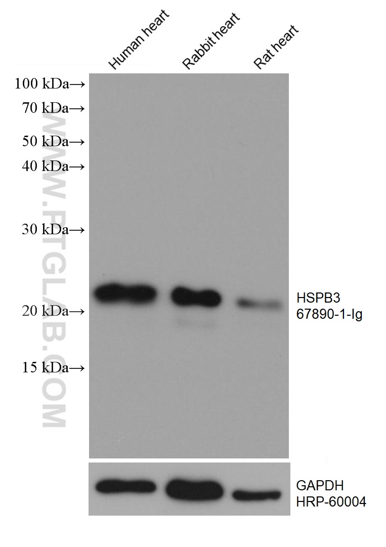
Various lysates were subjected to SDS PAGE followed by western blot with 67890-1-Ig (HSPB3 antibody) at dilution of 1:10000 incubated at room temperature for 1.5 hours. The membrane was stripped and reblotted with HRP-conjugated GAPDH Monoclonal antibody (HRP-60004) as loading control.
Various lysates were subjected to SDS PAGE followed by western blot with 67890-1-Ig (HSPB3 antibody) at dilution of 1:10000 incubated at room temperature for 1.5 hours. The membrane was stripped and reblotted with HRP-conjugated GAPDH Monoclonal antibody (HRP-60004) as loading control.
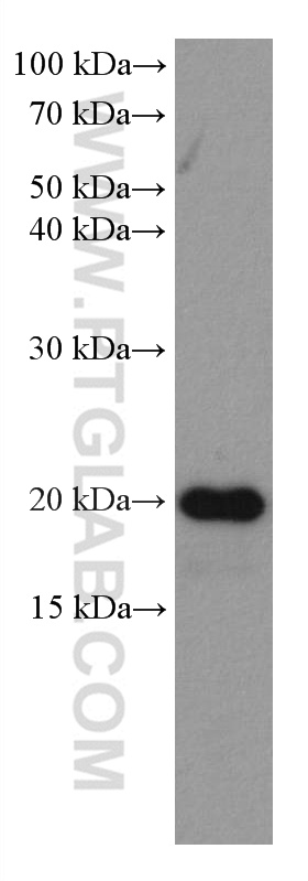
WB analysis of human heart using 67890-1-Ig
human heart tissue were subjected to SDS PAGE followed by western blot with 67890-1-Ig (HSPB3 antibody) at dilution of 1:5000 incubated at room temperature for 1.5 hours.
human heart tissue were subjected to SDS PAGE followed by western blot with 67890-1-Ig (HSPB3 antibody) at dilution of 1:5000 incubated at room temperature for 1.5 hours.
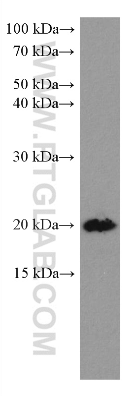
WB analysis of mouse heart using 67890-1-Ig
mouse heart tissue were subjected to SDS PAGE followed by western blot with 67890-1-Ig (HSPB3 antibody) at dilution of 1:5000 incubated at room temperature for 1.5 hours.
mouse heart tissue were subjected to SDS PAGE followed by western blot with 67890-1-Ig (HSPB3 antibody) at dilution of 1:5000 incubated at room temperature for 1.5 hours.
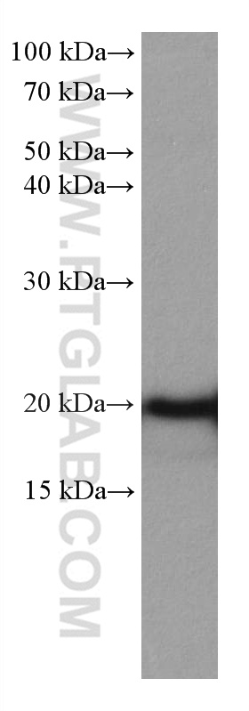
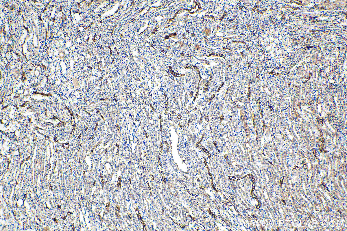
WB analysis of rabbit heart using 67890-1-Ig
rabbit heart tissue were subjected to SDS PAGE followed by western blot with 67890-1-Ig (HSPB3 antibody) at dilution of 1:5000 incubated at room temperature for 1.5 hours.
rabbit heart tissue were subjected to SDS PAGE followed by western blot with 67890-1-Ig (HSPB3 antibody) at dilution of 1:5000 incubated at room temperature for 1.5 hours.
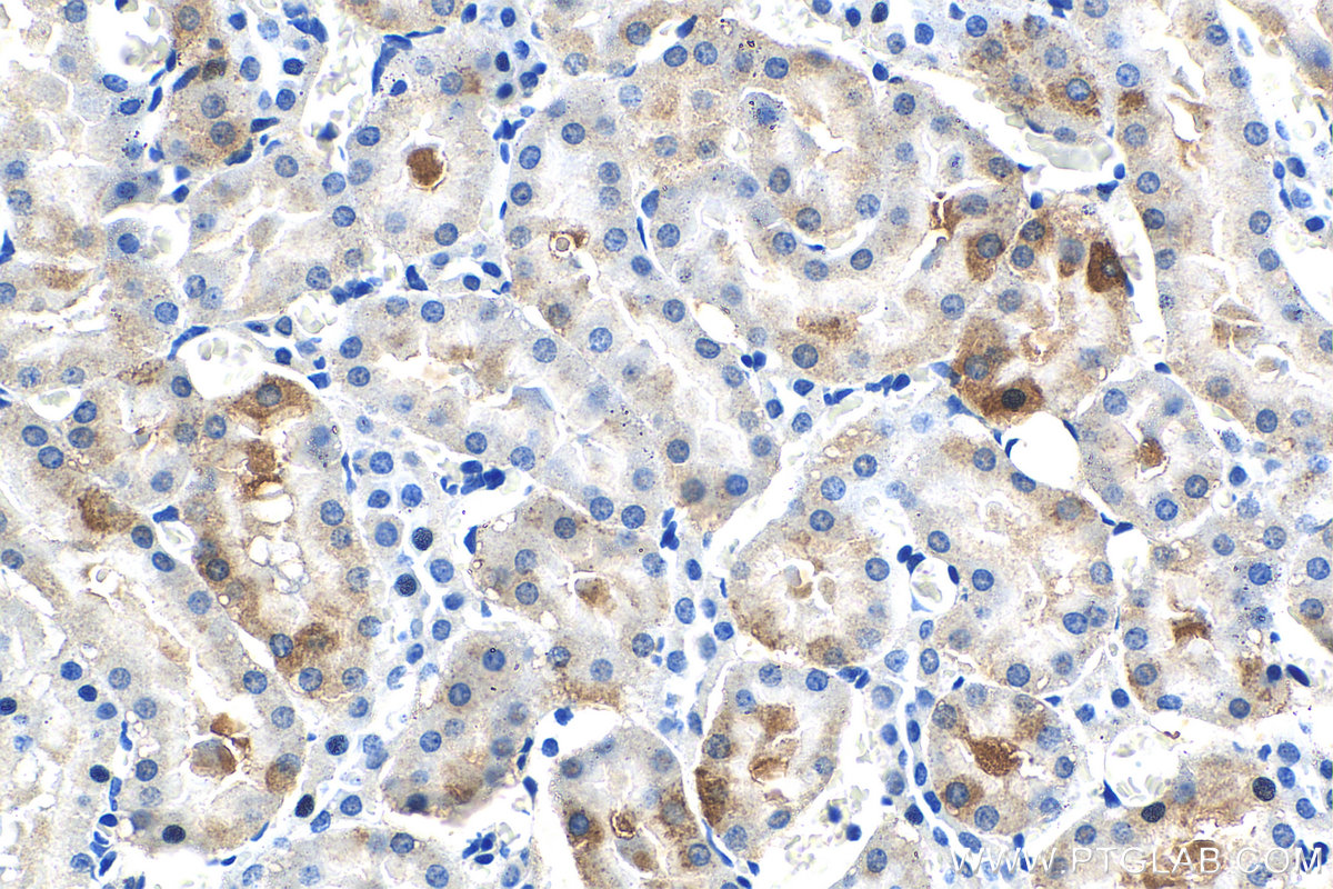
IHC staining of rat kidney using 67890-1-Ig
Immunohistochemical analysis of paraffin-embedded rat kidney tissue slide using 67890-1-Ig (HSPB3 antibody) at dilution of 1:500 (under 40x lens). Heat mediated antigen retrieval with Tris-EDTA buffer (pH 9.0).
Immunohistochemical analysis of paraffin-embedded rat kidney tissue slide using 67890-1-Ig (HSPB3 antibody) at dilution of 1:500 (under 40x lens). Heat mediated antigen retrieval with Tris-EDTA buffer (pH 9.0).
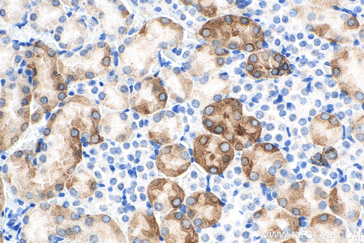
IHC staining of mouse kidney using 67890-1-Ig
Immunohistochemical analysis of paraffin-embedded mouse kidney tissue slide using 67890-1-Ig (HSPB3 antibody) at dilution of 1:500 (under 10x lens). Heat mediated antigen retrieval with Tris-EDTA buffer (pH 9.0).
Immunohistochemical analysis of paraffin-embedded mouse kidney tissue slide using 67890-1-Ig (HSPB3 antibody) at dilution of 1:500 (under 40x lens). Heat mediated antigen retrieval with Tris-EDTA buffer (pH 9.0).
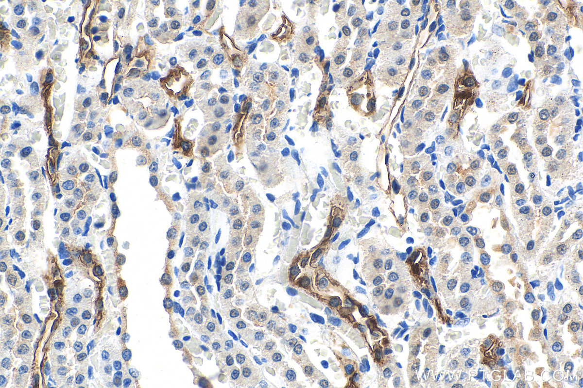
IHC staining of rat kidney using 67890-1-Ig
Immunohistochemical analysis of paraffin-embedded rat kidney tissue slide using 67890-1-Ig (HSPB3 antibody) at dilution of 1:500 (under 10x lens). Heat mediated antigen retrieval with Tris-EDTA buffer (pH 9.0).
Immunohistochemical analysis of paraffin-embedded rat kidney tissue slide using 67890-1-Ig (HSPB3 antibody) at dilution of 1:500 (under 10x lens). Heat mediated antigen retrieval with Tris-EDTA buffer (pH 9.0).
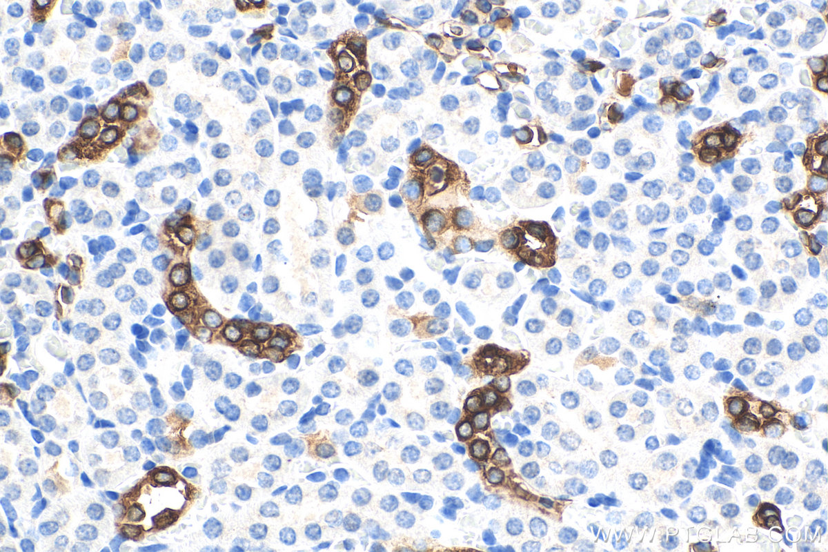
IHC staining of rat kidney using 67890-1-Ig
Immunohistochemical analysis of paraffin-embedded rat kidney tissue slide using 67890-1-Ig (HSPB3 antibody) at dilution of 1:500 (under 40x lens). Heat mediated antigen retrieval with Tris-EDTA buffer (pH 9.0).
Immunohistochemical analysis of paraffin-embedded rat kidney tissue slide using 67890-1-Ig (HSPB3 antibody) at dilution of 1:500 (under 40x lens). Heat mediated antigen retrieval with Tris-EDTA buffer (pH 9.0).
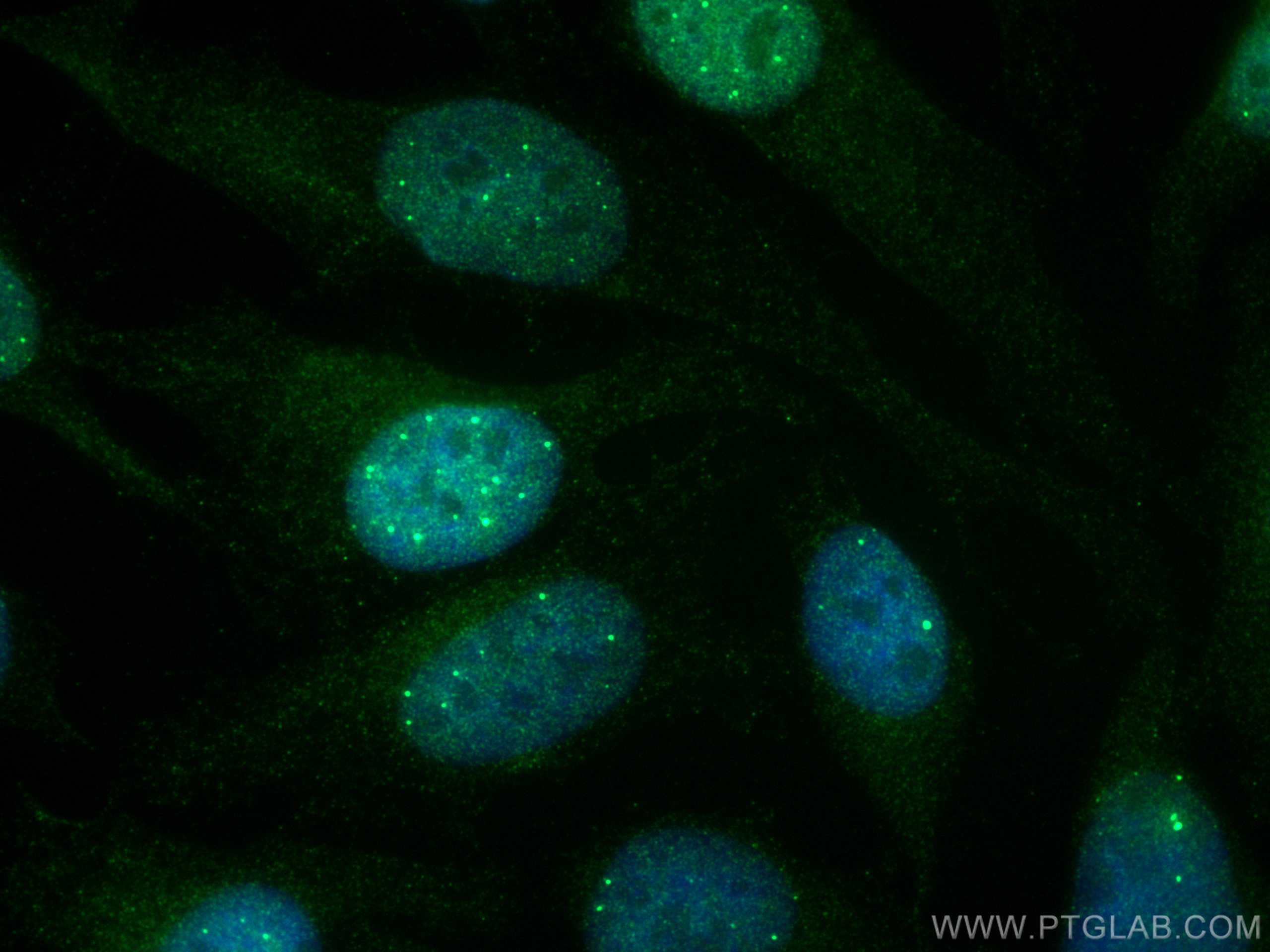
IF Staining of HeLa using 67890-1-Ig
Immunofluorescent analysis of (4% PFA) fixed HeLa cells using HSPB3 antibody (67890-1-Ig, Clone: 2A10B5 ) at dilution of 1:400 and CoraLite®488-Conjugated Goat Anti-Mouse IgG(H+L).
Immunofluorescent analysis of (4% PFA) fixed HeLa cells using HSPB3 antibody (67890-1-Ig, Clone: 2A10B5 ) at dilution of 1:400 and CoraLite®488-Conjugated Goat Anti-Mouse IgG(H+L).
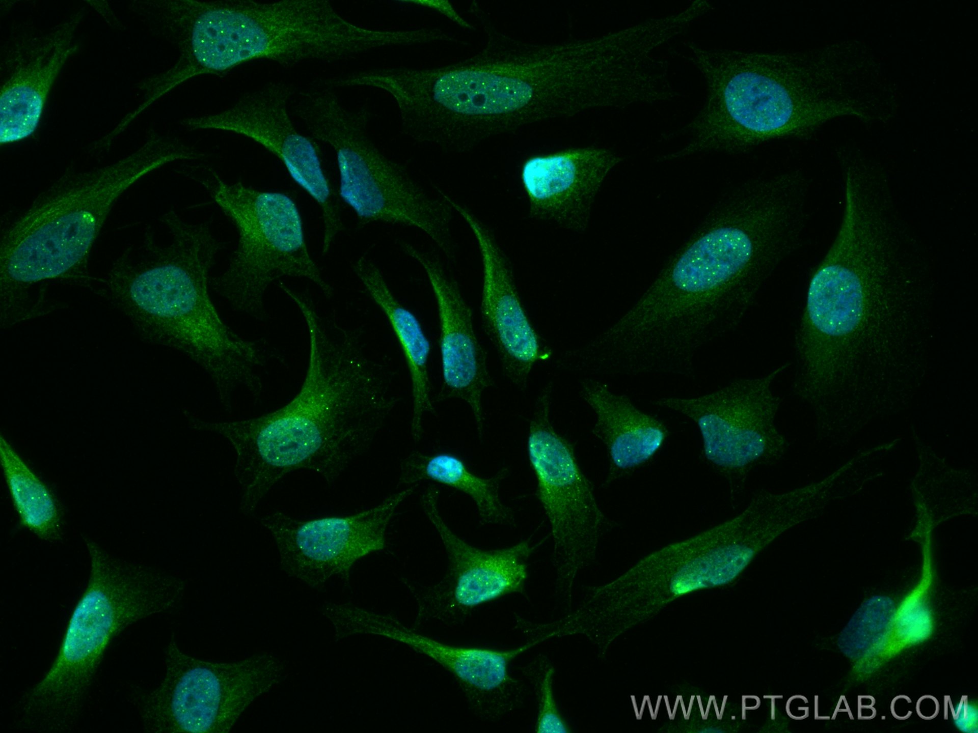
IF Staining of HeLa using 67890-1-Ig
Immunofluorescent analysis of (4% PFA) fixed HeLa cells using HSPB3 antibody (67890-1-Ig, Clone: 2A10B5 ) at dilution of 1:800 and CoraLite®488-Conjugated Goat Anti-Mouse IgG(H+L).
Immunofluorescent analysis of (4% PFA) fixed HeLa cells using HSPB3 antibody (67890-1-Ig, Clone: 2A10B5 ) at dilution of 1:800 and CoraLite®488-Conjugated Goat Anti-Mouse IgG(H+L).
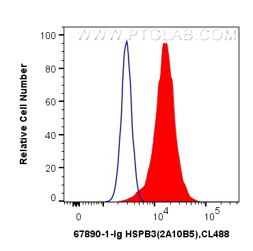
FC experiment of HeLa using 67890-1-Ig
1X10^6 HeLa cells were intracellularly stained with 0.5 ug Anti-Human HSPB3 (67890-1-Ig, Clone:2A10B5) and CoraLite®488-Conjugated Goat Anti-Mouse IgG(H+L) at dilution 1:1000 (red), or 0.5 ug Mouse IgG2b Isotype Control (66360-3-Ig, Clone: K11B8C4B5) (blue). Cells were fixed with 4% PFA and permeabilized with Flow Cytometry Perm Buffer (PF00011-C).
1X10^6 HeLa cells were intracellularly stained with 0.5 ug Anti-Human HSPB3 (67890-1-Ig, Clone:2A10B5) and CoraLite®488-Conjugated Goat Anti-Mouse IgG(H+L) at dilution 1:1000 (red), or 0.5 ug Mouse IgG2b Isotype Control (66360-3-Ig, Clone: K11B8C4B5) (blue). Cells were fixed with 4% PFA and permeabilized with Flow Cytometry Perm Buffer (PF00011-C).
The Proteintech guarantee covers Proteintech antibodies in any species and any application, including those not listed on the datasheet. If the antibody doesn’t perform, you can receive a hassle-free refund or credit note.
human heart tissue, mouse heart tissue, rabbit heart tissue, rat heart tissue
Positive IHC detected in
rat kidney tissue, mouse kidney tissue Note: suggested antigen retrieval with TE buffer pH 9.0; (*) Alternatively, antigen retrieval may be performed with citrate buffer pH 6.0
Positive IF/ICC detected in
HeLa cells
Positive FC (Intra) detected in
HeLa cells
Recommended dilution
Application
Dilution
Western Blot (WB)
WB : 1:5000-1:50000
Immunohistochemistry (IHC)
IHC : 1:250-1:1000
Immunofluorescence (IF)/ICC
IF/ICC : 1:400-1:1600
Flow Cytometry (FC) (INTRA)
FC (INTRA) : 0.50 ug per 10^6 cells in a 100 µl suspension
It is recommended that this reagent should be titrated in each testing system to obtain optimal results.
Sample-dependent, Check data in validation data gallery.
Product Information
67890-1-Ig targets HSPB3 in WB, IHC, IF/ICC, FC (Intra), ELISA applications and shows reactivity with human, mouse, rat, rabbit samples.
PBS with 0.02% sodium azide and 50% glycerol , pH 7.3
Storage Conditions
Store at -20°C. Stable for one year after shipment. Aliquoting is unnecessary for -20oC storage. 20ul sizes contain 0.1% BSA.
Background Information
Heat shock protein family B (small) member 3 (HSPB3) is the third member of the sHSP family in human and is mainly expressed in skeletal and smooth muscles, and can associate with HSPB2, which is another sHSP prominently expressed in cardiac and skeletal muscles. HSPB3 is involved in several cellular functions such as cell signaling, differentiation, and apoptosis, and mutations of HSPB3 are associated with neuromuscular diseases, such as axonal Charcot‐Marie‐Tooth disease (PMID: 31709619) (PMID: 32093037). The molecular weight of HSPB3 is about 17 kDa.
Various lysates were subjected to SDS PAGE followed by western blot with 67890-1-Ig (HSPB3 antibody) at dilution of 1:10000 incubated at room temperature for 1.5 hours. The membrane was stripped and reblotted with HRP-conjugated GAPDH Monoclonal antibody (HRP-60004) as loading control.
WB analysis of human heart using 67890-1-Ig
human heart tissue were subjected to SDS PAGE followed by western blot with 67890-1-Ig (HSPB3 antibody) at dilution of 1:5000 incubated at room temperature for 1.5 hours.
WB analysis of mouse heart using 67890-1-Ig
mouse heart tissue were subjected to SDS PAGE followed by western blot with 67890-1-Ig (HSPB3 antibody) at dilution of 1:5000 incubated at room temperature for 1.5 hours.
WB analysis of rabbit heart using 67890-1-Ig
rabbit heart tissue were subjected to SDS PAGE followed by western blot with 67890-1-Ig (HSPB3 antibody) at dilution of 1:5000 incubated at room temperature for 1.5 hours.
IHC Figures
IHC staining of rat kidney using 67890-1-Ig
Immunohistochemical analysis of paraffin-embedded rat kidney tissue slide using 67890-1-Ig (HSPB3 antibody) at dilution of 1:500 (under 40x lens). Heat mediated antigen retrieval with Tris-EDTA buffer (pH 9.0).
IHC staining of mouse kidney using 67890-1-Ig
Immunohistochemical analysis of paraffin-embedded mouse kidney tissue slide using 67890-1-Ig (HSPB3 antibody) at dilution of 1:500 (under 10x lens). Heat mediated antigen retrieval with Tris-EDTA buffer (pH 9.0).
IHC staining of mouse kidney using 67890-1-Ig
Immunohistochemical analysis of paraffin-embedded mouse kidney tissue slide using 67890-1-Ig (HSPB3 antibody) at dilution of 1:500 (under 40x lens). Heat mediated antigen retrieval with Tris-EDTA buffer (pH 9.0).
IHC staining of mouse kidney using 67890-1-Ig
Immunohistochemical analysis of paraffin-embedded mouse kidney tissue slide using 67890-1-Ig (HSPB3 antibody) at dilution of 1:500 (under 40x lens). Heat mediated antigen retrieval with Tris-EDTA buffer (pH 9.0).
IHC staining of rat kidney using 67890-1-Ig
Immunohistochemical analysis of paraffin-embedded rat kidney tissue slide using 67890-1-Ig (HSPB3 antibody) at dilution of 1:500 (under 10x lens). Heat mediated antigen retrieval with Tris-EDTA buffer (pH 9.0).
IHC staining of rat kidney using 67890-1-Ig
Immunohistochemical analysis of paraffin-embedded rat kidney tissue slide using 67890-1-Ig (HSPB3 antibody) at dilution of 1:500 (under 40x lens). Heat mediated antigen retrieval with Tris-EDTA buffer (pH 9.0).
IF/ICC Figures
IF Staining of HeLa using 67890-1-Ig
Immunofluorescent analysis of (4% PFA) fixed HeLa cells using HSPB3 antibody (67890-1-Ig, Clone: 2A10B5 ) at dilution of 1:400 and CoraLite®488-Conjugated Goat Anti-Mouse IgG(H+L).
IF Staining of HeLa using 67890-1-Ig
Immunofluorescent analysis of (4% PFA) fixed HeLa cells using HSPB3 antibody (67890-1-Ig, Clone: 2A10B5 ) at dilution of 1:800 and CoraLite®488-Conjugated Goat Anti-Mouse IgG(H+L).
FC (INTRA) Figures
FC experiment of HeLa using 67890-1-Ig
1X10^6 HeLa cells were intracellularly stained with 0.5 ug Anti-Human HSPB3 (67890-1-Ig, Clone:2A10B5) and CoraLite®488-Conjugated Goat Anti-Mouse IgG(H+L) at dilution 1:1000 (red), or 0.5 ug Mouse IgG2b Isotype Control (66360-3-Ig, Clone: K11B8C4B5) (blue). Cells were fixed with 4% PFA and permeabilized with Flow Cytometry Perm Buffer (PF00011-C).
The species listed in Tested Reactivity are in-house verified and applicable species. For unlisted species, please refer to the homology analysis of the immunogen sequence and related species. For rabbit polyclonal antibodies, homology >70% is recommended. For mouse monoclonal antibodies and rabbit recombinant antibodies, homology >90% is recommended. Generally, the higher the homology, the greater the applicability. However, there will be certain differences in protein expression in different species, tissues or cells. Therefore, the homology analysis results are for reference only and do not serve as a guarantee.
At Proteintech, we pride ourselves on our antibody quality, customer service and transparency. As such, we are comparing our antibodies with other vendors, enabling easy identification and comparisons of key data to help you choose the suitable antibody for your needs.
We have selected the top cited antibodies from these vendors for you to compare.
Proteintech
HSPB3 Monoclonal antibody
Catalog Number
67890-1-Ig
Citations
-
Dilutions
WB : 1:5000-1:50000 IHC : 1:250-1:1000 IF/ICC : 1:400-1:1600 FC (INTRA) : 0.50 ug per 10^6 cells in a 100 µl suspension
Applications
WB, IHC, IF/ICC, FC (Intra), ELISA
Reactivity
human, mouse, rat, rabbit
Product Guarantee
Covers any species including not listed on datasheet
Covers any applications including not listed on datasheet
 at dilution of 1:10000 incubated at room temperature for 1.5 hours. The membrane was stripped and reblotted with HRP-conjugated GAPDH Monoclonal antibody (HRP-60004) as loading control.")
 at dilution of 1:5000 incubated at room temperature for 1.5 hours.")
 at dilution of 1:5000 incubated at room temperature for 1.5 hours.")
 at dilution of 1:5000 incubated at room temperature for 1.5 hours.")
 at dilution of 1:500 (under 40x lens). Heat mediated antigen retrieval with Tris-EDTA buffer (pH 9.0).")
 at dilution of 1:500 (under 10x lens). Heat mediated antigen retrieval with Tris-EDTA buffer (pH 9.0).")
 at dilution of 1:500 (under 40x lens). Heat mediated antigen retrieval with Tris-EDTA buffer (pH 9.0).")
 at dilution of 1:500 (under 40x lens). Heat mediated antigen retrieval with Tris-EDTA buffer (pH 9.0).")
 at dilution of 1:500 (under 10x lens). Heat mediated antigen retrieval with Tris-EDTA buffer (pH 9.0).")
 at dilution of 1:500 (under 40x lens). Heat mediated antigen retrieval with Tris-EDTA buffer (pH 9.0).")
 fixed HeLa cells using HSPB3 antibody (67890-1-Ig, Clone: 2A10B5 ) at dilution of 1:400 and CoraLite®488-Conjugated Goat Anti-Mouse IgG(H+L).")
 fixed HeLa cells using HSPB3 antibody (67890-1-Ig, Clone: 2A10B5 ) at dilution of 1:800 and CoraLite®488-Conjugated Goat Anti-Mouse IgG(H+L).")
 and CoraLite®488-Conjugated Goat Anti-Mouse IgG(H+L) at dilution 1:1000 (red), or 0.5 ug Mouse IgG2b Isotype Control (66360-3-Ig, Clone: K11B8C4B5) (blue). Cells were fixed with 4% PFA and permeabilized with Flow Cytometry Perm Buffer (PF00011-C).")