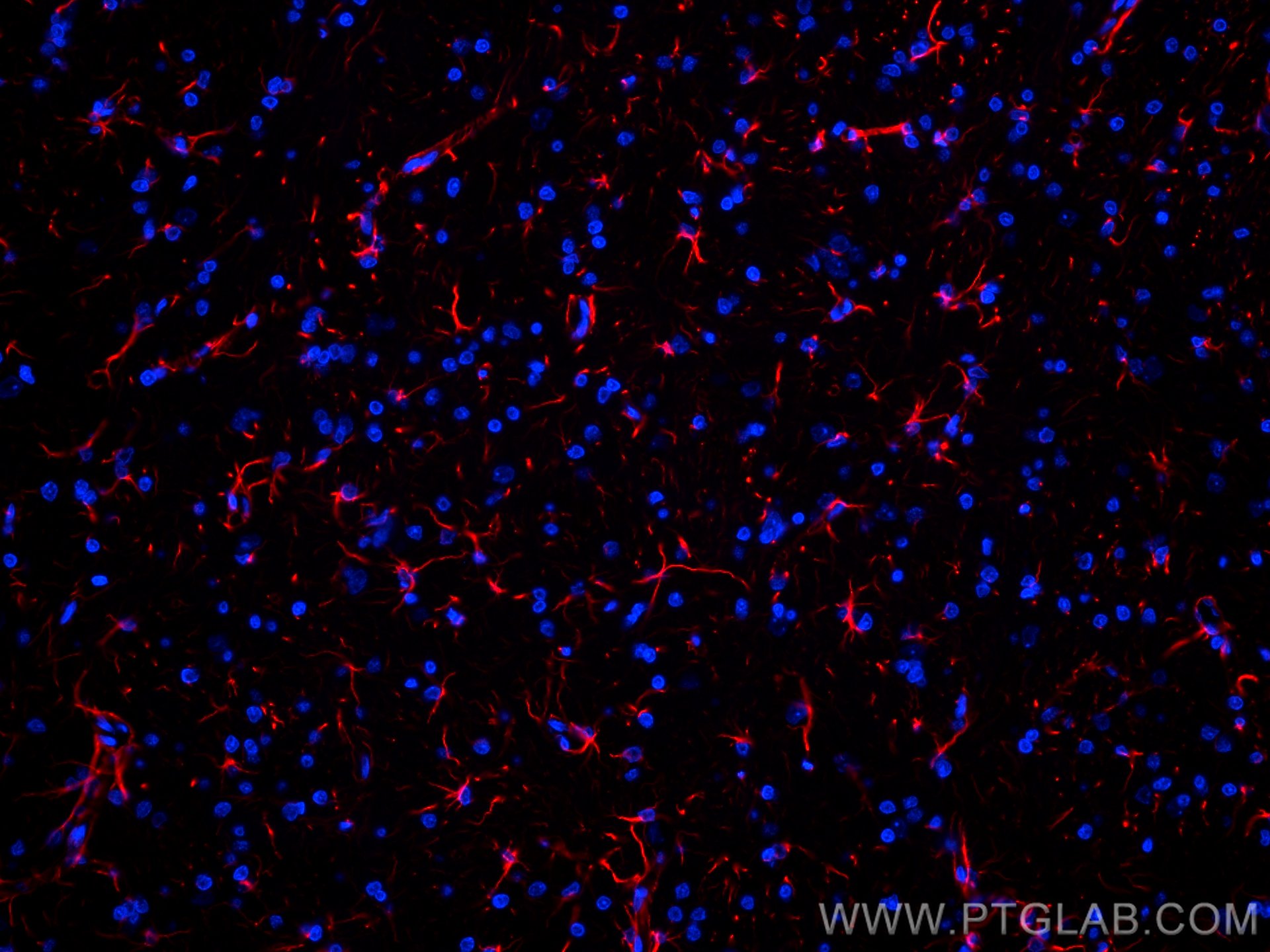
fixed rat brain tissue using CoraLite®594 GFAP antibody (CL594-60190, Clone: 4B2E10 ) at dilution of 1:200.")
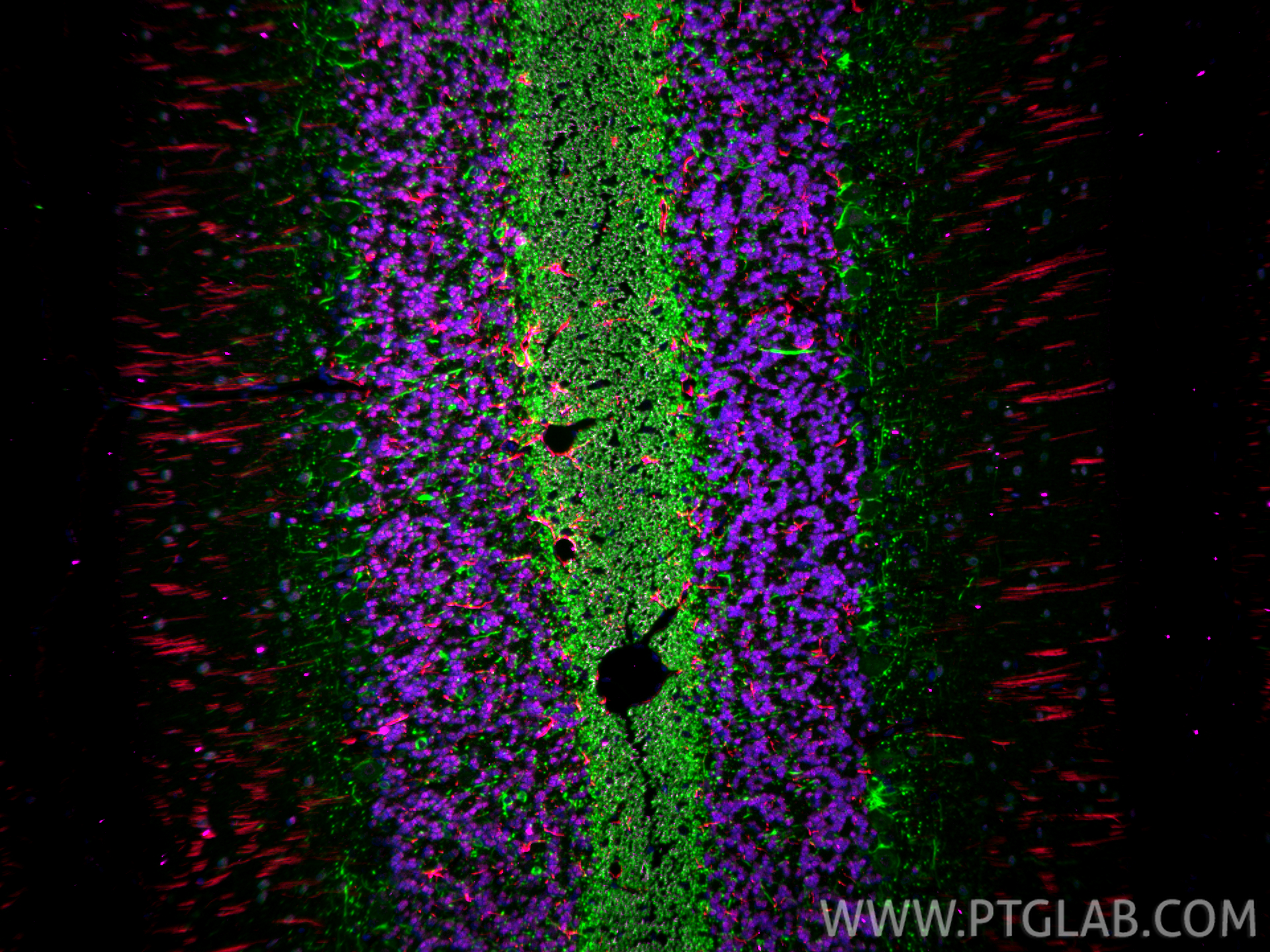
 fixed paraffin-embedded rat cerebellum tissue using CoraLite®594 GFAP antibody (CL594-60190, Clone: 4B2E10 ) at dilution of 1:200, NeuN antibody (66836-1-Ig, Clone: 3A4C1, Magenta), NF-H/NF200 antibody (18934-1-AP, green). Heat mediated antigen retrieval with Tris-EDTA buffer (pH 9.0).")
Tested Applications
| Positive IF-P detected in | rat brain tissue, rat cerebellum tissue |
This antibody is not recommended for immunocytofluorescent assays. It is not suitable for frozen sections.
Recommended dilution
| Application | Dilution |
|---|---|
| Immunofluorescence (IF)-P | IF-P : 1:50-1:500 |
| It is recommended that this reagent should be titrated in each testing system to obtain optimal results. | |
| Sample-dependent, Check data in validation data gallery. | |
This antibody is not recommended for immunocytofluorescent assays. It is not suitable for frozen sections.
Published Applications
| IF | See 4 publications below |
Product Information
CL594-60190 targets GFAP in IF-P applications and shows reactivity with human, mouse, rat, pig samples.
| Tested Reactivity | human, mouse, rat, pig |
| Cited Reactivity | mouse |
| Host / Isotype | Mouse / IgG2a |
| Class | Monoclonal |
| Type | Antibody |
| Immunogen |
CatNo: Ag10452 Product name: Recombinant human GFAP protein Source: e coli.-derived, PET28a Tag: 6*His Domain: 83-432 aa of BC013596 Sequence: YIEKVRFLEQQNKALAAELNQLRAKEPTKLADVYQAELRELRLRLDQLTANSARLEVERDNLAQDLATVRQKLQDETNLRLEAENNLAAYRQEADEATLARLDLERKIESLEEEIRFLRKIHEEEVRELQEQLARQQVHVELDVAKPDLTAALKEIRTQYEAMASSNMHEAEEWYRSKFADLTDAAARNAELLRQAKHEANDYRRQLQSLTCDLESLRGTNESLERQMREQEERHVREAASYQEALARLEEEGQSLKDEMARHLQEYQDLLNVKLALDIEIATYRKLLEGEENRITIPVQTFSNLQIRETSLDTKSVSEGHLKRNIVVKTVEMRDGEVIKESKQEHKDVM Predict reactive species |
| Full Name | glial fibrillary acidic protein |
| Calculated Molecular Weight | 432 aa, 50 kDa |
| GenBank Accession Number | BC013596 |
| Gene Symbol | GFAP |
| Gene ID (NCBI) | 2670 |
| RRID | AB_2883437 |
| Conjugate | CoraLite®594 Fluorescent Dye |
| Excitation/Emission Maxima Wavelengths | 588 nm / 604 nm |
| Excitation Laser | Yellow-Green Laser (561 nm) |
| Form | Liquid |
| Purification Method | Protein A purification |
| UNIPROT ID | P14136 |
| Storage Buffer | PBS with 50% glycerol, 0.05% Proclin300, 0.5% BSA, pH 7.3. |
| Storage Conditions | Store at -20°C. Avoid exposure to light. Stable for one year after shipment. Aliquoting is unnecessary for -20oC storage. |
Background Information
Function
GFAP (Glial fibrillary acidic protein) is a type III intermediate filament (IF) protein specific to the central nervous system (CNS). GFAP is one of the main components of the intermediate filament network in astrocytes and has been proposed as playing a role in cell migration, cell motility, maintaining mechanical strength, and in mitosis.Tissue specificity
GFAP is expressed in central nervous system cells, predominantly in astrocytes. GFAP is commonly used as an astrocyte marker. However, GFAP is also present in peripheral glia and in non-CNS cells, including fibroblasts, chondrocytes, lymphocytes, and liver stellate cells (PMID: 21219963).Involvement in disease
- Mutations in GFAP lead to Alexander disease (OMIM: 203450), an autosomal dominant CNS disorder. The mutations present in affected individuals are thought to be gain-of-function.
- Upregulation of GFAP is a hallmark of reactive astrocytes, in which GFAP is present in hypertrophic cellular processes. Reactive astrogliosis is present in many neurological disorders, such as stroke, various neurodegenerative diseases (including Alzheimer's and Parkinson's disease), and neurotrauma.
Isoforms
Astrocytes express 10 different isoforms of GFAP that differ in the rod and tail domains (PMID: 25726916), which means that they differ in molecular size. Isoform expression varies during the development and across different subtypes of astrocytes. Not all isoforms are upregulated in reactive astrocytes.Post-translational modifications
Intermediate filament proteins are regulated by phosphorylation. Six phosphorylation sites have been identified in GFAP protein, at least some of which are reported to control filament assembly (PMID: 21219963).Cellular localization
GFAP localizes to intermediate filaments and stains well in astrocyte cellular processes.
Protocols
| Product Specific Protocols | |
|---|---|
| IF protocol for CL594 GFAP antibody CL594-60190 | Download protocol |
| Standard Protocols | |
|---|---|
| Click here to view our Standard Protocols |
Publications
| Species | Application | Title |
|---|---|---|
Glia Mechanosensitive channel Piezo1 is an essential regulator in cell cycle progression of optic nerve head astrocytes | ||
Neoplasia ZC3H15 suppression ameliorates bone cancer pain through inhibiting neuronal oxidative stress and microglial inflammation | ||
Neuron Metabolic reprogramming through histone lactylation in microglia and macrophages recruits CD8+ T lymphocytes and aggravates spinal cord injury | ||
Cell Death Differ Metabolic reprogramming in astrocytes prevents neuronal death through a UCHL1/PFKFB3/H4K8la positive feedback loop |