Anticorps Polyclonal de lapin anti-uPAR
uPAR Polyclonal Antibody for WB, ELISA
Hôte / Isotype
Lapin / IgG
Réactivité testée
Humain, souris et plus (1)
Applications
WB, IHC, CoIP, ELISA, IF
Conjugaison
Non conjugué
N° de cat : 10286-1-AP
Synonymes
Galerie de données de validation
Applications testées
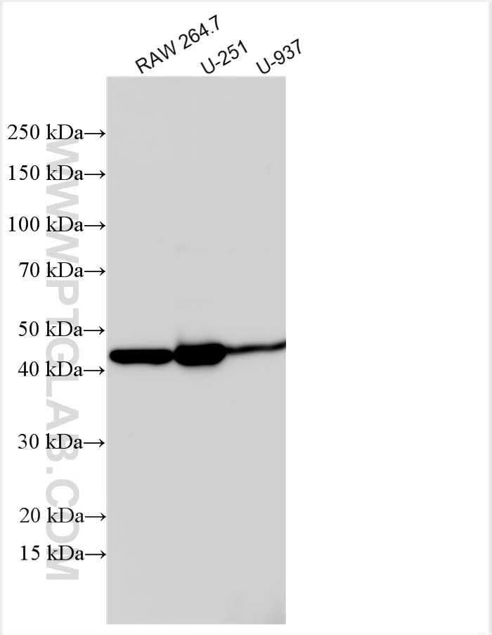
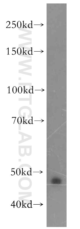
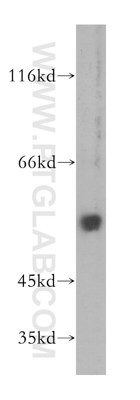
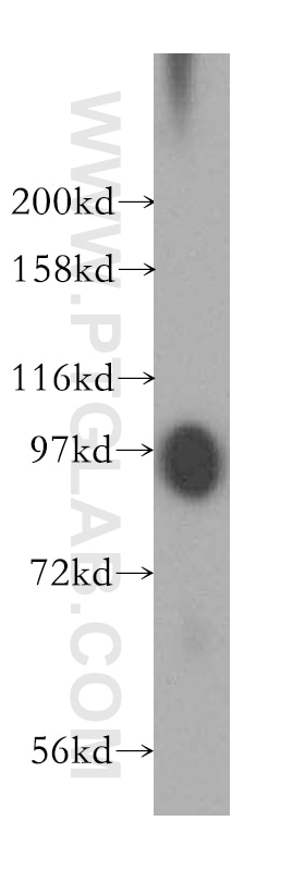
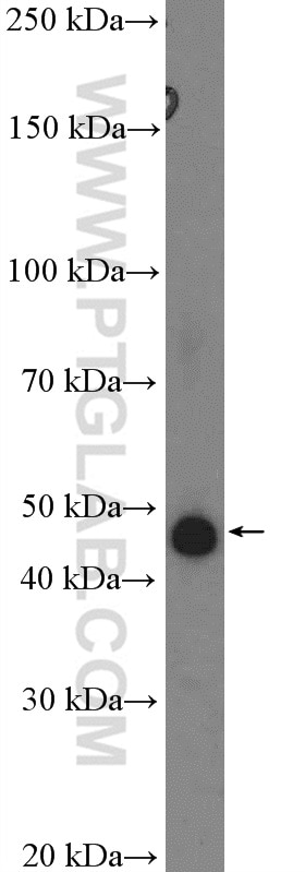
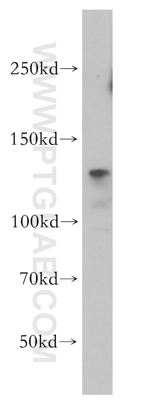
| Résultats positifs en WB | cellules RAW 264.7, cellules A2780, cellules U-251, cellules U-937 |
Dilution recommandée
| Application | Dilution |
|---|---|
| Western Blot (WB) | WB : 1:1000-1:8000 |
| It is recommended that this reagent should be titrated in each testing system to obtain optimal results. | |
| Sample-dependent, check data in validation data gallery | |
Applications publiées
| KD/KO | See 1 publications below |
| WB | See 12 publications below |
| IHC | See 6 publications below |
| IF | See 4 publications below |
| CoIP | See 1 publications below |
Informations sur le produit
10286-1-AP cible uPAR dans les applications de WB, IHC, CoIP, ELISA, IF et montre une réactivité avec des échantillons Humain, souris
| Réactivité | Humain, souris |
| Réactivité citée | bovin, Humain, souris |
| Hôte / Isotype | Lapin / IgG |
| Clonalité | Polyclonal |
| Type | Anticorps |
| Immunogène | uPAR Protéine recombinante Ag0187 |
| Nom complet | PLAUR |
| Masse moléculaire calculée | 37 kDa |
| Poids moléculaire observé | 35-70 kDa |
| Numéro d’acquisition GenBank | BC002788 |
| Symbole du gène | uPAR |
| Identification du gène (NCBI) | 5329 |
| Conjugaison | Non conjugué |
| Forme | Liquide |
| Méthode de purification | Purification par affinité contre l'antigène |
| Tampon de stockage | PBS avec azoture de sodium à 0,02 % et glycérol à 50 % pH 7,3 |
| Conditions de stockage | Stocker à -20°C. Stable pendant un an après l'expédition. L'aliquotage n'est pas nécessaire pour le stockage à -20oC Les 20ul contiennent 0,1% de BSA. |
Informations générales
uPAR is a 45-65 kDa, highly glycosylated, GPI-anchored membrane protein. In addition to the membrane-anchored form, uPAR is released from the plasma membrane by cleavage of the GPI anchor and can be found as a soluble form (suPAR). uPAR contains three homologous domains (D1-D3) of which the N-terminal one (D1) represents the uPA-binding domain. After binding to uPAR, uPA cleaves plasminogen, generating the active protease plasmin which is involved in a wide variety of physiologic and pathologic processes. In addition to regulating proteolysis, uPAR has important function in cell adhesion, migration and proliferation. Studies reveal that uPAR expression is elevated during inflammation and tissue remodelling and in many human cancers, in which it frequently indicates poor prognosis. (PMID 20027185; 12461559)
Protocole
| Product Specific Protocols | |
|---|---|
| WB protocol for uPAR antibody 10286-1-AP | Download protocol |
| FC protocol for uPAR antibody 10286-1-AP | Download protocol |
| Standard Protocols | |
|---|---|
| Click here to view our Standard Protocols |
Publications
| Species | Application | Title |
|---|---|---|
Mol Cell A transient transcriptional activation governs unpolarized-to-polarized morphogenesis during embryo implantation | ||
Nat Commun Sirt6 deficiency exacerbates podocyte injury and proteinuria through targeting Notch signaling. | ||
Cancer Res Bcl-xL enforces a slow-cycling state necessary for survival in the nutrient-deprived microenvironment of pancreatic cancer. | ||
Nucleic Acids Res Regulation of u-PAR gene expression by H2A.Z is modulated by the MEK-ERK/AP-1 pathway. | ||
J Inflamm Res PLAUR as a Potential Biomarker Associated with Immune Infiltration in Bladder Urothelial Carcinoma. |
Avis
The reviews below have been submitted by verified Proteintech customers who received an incentive forproviding their feedback.
FH Ruchi (Verified Customer) (07-26-2022) | IF on mouse skin
 |
