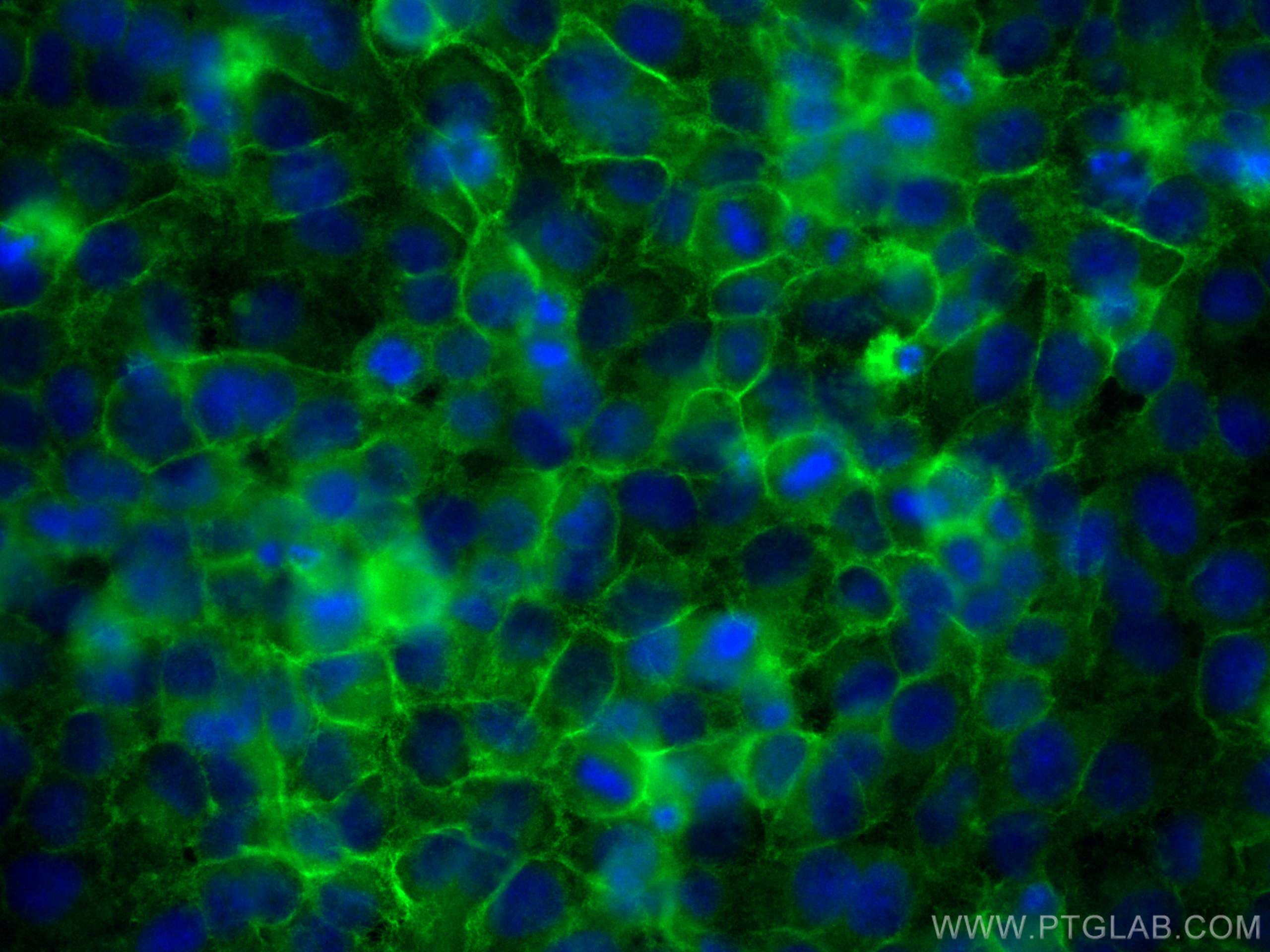
Tested Applications
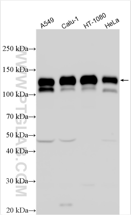
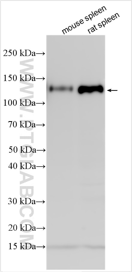
| Positive WB detected in | A549 cells, mouse spleen tissue, Calu-1 cells, HT-1080 cells, HeLa cells, rat spleen tissue |
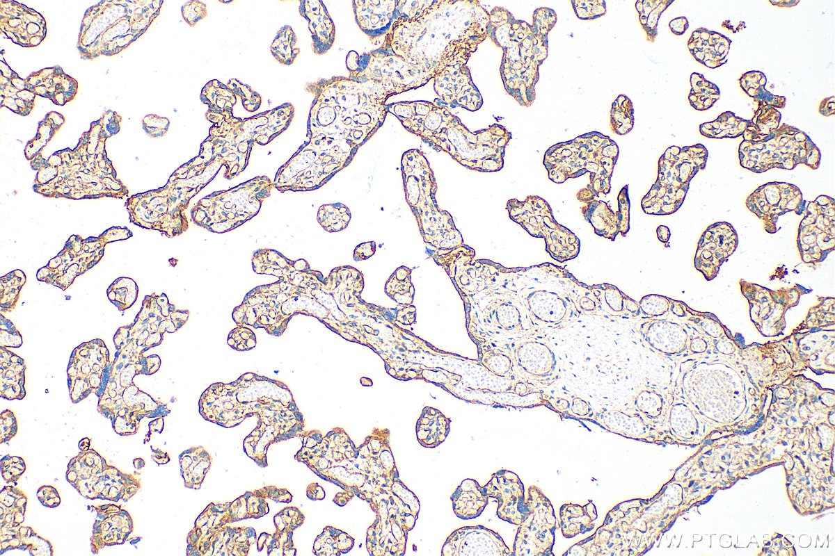
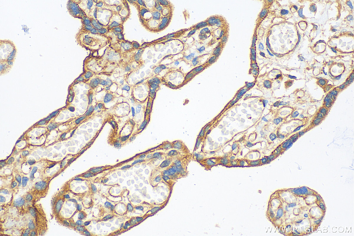
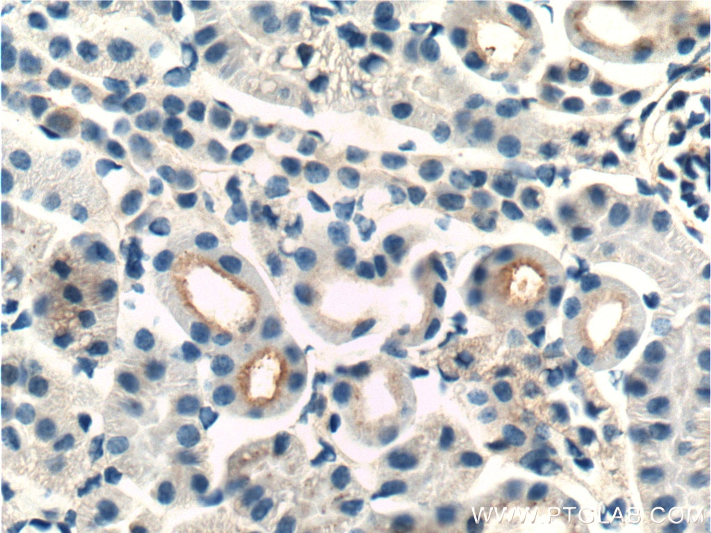
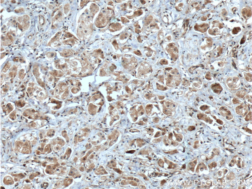
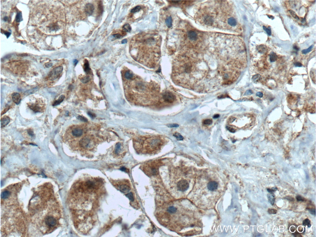
| Positive IHC detected in | human placenta tissue, mouse kidney tissue, human breast cancer tissue Note: suggested antigen retrieval with TE buffer pH 9.0; (*) Alternatively, antigen retrieval may be performed with citrate buffer pH 6.0 |
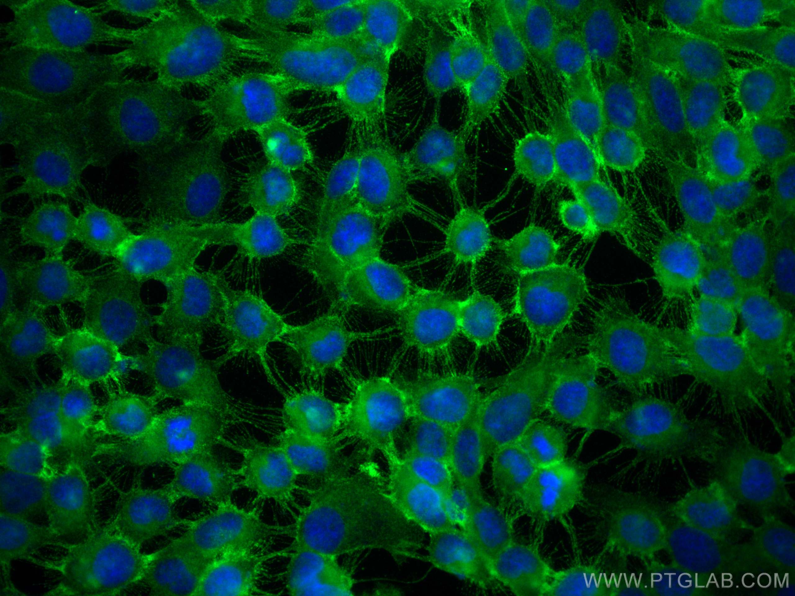
| Positive IF/ICC detected in | HeLa cells, A431 cells |
Recommended dilution
| Application | Dilution |
|---|---|
| Western Blot (WB) | WB : 1:5000-1:50000 |
| Immunohistochemistry (IHC) | IHC : 1:1000-1:4000 |
| Immunofluorescence (IF)/ICC | IF/ICC : 1:50-1:500 |
| It is recommended that this reagent should be titrated in each testing system to obtain optimal results. | |
| Sample-dependent, Check data in validation data gallery. | |
Published Applications
| KD/KO | See 5 publications below |
| WB | See 81 publications below |
| IHC | See 21 publications below |
| IF | See 34 publications below |
| IP | See 4 publications below |
| CoIP | See 1 publications below |
Product Information
12594-1-AP targets Integrin Beta 1/CD29 in WB, IHC, IF/ICC, IP, CoIP, ELISA applications and shows reactivity with human, mouse, rat samples.
| Tested Reactivity | human, mouse, rat |
| Cited Reactivity | human, mouse, rat, rabbit, chicken, bovine, sika deer |
| Host / Isotype | Rabbit / IgG |
| Class | Polyclonal |
| Type | Antibody |
| Immunogen |
CatNo: Ag3078 Product name: Recombinant human Integrin beta-1 protein Source: e coli.-derived, PGEX-4T Tag: GST Domain: 30-359 aa of BC020057 Sequence: ANAKSCGECIQAGPNCGWCTNSTFLQEGMPTSARCDDLEALKKKGCPPDDIENPRGSKDIKKNKNVTNRSKGTAEKLKPEDITQIQPQQLVLRLRSGEPQTFTLKFKRAEDYPIDLYYLMDLSYSMKDDLENVKSLGTDLMNEMRRITSDFRIGFGSFVEKTVMPYISTTPAKLRNPCTSEQNCTSPFSYKNVLSLTNKGEVFNELVGKQRISGNLDSPEGGFDAIMQVAVCGSLIGWRNVTRLLVFSTDAGFHFAGDGKLGGIVLPNDGQCHLENNMYTMSHYYDYPSIAHLVQKLSENNIQTIFAVTEEFQPVYKELKNLIPKSAVGT Predict reactive species |
| Full Name | integrin, beta 1 (fibronectin receptor, beta polypeptide, antigen CD29 includes MDF2, MSK12) |
| Calculated Molecular Weight | 88 kDa |
| Observed Molecular Weight | 100-130 kDa |
| GenBank Accession Number | BC020057 |
| Gene Symbol | Integrin beta 1 |
| Gene ID (NCBI) | 3688 |
| ENSEMBL Gene ID | ENSG00000150093 |
| RRID | AB_2130085 |
| Conjugate | Unconjugated |
| Form | Liquid |
| Purification Method | Antigen affinity purification |
| UNIPROT ID | P05556 |
| Storage Buffer | PBS with 0.02% sodium azide and 50% glycerol, pH 7.3. |
| Storage Conditions | Store at -20°C. Stable for one year after shipment. Aliquoting is unnecessary for -20oC storage. 20ul sizes contain 0.1% BSA. |
Background Information
Integrin beta-1 (ITGB1), also named as CD29, is a single chain type I glycoprotein that is expressed in a heterodimeric complex with one of six distinct α subunits, comprising the very late activation antigen (VLA) subfamily of adhesion receptors. It is one of the essential surface molecules expressed on human MSC from bone marrow and other sources. The β1 subunit is also broadly expressed on lymphocytes and monocytes, weakly expressed on granulocytes, and not expressed on erythrocytes. These receptors are involved in a variety of cell-cell and cell-matrix interactions.
Protocols
| Product Specific Protocols | |
|---|---|
| IF protocol for Integrin Beta 1/CD29 antibody 12594-1-AP | Download protocol |
| IHC protocol for Integrin Beta 1/CD29 antibody 12594-1-AP | Download protocol |
| WB protocol for Integrin Beta 1/CD29 antibody 12594-1-AP | Download protocol |
| Standard Protocols | |
|---|---|
| Click here to view our Standard Protocols |
Publications
| Species | Application | Title |
|---|---|---|
Sci Adv Decellularized extracellular matrix scaffolds identify full-length collagen VI as a driver of breast cancer cell invasion in obesity and metastasis. | ||
Nat Commun Homophilic ATP1A1 binding induces activin A secretion to promote EMT of tumor cells and myofibroblast activation. | ||
Adv Healthc Mater Modification of PLGA Scaffold by MSC-Derived Extracellular Matrix Combats Macrophage Inflammation to Initiate Bone Regeneration via TGF-β-Induced Protein.
| ||
Sci Bull (Beijing) The Rhinolophus affinis bat ACE2 and multiple animal orthologs are functional receptors for bat coronavirus RaTG13 and SARS-CoV-2. |
Reviews
The reviews below have been submitted by verified Proteintech customers who received an incentive for providing their feedback.
FH GAURAV (Verified Customer) (10-27-2025) | Very good antibody that works on higher dilution also
|
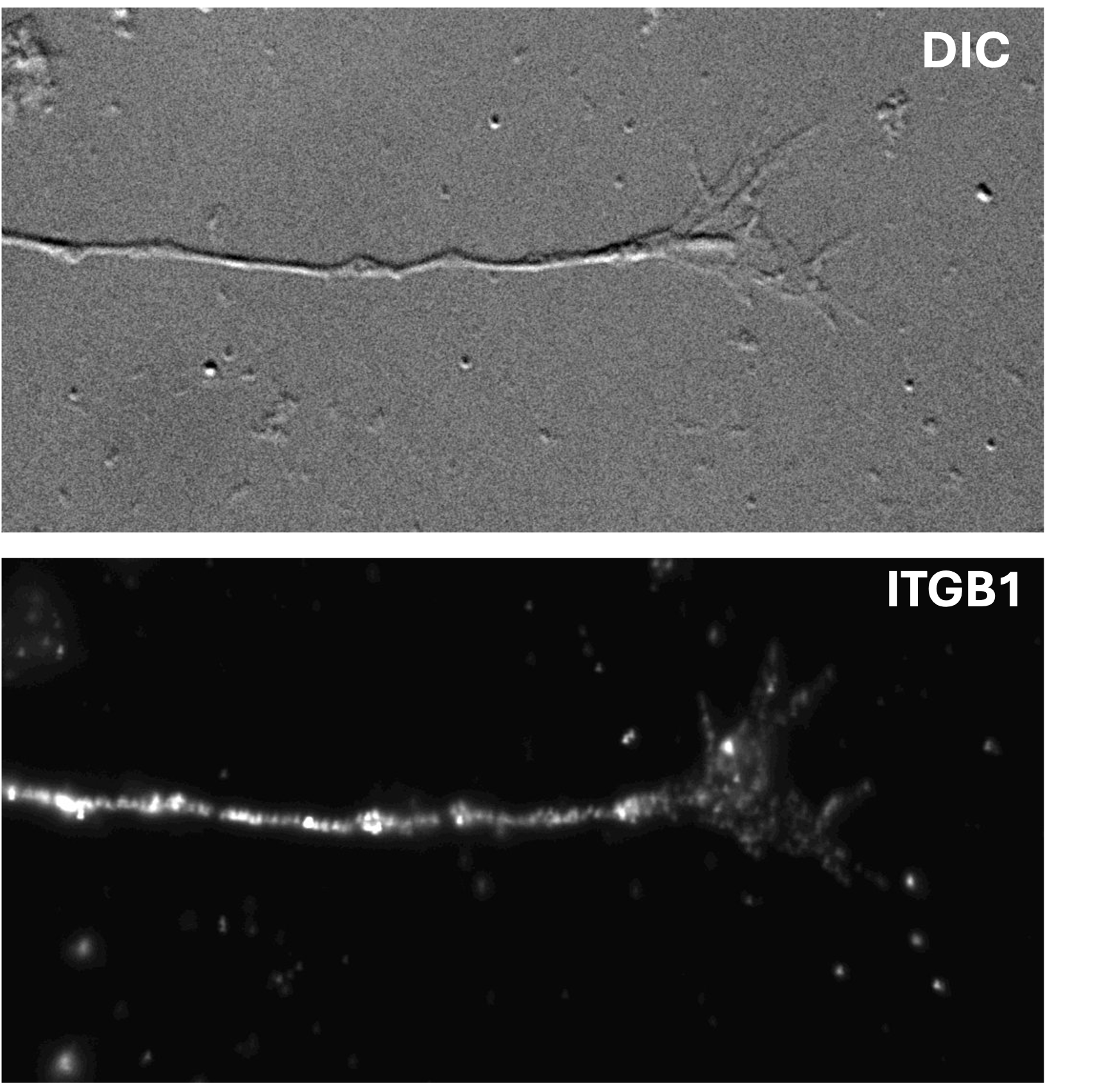
FH Nikita (Verified Customer) (07-23-2025) | I have used it for both ICC and Western blot. I haven't been successful with the blot yet, but my ICC images looked beautiful on embryonic day 17 mouse cortical neurons cultured in vitro for 2 days.
 |
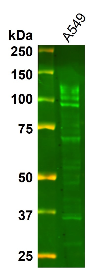
FH Angie (Verified Customer) (08-22-2024) | A549 lysate was subjected to western blot with Integrin beta 1antibody used at dilution of 1:5000 and incubated at room temperature for 1.5 hours. Secondary antibody: Donkey-anti-rabbit (Alexa Fluor 800) at 1:20000 dilution incubated at room temperature for 1 hour. Several bands were observed.
 |
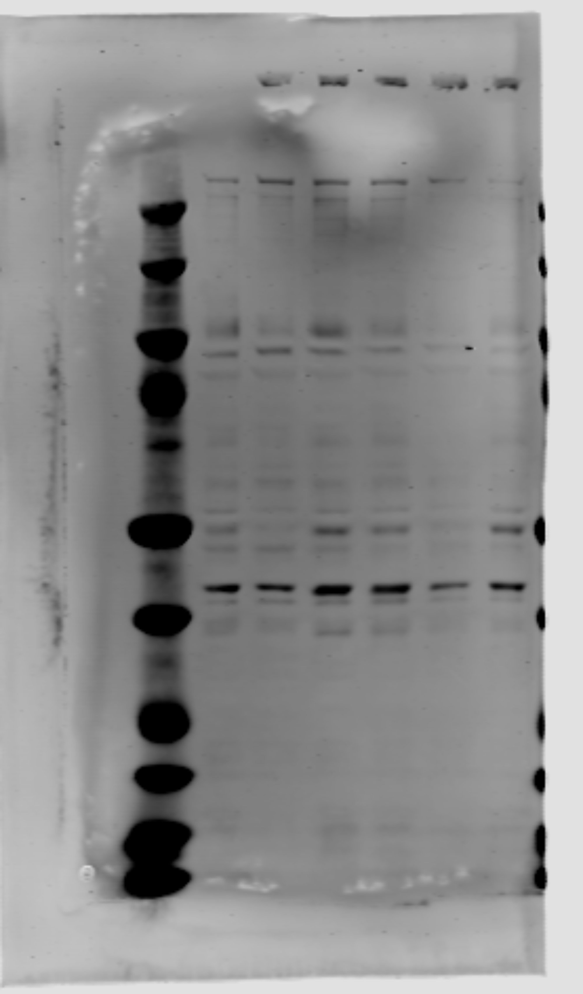
FH Alberto (Verified Customer) (01-16-2024) | Multiple bands, very different compared to advertised picture. I want my money back.
 |
FH Udesh (Verified Customer) (07-10-2022) | The antibody worked great in Co-IP and WB. Great chunky band close to 130 kD. For Co-IP: 6uL/200ug protein and 1:2000 in 3%BSA for WB
|
FH Guorong (Verified Customer) (05-23-2022) | works better in ARPE-19 cells than 293T cells
 |
FH Boyan (Verified Customer) (03-29-2019) | Good for WB - recognises a clean band around the expected molecular weight.
|

