Anticorps Polyclonal de lapin anti-LC3B-Specific
LC3B-Specific Polyclonal Antibody for WB, IHC, IF/ICC, FC (Intra), ELISA
Hôte / Isotype
Lapin / IgG
Réactivité testée
Humain, rat, souris et plus (2)
Applications
WB, IHC, IF/ICC, FC (Intra), IP, ELISA
Conjugaison
Non conjugué
288
N° de cat : 18725-1-AP
Synonymes
Galerie de données de validation
Applications testées
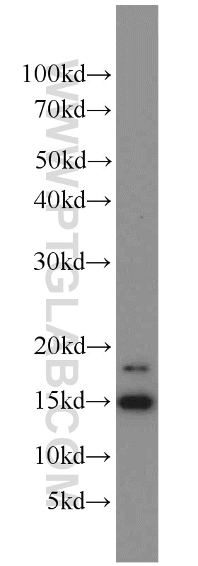
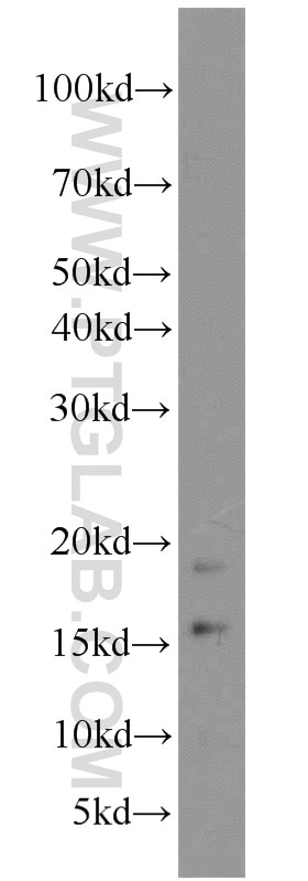
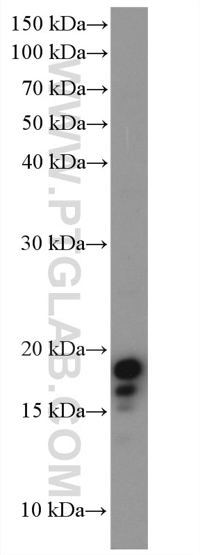
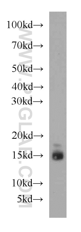
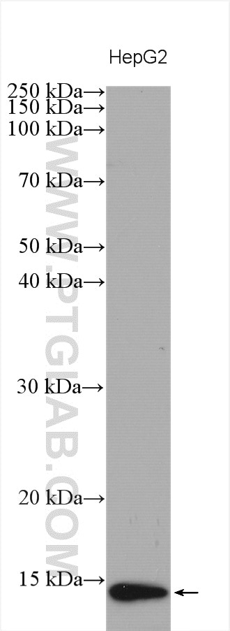
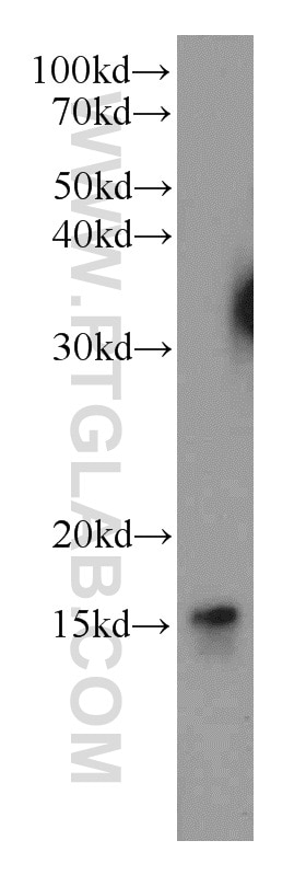
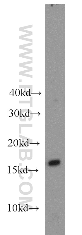
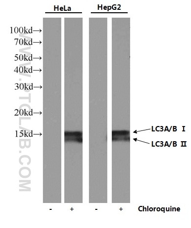
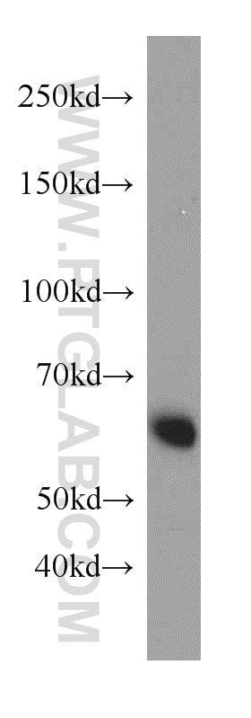
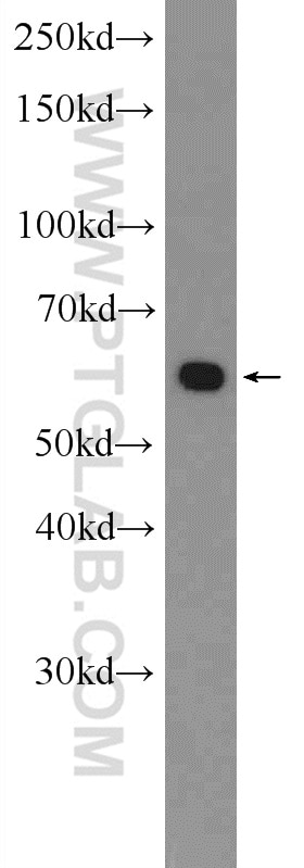
| Résultats positifs en WB | tissu cérébral humain, cellules A549, cellules HepG2, cellules MCF-7, HEK-293 traitées aux UV, Hela traitées au TN, tissu cérébral de souris |
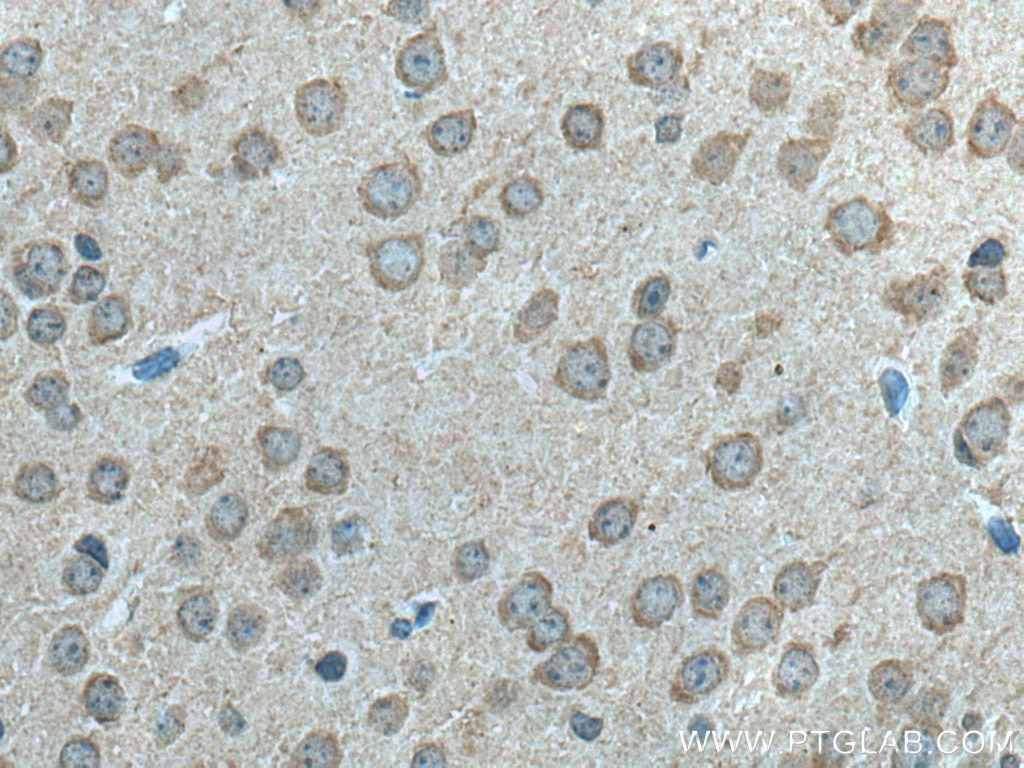
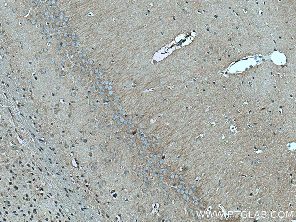
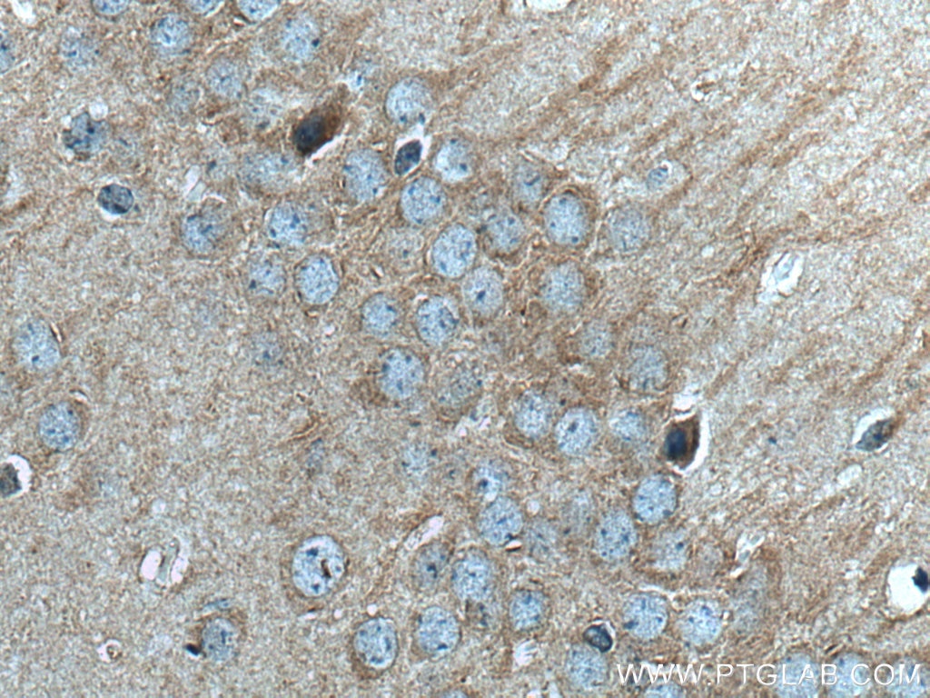
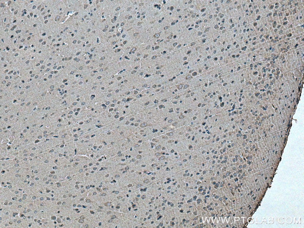
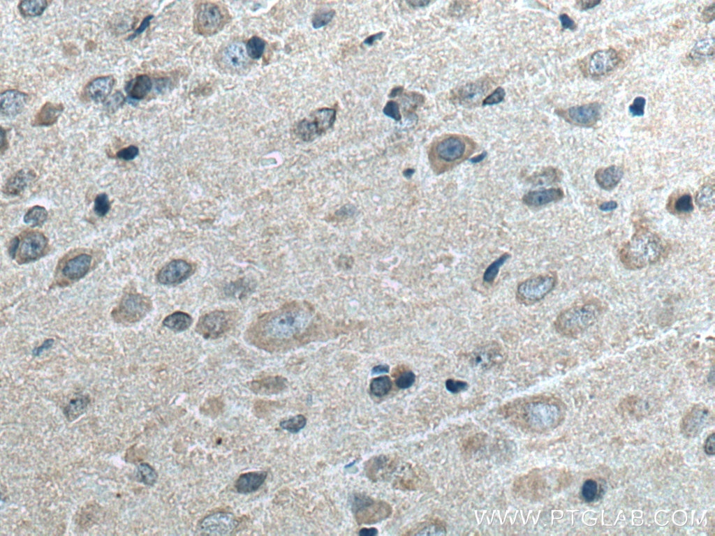
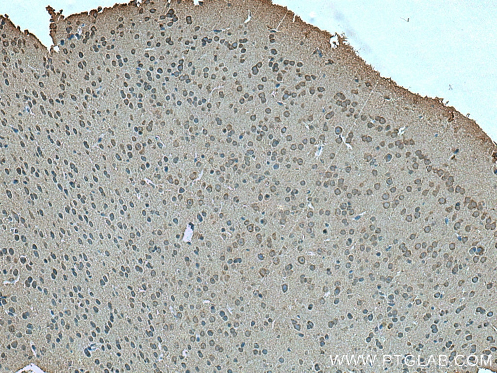
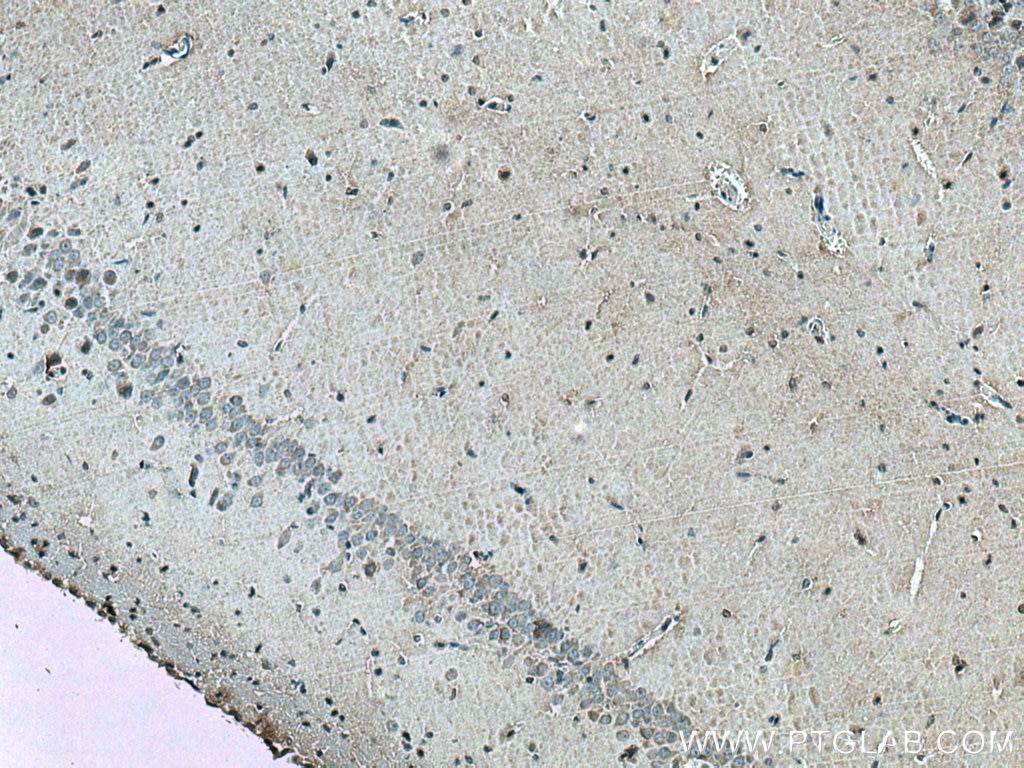
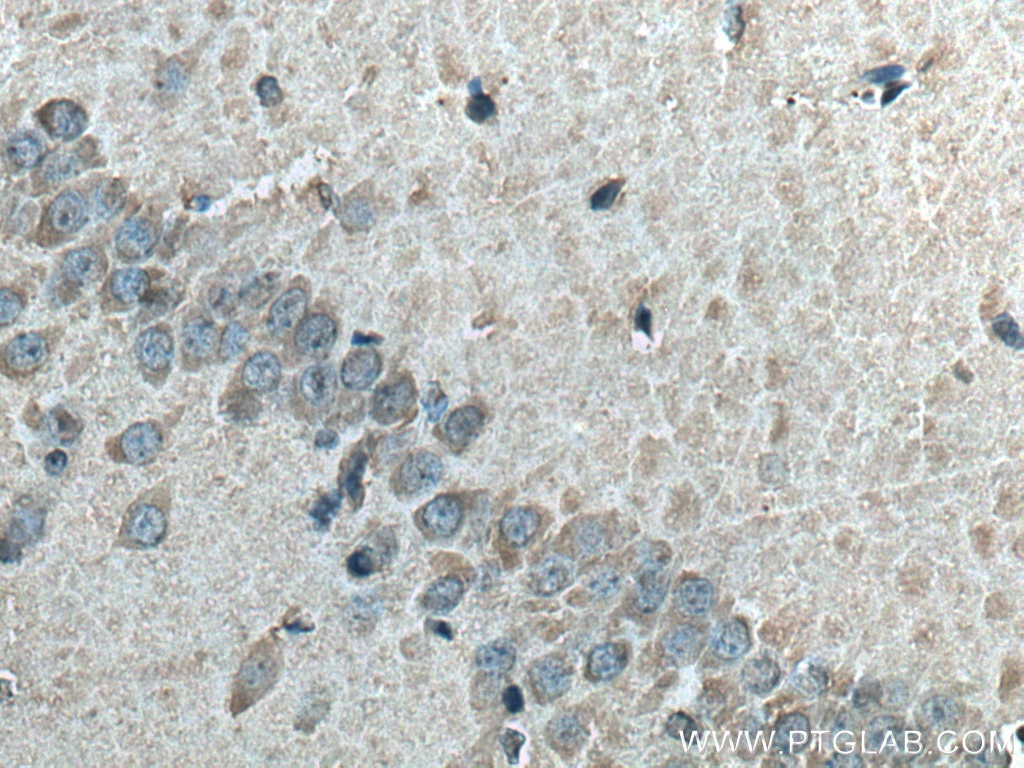
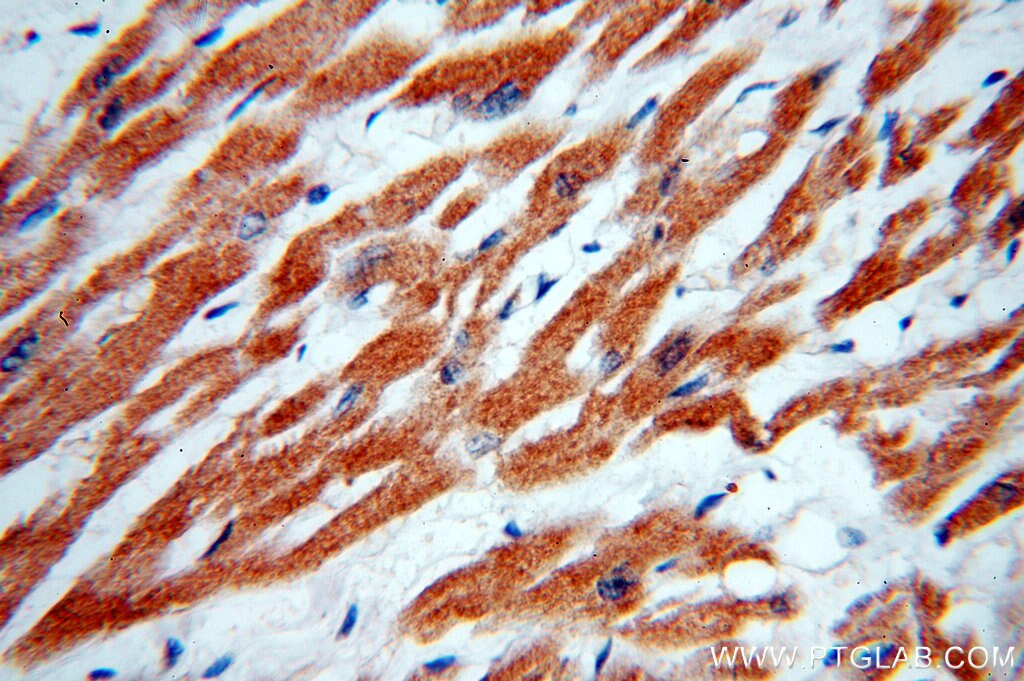
| Résultats positifs en IHC | tissu cérébral de souris, tissu cérébral de rat il est suggéré de démasquer l'antigène avec un tampon de TE buffer pH 9.0; (*) À défaut, 'le démasquage de l'antigène peut être 'effectué avec un tampon citrate pH 6,0. |
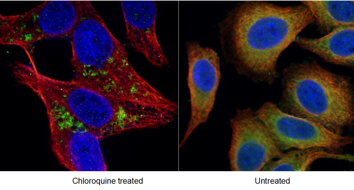
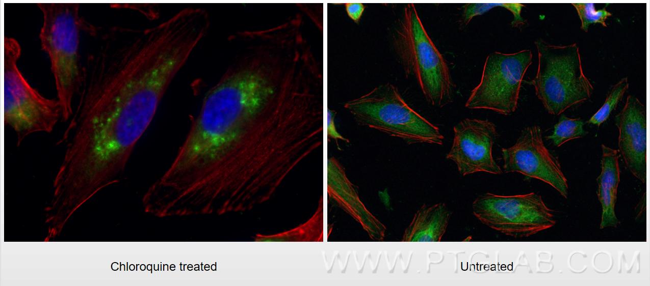
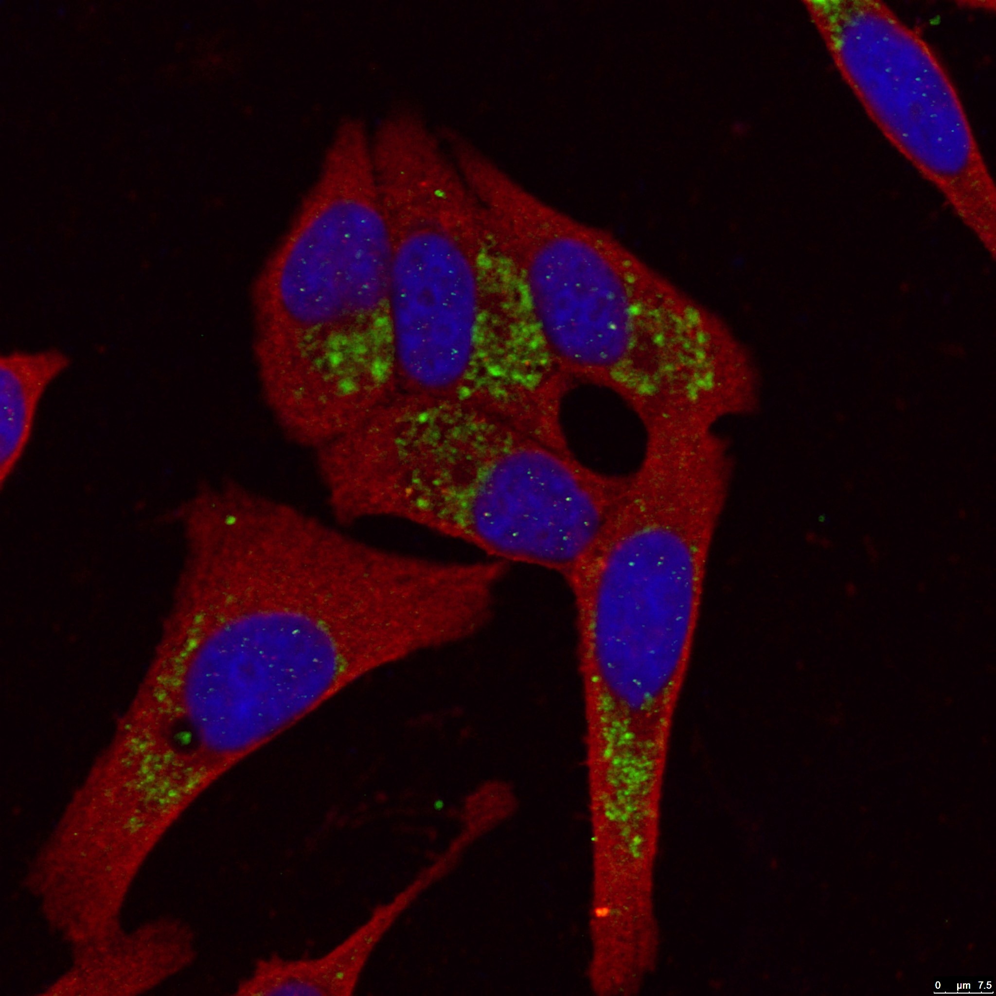
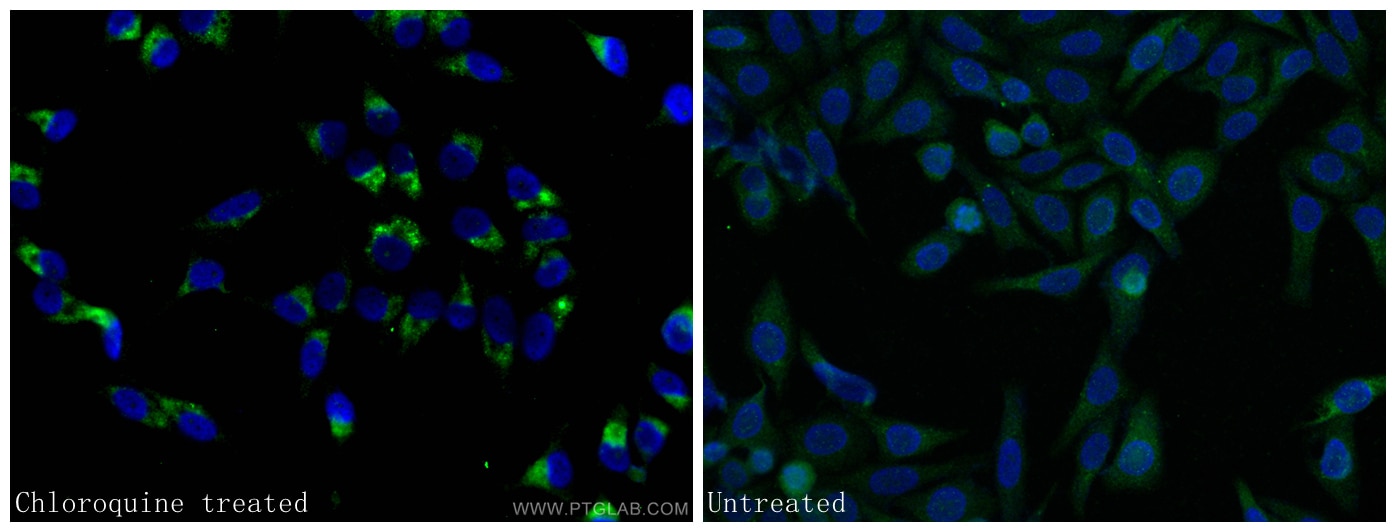
| Résultats positifs en IF/ICC | cellules HeLa traitées à la chloroquine, cellules HepG2 traitées à la chloroquine |
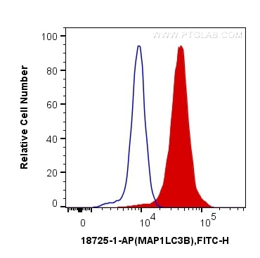
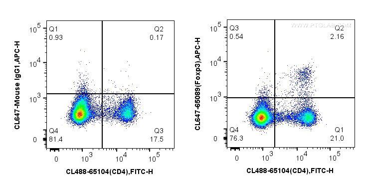
| Résultats positifs en FC (Intra) | cellules HeLa, |
Dilution recommandée
| Application | Dilution |
|---|---|
| Western Blot (WB) | WB : 1:300-1:1000 |
| Immunohistochimie (IHC) | IHC : 1:50-1:500 |
| Immunofluorescence (IF)/ICC | IF/ICC : 1:50-1:500 |
| Flow Cytometry (FC) (INTRA) | FC (INTRA) : 0.40 ug per 10^6 cells in a 100 µl suspension |
| It is recommended that this reagent should be titrated in each testing system to obtain optimal results. | |
| Sample-dependent, check data in validation data gallery | |
Applications publiées
| WB | See 236 publications below |
| IHC | See 41 publications below |
| IF | See 80 publications below |
| IP | See 3 publications below |
Informations sur le produit
18725-1-AP cible LC3B-Specific dans les applications de WB, IHC, IF/ICC, FC (Intra), IP, ELISA et montre une réactivité avec des échantillons Humain, rat, souris
| Réactivité | Humain, rat, souris |
| Réactivité citée | rat, bovin, Humain, porc, souris |
| Hôte / Isotype | Lapin / IgG |
| Clonalité | Polyclonal |
| Type | Anticorps |
| Immunogène | Peptide |
| Nom complet | microtubule-associated protein 1 light chain 3 beta |
| Masse moléculaire calculée | 15 kDa |
| Poids moléculaire observé | 15 kDa, 18 kDa |
| Numéro d’acquisition GenBank | NM_022818 |
| Symbole du gène | LC3B |
| Identification du gène (NCBI) | 81631 |
| Conjugaison | Non conjugué |
| Forme | Liquide |
| Méthode de purification | Purification par affinité contre l'antigène |
| Tampon de stockage | PBS avec azoture de sodium à 0,02 % et glycérol à 50 % pH 7,3 |
| Conditions de stockage | Stocker à -20°C. Stable pendant un an après l'expédition. L'aliquotage n'est pas nécessaire pour le stockage à -20oC Les 20ul contiennent 0,1% de BSA. |
Informations générales
LC3B, also named as MAP1LC3B, MAP1A/1BLC3, belongs to the MAP1 LC3 family. It is a subunit of neuronal microtubule-associated MAP1A and MAP1B proteins, which are involved in microtubule assembly and important for neurogenesis. In cell biology, autophagy, or autophagocytosis, is a catabolic process involving the degradation of a cell's own components through the lysosomalmachinery. It is a major mechanism by which a starving cell reallocates nutrients from unnecessary processes to more-essential processes. Two forms of LC3, called LC3-I (17-19kd) and -II(14-16kd), were produced post-translationally in various cells. LC3-I is cytosolic, whereas LC3-II is membrane bound. The precursor molecule is cleaved by APG4B/ATG4B to form the cytosolic form, LC3-I. This is activated by APG7L/ATG7, transferred to ATG3 and conjugated to phospholipid to form the membrane-bound form, LC3-II. The amount of LC3-II is correlated with the extent of autophagosome formation. LC3-II is the first mammalian protein identified that specifically associates with autophagosome membranes. MAP1LC3 has 3 isoforms MAP1LC3A, MAP1LC3B and MAP1LC3C. MAP1LC3A and MAP1LC3C are produced by the proteolytic cleavage after the conserved C-terminal Gly residue, like their rat counterpart, MAP1LC3B does not undergo C-terminal cleavage and exists in a single modified form. This antibody is specific to LC3B.
Protocole
| Product Specific Protocols | |
|---|---|
| WB protocol for LC3B-Specific antibody 18725-1-AP | Download protocol |
| IHC protocol for LC3B-Specific antibody 18725-1-AP | Download protocol |
| IF protocol for LC3B-Specific antibody 18725-1-AP | Download protocol |
| FC protocol for LC3B-Specific antibody 18725-1-AP | Download protocol |
| Standard Protocols | |
|---|---|
| Click here to view our Standard Protocols |
Publications
| Species | Application | Title |
|---|---|---|
Am J Respir Crit Care Med Acid Sphingomyelinase Inhibition Attenuates Cell Death in Mechanically-Ventilated Newborn Rat Lung. | ||
Nat Microbiol Chloroquine ameliorates carbon tetrachloride-induced acute liver injury in mice via the concomitant inhibition of inflammation and induction of apoptosis | ||
ACS Nano Nanoenabled Disruption of Multiple Barriers in Antigen Cross-Presentation of Dendritic Cells via Calcium Interference for Enhanced Chemo-Immunotherapy. | ||
Autophagy TRIM59 regulates autophagy through modulating both the transcription and the ubiquitination of BECN1. | ||
Autophagy STYK1 promotes autophagy through enhancing the assembly of autophagy-specific class III phosphatidylinositol 3-kinase complex I. | ||
Mater Today Bio Carbon dots enhance extracellular matrix secretion for dentin-pulp complex regeneration through PI3K/Akt/mTOR pathway-mediated activation of autophagy. |
Avis
The reviews below have been submitted by verified Proteintech customers who received an incentive forproviding their feedback.
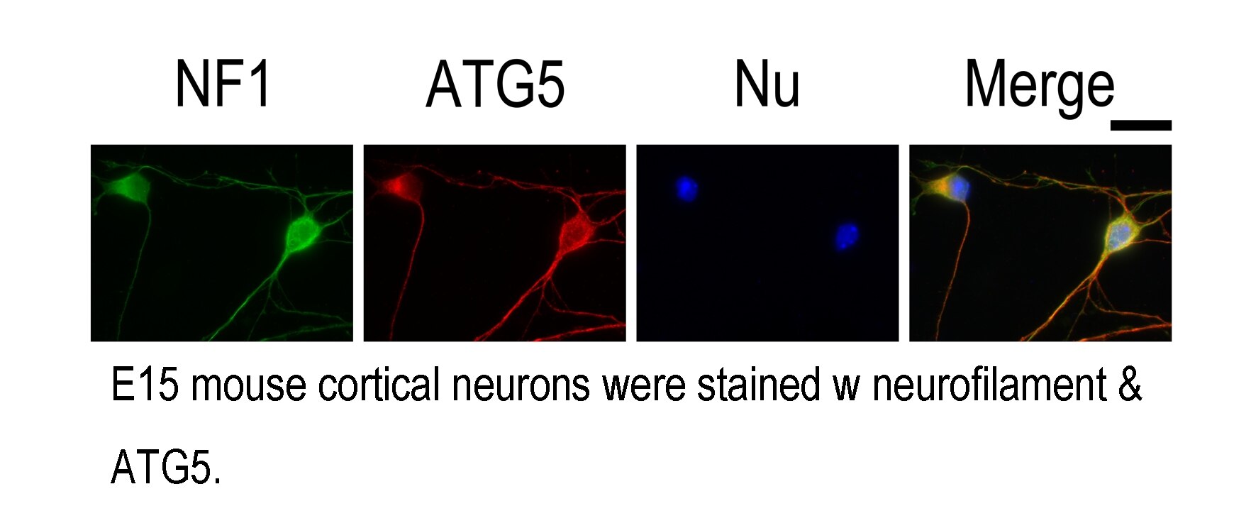
FH Azita (Verified Customer) (06-02-2021) | Western blot analysis using LC3B-Specific Polyclonal antibody in NSC34 cell line at dilution of 1:300. Immunohistochemistry labelling of primary rat cortical neurons using LC3B-Specific Polyclonal antibody at dilution of 1:500 showed strong labelling.
|
FH Tanusree (Verified Customer) (12-03-2019) | The antibody works great in Western blotting analysis.
|
