Anticorps Polyclonal de lapin anti-Centrin 1
Centrin 1 Polyclonal Antibody for WB, IF, ELISA
Hôte / Isotype
Lapin / IgG
Réactivité testée
Humain et plus (2)
Applications
WB, IF/ICC, ELISA
Conjugaison
Non conjugué
N° de cat : 12794-1-AP
Synonymes
Galerie de données de validation
Applications testées
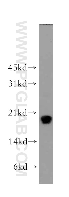
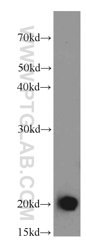
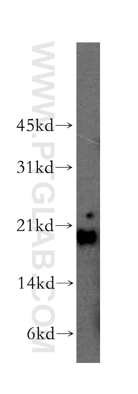
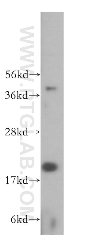
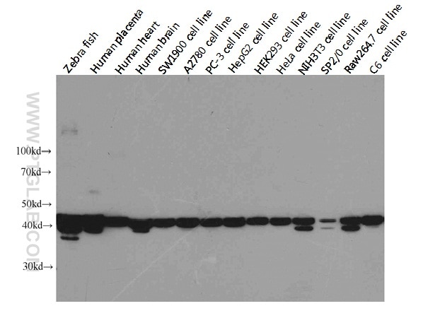
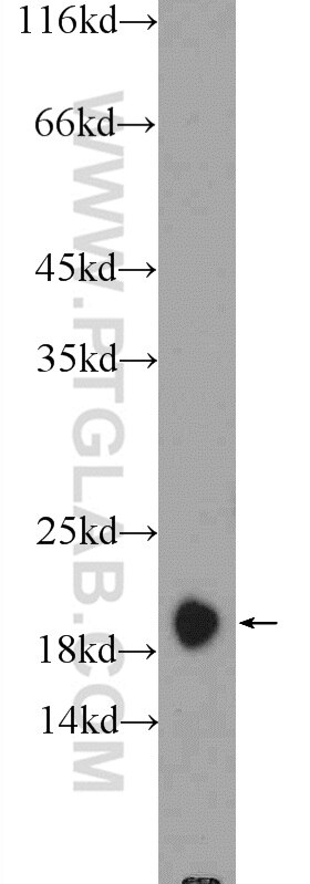
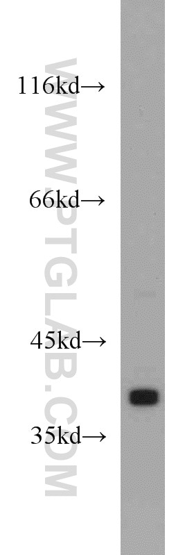
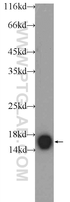
| Résultats positifs en WB | cellules HL-60, tissu cardiaque humain |
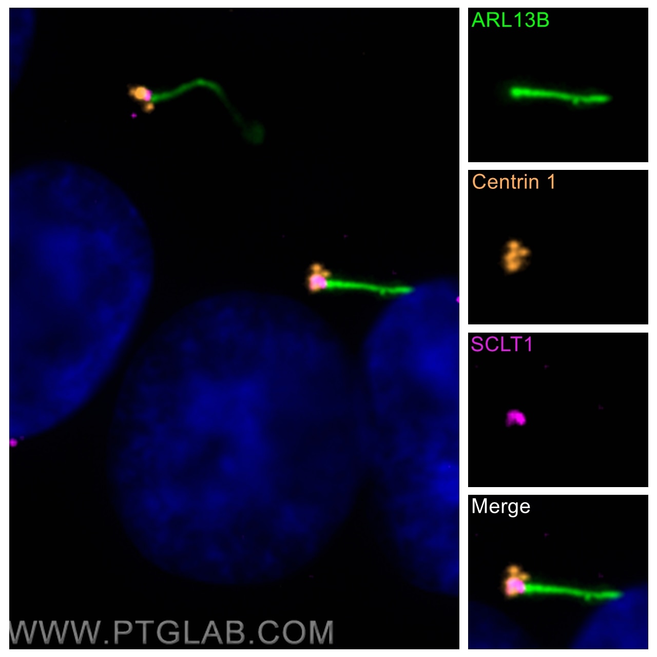
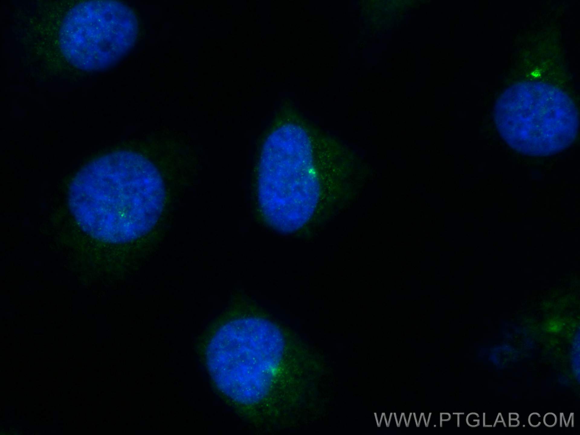
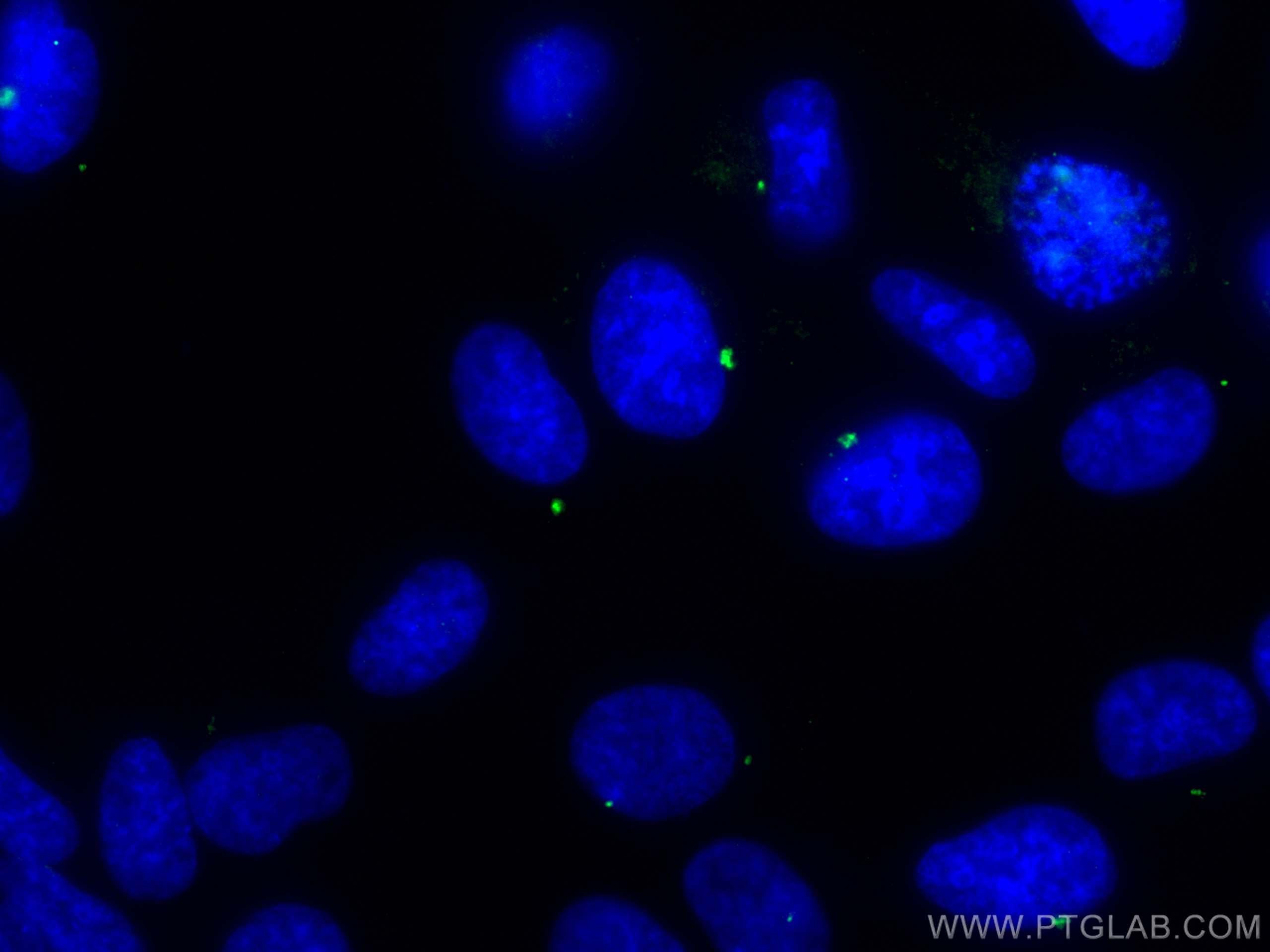
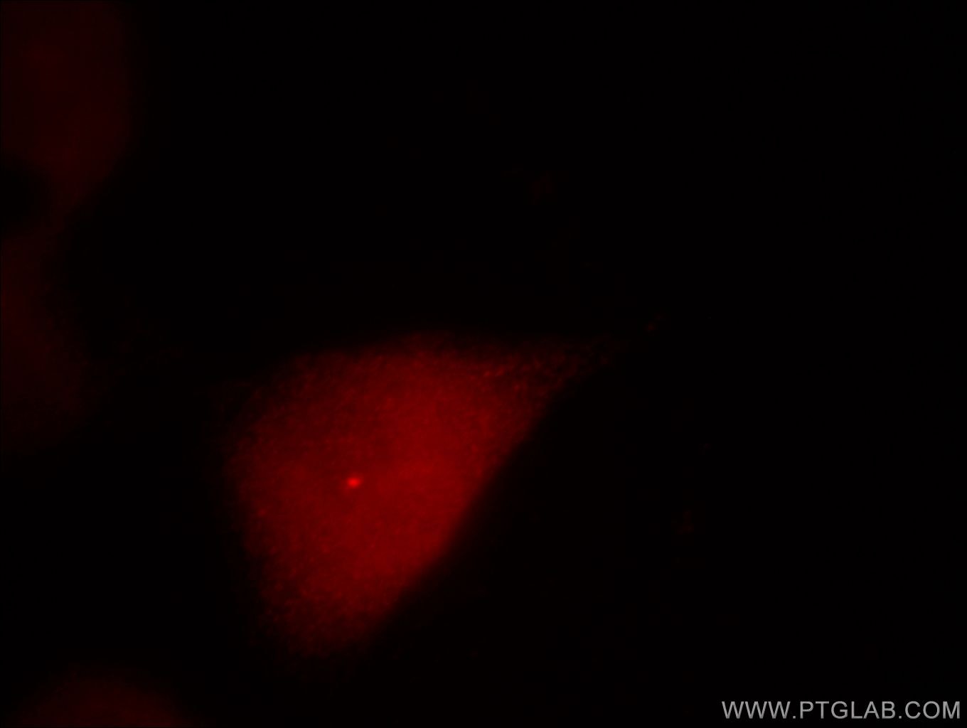
| Résultats positifs en IF/ICC | cellules hTERT-RPE1, cellules HeLa |
Dilution recommandée
| Application | Dilution |
|---|---|
| Western Blot (WB) | WB : 1:500-1:2000 |
| Immunofluorescence (IF)/ICC | IF/ICC : 1:50-1:500 |
| It is recommended that this reagent should be titrated in each testing system to obtain optimal results. | |
| Sample-dependent, check data in validation data gallery | |
Applications publiées
| WB | See 5 publications below |
| IF | See 29 publications below |
Informations sur le produit
12794-1-AP cible Centrin 1 dans les applications de WB, IF/ICC, ELISA et montre une réactivité avec des échantillons Humain
| Réactivité | Humain |
| Réactivité citée | Humain, poisson-zèbre, souris |
| Hôte / Isotype | Lapin / IgG |
| Clonalité | Polyclonal |
| Type | Anticorps |
| Immunogène | Centrin 1 Protéine recombinante Ag3529 |
| Nom complet | centrin, EF-hand protein, 1 |
| Masse moléculaire calculée | 172 aa, 20 kDa |
| Poids moléculaire observé | 20 kDa |
| Numéro d’acquisition GenBank | BC029515 |
| Symbole du gène | Centrin 1 |
| Identification du gène (NCBI) | 1068 |
| Conjugaison | Non conjugué |
| Forme | Liquide |
| Méthode de purification | Purification par affinité contre l'antigène |
| Tampon de stockage | PBS avec azoture de sodium à 0,02 % et glycérol à 50 % pH 7,3 |
| Conditions de stockage | Stocker à -20°C. Stable pendant un an après l'expédition. L'aliquotage n'est pas nécessaire pour le stockage à -20oC Les 20ul contiennent 0,1% de BSA. |
Informations générales
EF-hand type Ca2+-binding proteins consists of several family members, including Centrin-1, Centrin-2 and Centrin-3. The Centrin proteins are ubiquitously expressed cytoskeletal components that show increased expression during cell differentiation. Centrin-1 plays important roles in the determination of centrosome position and segregation, and in the process of microtubule severing. Centrin-1 is localized to the centrosome of interphase cells, and redistributes to the region of the spindle poles during mitosis, reflecting the dynamic behavior of the centrosome during the cell cycle.
Protocole
| Product Specific Protocols | |
|---|---|
| WB protocol for Centrin 1 antibody 12794-1-AP | Download protocol |
| IF protocol for Centrin 1 antibody 12794-1-AP | Download protocol |
| Standard Protocols | |
|---|---|
| Click here to view our Standard Protocols |
Publications
| Species | Application | Title |
|---|---|---|
Nat Cell Biol The Cep63 paralogue Deup1 enables massive de novo centriole biogenesis for vertebrate multiciliogenesis. | ||
Neuron Posterior Neocortex-Specific Regulation of Neuronal Migration by CEP85L Identifies Maternal Centriole-Dependent Activation of CDK5. | ||
Nat Commun Cytoplasmic E2f4 forms organizing centres for initiation of centriole amplification during multiciliogenesis. | ||
Nat Commun DNA replication licensing factor Cdc6 and Plk4 kinase antagonistically regulate centrosome duplication via Sas-6. |
Avis
The reviews below have been submitted by verified Proteintech customers who received an incentive forproviding their feedback.
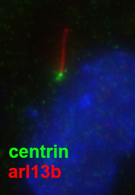
FH Julie (Verified Customer) (02-14-2019) | Nice staining of ciliated human fibroblasts.
 |
