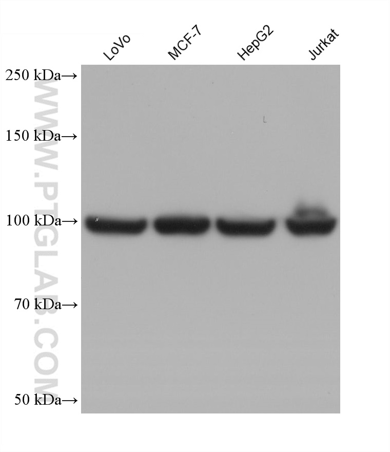
Various lysates were subjected to SDS PAGE followed by western blot with 67550-1-Ig (EEF2 antibody) at dilution of 1:40000 incubated at room temperature for 1.5 hours.
Various lysates were subjected to SDS PAGE followed by western blot with 67550-1-Ig (EEF2 antibody) at dilution of 1:40000 incubated at room temperature for 1.5 hours.
WB analysis using 67550-1-Ig
Various lysates were subjected to SDS PAGE followed by western blot with 67550-1-Ig (EEF2 antibody) at dilution of 1:50000 incubated at room temperature for 1.5 hours.
Various lysates were subjected to SDS PAGE followed by western blot with 67550-1-Ig (EEF2 antibody) at dilution of 1:50000 incubated at room temperature for 1.5 hours.
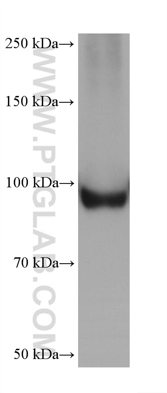
WB analysis of HSC-T6 using 67550-1-Ig
HSC-T6 cells were subjected to SDS PAGE followed by western blot with 67550-1-Ig (EEF2 antibody) at dilution of 1:50000 incubated at room temperature for 1.5 hours.
HSC-T6 cells were subjected to SDS PAGE followed by western blot with 67550-1-Ig (EEF2 antibody) at dilution of 1:50000 incubated at room temperature for 1.5 hours.
WB analysis using 67550-1-Ig
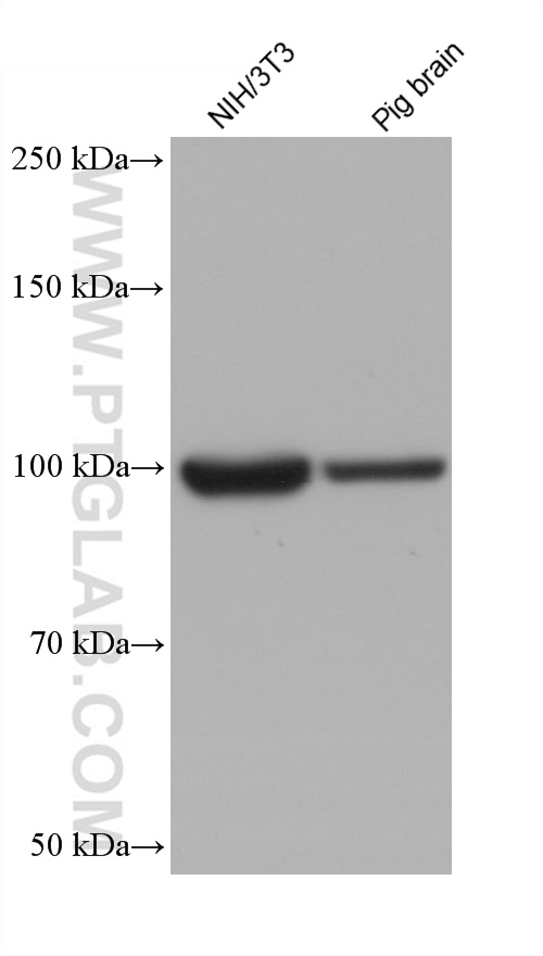
Various lysates were subjected to SDS PAGE followed by western blot with 67550-1-Ig (EEF2 antibody) at dilution of 1:50000 incubated at room temperature for 1.5 hours.
Various lysates were subjected to SDS PAGE followed by western blot with 67550-1-Ig (EEF2 antibody) at dilution of 1:50000 incubated at room temperature for 1.5 hours.
WB analysis using 67550-1-Ig
Various lysates were subjected to SDS PAGE followed by western blot with 67550-1-Ig (EEF2 antibody) at dilution of 1:10000 incubated at room temperature for 1.5 hours.
Various lysates were subjected to SDS PAGE followed by western blot with 67550-1-Ig (EEF2 antibody) at dilution of 1:10000 incubated at room temperature for 1.5 hours.
IHC staining of human breast cancer using 67550-1-Ig
Immunohistochemical analysis of paraffin-embedded human breast cancer tissue slide using 67550-1-Ig (EEF2 antibody) at dilution of 1:1000 (under 40x lens). Heat mediated antigen retrieval with Tris-EDTA buffer (pH 9.0).
Immunohistochemical analysis of paraffin-embedded human breast cancer tissue slide using 67550-1-Ig (EEF2 antibody) at dilution of 1:1000 (under 40x lens). Heat mediated antigen retrieval with Tris-EDTA buffer (pH 9.0).
IHC staining of human colon cancer using 67550-1-Ig
Immunohistochemical analysis of paraffin-embedded human colon cancer tissue slide using 67550-1-Ig (EEF2 antibody) at dilution of 1:1000 (under 10x lens). Heat mediated antigen retrieval with Tris-EDTA buffer (pH 9.0).
Immunohistochemical analysis of paraffin-embedded human colon cancer tissue slide using 67550-1-Ig (EEF2 antibody) at dilution of 1:1000 (under 10x lens). Heat mediated antigen retrieval with Tris-EDTA buffer (pH 9.0).
IHC staining of human colon cancer using 67550-1-Ig
Immunohistochemical analysis of paraffin-embedded human colon cancer tissue slide using 67550-1-Ig (EEF2 antibody) at dilution of 1:1000 (under 40x lens). Heat mediated antigen retrieval with Tris-EDTA buffer (pH 9.0).
Immunohistochemical analysis of paraffin-embedded human colon cancer tissue slide using 67550-1-Ig (EEF2 antibody) at dilution of 1:1000 (under 40x lens). Heat mediated antigen retrieval with Tris-EDTA buffer (pH 9.0).
IHC staining of human breast cancer using 67550-1-Ig
Immunohistochemical analysis of paraffin-embedded human breast cancer tissue slide using 67550-1-Ig (EEF2 antibody) at dilution of 1:1000 (under 10x lens). Heat mediated antigen retrieval with Tris-EDTA buffer (pH 9.0).
Immunohistochemical analysis of paraffin-embedded human breast cancer tissue slide using 67550-1-Ig (EEF2 antibody) at dilution of 1:1000 (under 10x lens). Heat mediated antigen retrieval with Tris-EDTA buffer (pH 9.0).
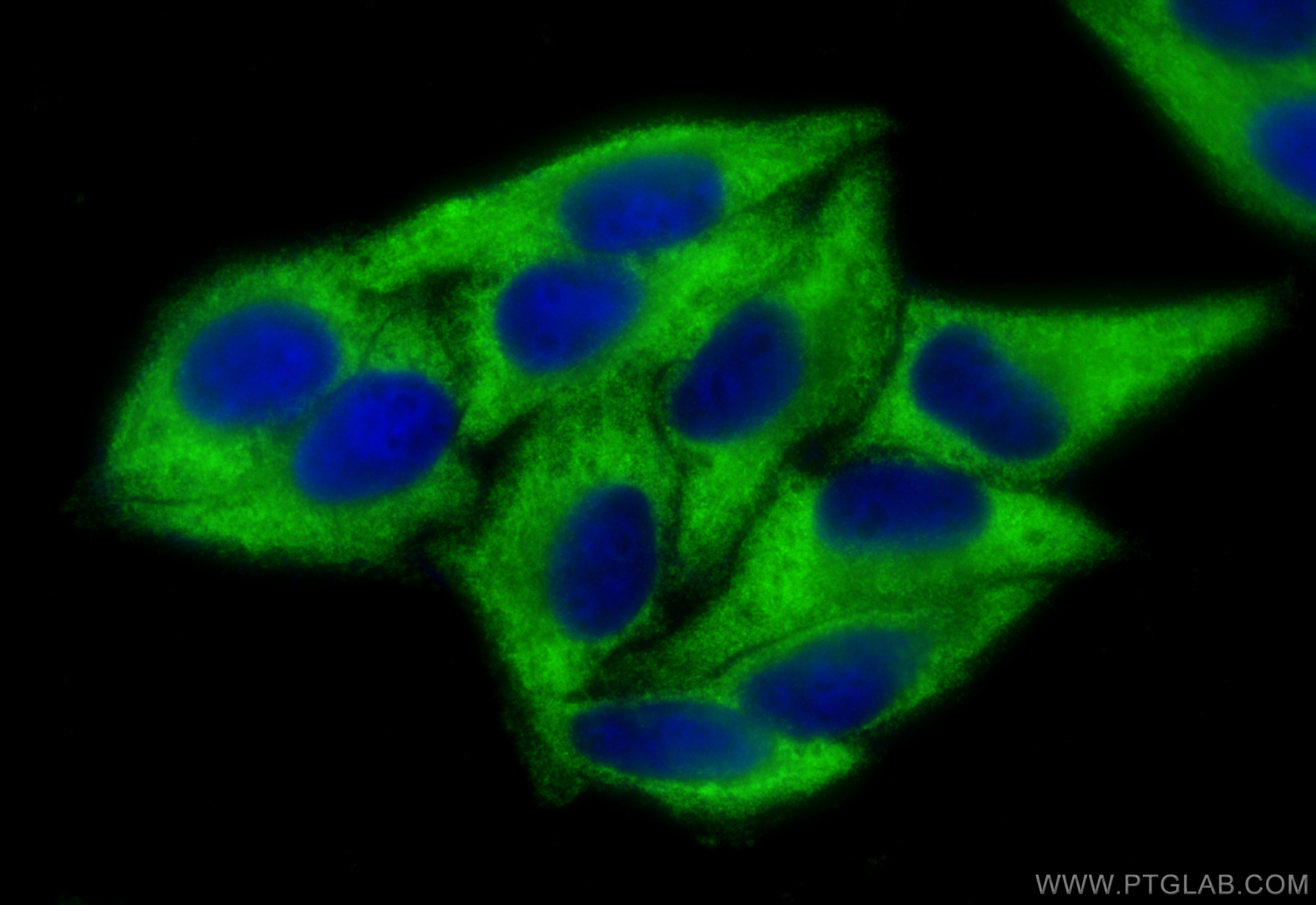
IF Staining of HepG2 using 67550-1-Ig
Immunofluorescent analysis of (-20°C Ethanol) fixed HepG2 cells using 67550-1-Ig (EEF2 antibody) at dilution of 1:100 and CoraLite488-Conjugated AffiniPure Goat Anti-Mouse IgG(H+L).
Immunofluorescent analysis of (-20°C Ethanol) fixed HepG2 cells using 67550-1-Ig (EEF2 antibody) at dilution of 1:100 and CoraLite488-Conjugated AffiniPure Goat Anti-Mouse IgG(H+L).
IF Staining of HepG2 using 67550-1-Ig
Immunofluorescent analysis of (-20°C Ethanol) fixed HepG2 cells using EEF2 antibody (67550-1-Ig, Clone: 1E4D5 ) at dilution of 1:800 and CoraLite®488-Conjugated Goat Anti-Mouse IgG(H+L) (SA00013-1).
Immunofluorescent analysis of (-20°C Ethanol) fixed HepG2 cells using EEF2 antibody (67550-1-Ig, Clone: 1E4D5 ) at dilution of 1:800 and CoraLite®488-Conjugated Goat Anti-Mouse IgG(H+L) (SA00013-1).
The Proteintech guarantee covers Proteintech antibodies in any species and any application, including those not listed on the datasheet. If the antibody doesn’t perform, you can receive a hassle-free refund or credit note.
human breast cancer tissue, human colon cancer tissue Note: suggested antigen retrieval with TE buffer pH 9.0; (*) Alternatively, antigen retrieval may be performed with citrate buffer pH 6.0
Positive IF/ICC detected in
HepG2 cells
Recommended dilution
Application
Dilution
Western Blot (WB)
WB : 1:5000-1:50000
Immunohistochemistry (IHC)
IHC : 1:500-1:2000
Immunofluorescence (IF)/ICC
IF/ICC : 1:400-1:1600
It is recommended that this reagent should be titrated in each testing system to obtain optimal results.
Sample-dependent, Check data in validation data gallery.
Product Information
67550-1-Ig targets EEF2 in WB, IHC, IF/ICC, ELISA applications and shows reactivity with human, mouse, rat, pig samples.
PBS with 0.02% sodium azide and 50% glycerol pH 7.3.
Storage Conditions
Store at -20°C. Stable for one year after shipment. Aliquoting is unnecessary for -20oC storage. 20ul sizes contain 0.1% BSA.
Background Information
EEF2 (Eukaryotic Elongation Factor 2), also known as EF-2 and EF2, is a crucial protein involved in the process of protein synthesis in eukaryotic cells. It plays a pivotal role in the elongation phase of translation, where it facilitates the movement of the ribosome along the mRNA strand. This movement is essential for the addition of amino acids to the growing polypeptide chain.
Various lysates were subjected to SDS PAGE followed by western blot with 67550-1-Ig (EEF2 antibody) at dilution of 1:40000 incubated at room temperature for 1.5 hours.
WB analysis using 67550-1-Ig
Various lysates were subjected to SDS PAGE followed by western blot with 67550-1-Ig (EEF2 antibody) at dilution of 1:50000 incubated at room temperature for 1.5 hours.
WB analysis of HSC-T6 using 67550-1-Ig
HSC-T6 cells were subjected to SDS PAGE followed by western blot with 67550-1-Ig (EEF2 antibody) at dilution of 1:50000 incubated at room temperature for 1.5 hours.
WB analysis using 67550-1-Ig
Various lysates were subjected to SDS PAGE followed by western blot with 67550-1-Ig (EEF2 antibody) at dilution of 1:50000 incubated at room temperature for 1.5 hours.
WB analysis using 67550-1-Ig
Various lysates were subjected to SDS PAGE followed by western blot with 67550-1-Ig (EEF2 antibody) at dilution of 1:10000 incubated at room temperature for 1.5 hours.
IHC Figures
IHC staining of human breast cancer using 67550-1-Ig
Immunohistochemical analysis of paraffin-embedded human breast cancer tissue slide using 67550-1-Ig (EEF2 antibody) at dilution of 1:1000 (under 40x lens). Heat mediated antigen retrieval with Tris-EDTA buffer (pH 9.0).
IHC staining of human colon cancer using 67550-1-Ig
Immunohistochemical analysis of paraffin-embedded human colon cancer tissue slide using 67550-1-Ig (EEF2 antibody) at dilution of 1:1000 (under 10x lens). Heat mediated antigen retrieval with Tris-EDTA buffer (pH 9.0).
IHC staining of human colon cancer using 67550-1-Ig
Immunohistochemical analysis of paraffin-embedded human colon cancer tissue slide using 67550-1-Ig (EEF2 antibody) at dilution of 1:1000 (under 40x lens). Heat mediated antigen retrieval with Tris-EDTA buffer (pH 9.0).
IHC staining of human breast cancer using 67550-1-Ig
Immunohistochemical analysis of paraffin-embedded human breast cancer tissue slide using 67550-1-Ig (EEF2 antibody) at dilution of 1:1000 (under 10x lens). Heat mediated antigen retrieval with Tris-EDTA buffer (pH 9.0).
IF/ICC Figures
IF Staining of HepG2 using 67550-1-Ig
Immunofluorescent analysis of (-20°C Ethanol) fixed HepG2 cells using 67550-1-Ig (EEF2 antibody) at dilution of 1:100 and CoraLite488-Conjugated AffiniPure Goat Anti-Mouse IgG(H+L).
IF Staining of HepG2 using 67550-1-Ig
Immunofluorescent analysis of (-20°C Ethanol) fixed HepG2 cells using EEF2 antibody (67550-1-Ig, Clone: 1E4D5 ) at dilution of 1:800 and CoraLite®488-Conjugated Goat Anti-Mouse IgG(H+L) (SA00013-1).
The species listed in Tested Reactivity are in-house verified and applicable species. For unlisted species, please refer to the homology analysis of the immunogen sequence and related species. For rabbit polyclonal antibodies, homology >70% is recommended. For mouse monoclonal antibodies and rabbit recombinant antibodies, homology >90% is recommended. Generally, the higher the homology, the greater the applicability. However, there will be certain differences in protein expression in different species, tissues or cells. Therefore, the homology analysis results are for reference only and do not serve as a guarantee.
At Proteintech, we pride ourselves on our antibody quality, customer service and transparency. As such, we are comparing our antibodies with other vendors, enabling easy identification and comparisons of key data to help you choose the suitable antibody for your needs.
We have selected the top cited antibodies from these vendors for you to compare.
 at dilution of 1:40000 incubated at room temperature for 1.5 hours.")
 at dilution of 1:50000 incubated at room temperature for 1.5 hours.")
 at dilution of 1:50000 incubated at room temperature for 1.5 hours.")
 at dilution of 1:50000 incubated at room temperature for 1.5 hours.")
 at dilution of 1:10000 incubated at room temperature for 1.5 hours.")
 at dilution of 1:1000 (under 40x lens). Heat mediated antigen retrieval with Tris-EDTA buffer (pH 9.0).")
 at dilution of 1:1000 (under 10x lens). Heat mediated antigen retrieval with Tris-EDTA buffer (pH 9.0).")
 at dilution of 1:1000 (under 40x lens). Heat mediated antigen retrieval with Tris-EDTA buffer (pH 9.0).")
 at dilution of 1:1000 (under 10x lens). Heat mediated antigen retrieval with Tris-EDTA buffer (pH 9.0).")
 fixed HepG2 cells using 67550-1-Ig (EEF2 antibody) at dilution of 1:100 and CoraLite488-Conjugated AffiniPure Goat Anti-Mouse IgG(H+L).")
 fixed HepG2 cells using EEF2 antibody (67550-1-Ig, Clone: 1E4D5 ) at dilution of 1:800 and CoraLite®488-Conjugated Goat Anti-Mouse IgG(H+L) (SA00013-1).")