Multi-rAb
Goat Recombinant Secondary Antibodies
Multi-rAb Recombinant Secondary Antibodies are mixtures of precisely engineered recombinant monoclonal antibodies that recognize multiple complementary epitopes on the same IgG. Each recombinant clone in the final multiclonal secondary antibody mixture is carefully selected after rigorous characterization and screening to ensure the highest level of performance.
Multi-rAb Recombinant Secondary Antibodies are mixtures of precisely engineered recombinant monoclonal antibodies that recognize multiple complementary epitopes on the same IgG. Each recombinant clone in the final multiclonal secondary antibody mixture is carefully selected after rigorous characterization and screening to ensure the highest level of performance.
-
Combines the high sensitivity of polyclonals with the superior specificity of monoclonals
-
Minimal cross-reactivity with off-target species
-
Unlimited supply, high lot-to-lot consistency, and reproducibility of results throughout your project
-
Validated in WB, ELISA, IHC, ICC and Flow Cytometry
Available Multi-rAb Recombinant Secondary Antibodies
| Species Reactivity | Size | Conjugates |
|---|---|---|
| Rabbit | 200 uL, 1 mL | CoraLite® Plus 488, CoraLite® Plus 555, CoraLite® Plus 594, CoraLite® Plus 647, CoraLite® Plus 750, HRP, Poly-HRP New |
| Mouse | 200 uL, 1 mL | CoraLite® Plus 488, CoraLite® Plus 555, CoraLite® Plus 594, CoraLite® Plus 647, CoraLite® Plus 750, HRP, Poly-HRP New |
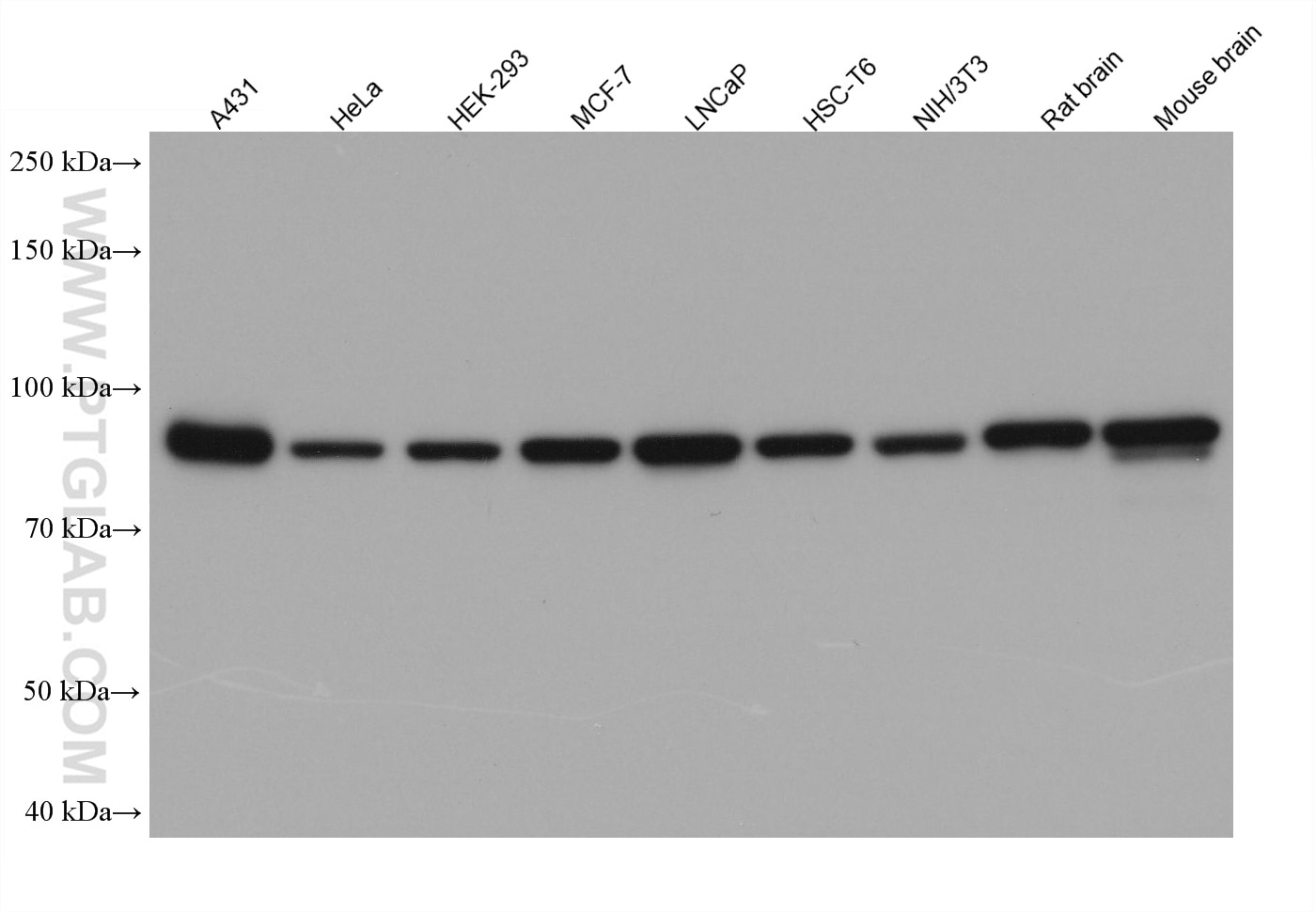
Various lysates were subjected to SDS-PAGE followed by western blot with anti-beta catenin rabbit polyclonal antibody (51067-2-AP) at a dilution of 1:50000. Multi-rAb HRP-Goat Anti-Rabbit Recombinant Secondary Antibody (H+L) (RGAR001) was used at a dilution of 1:20000 for detection.
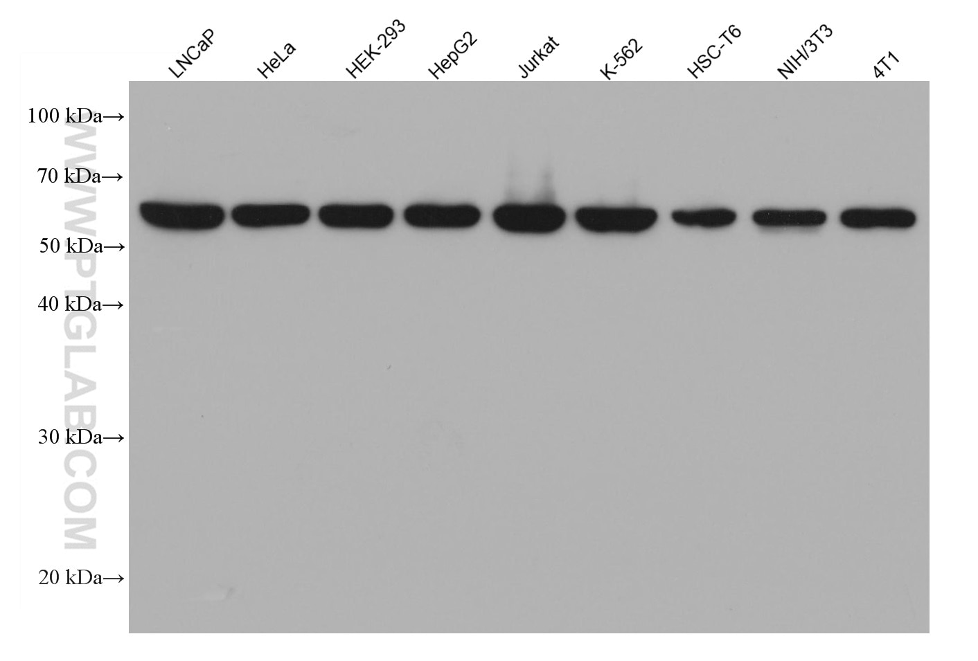
Various lysates were subjected to SDS-PAGE followed by western blot with U2AF2 mouse monoclonal antibody (68166-1-Ig) at a dilution of 1:20000. Multi-rAb HRP-Goat Anti-Mouse Recombinant Secondary Antibody (H+L) (RGAM001) was used at a dilution of 1:20000 for detection.
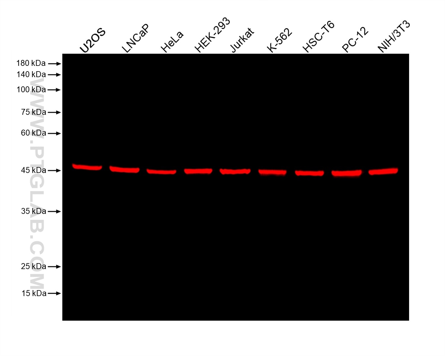
Various lysates were subjected to SDS-PAGE followed by western blot with anti-EIF3E mouse monoclonal antibody (67095-1-Ig, isotype IgG1) at a dilution of 1:50000. Multi-rAb CoraLite® Plus 750-Goat Anti-Mouse Recombinant Secondary Antibody (H+L) (RGAM006) was used at a dilution of 1:10000 for detection.
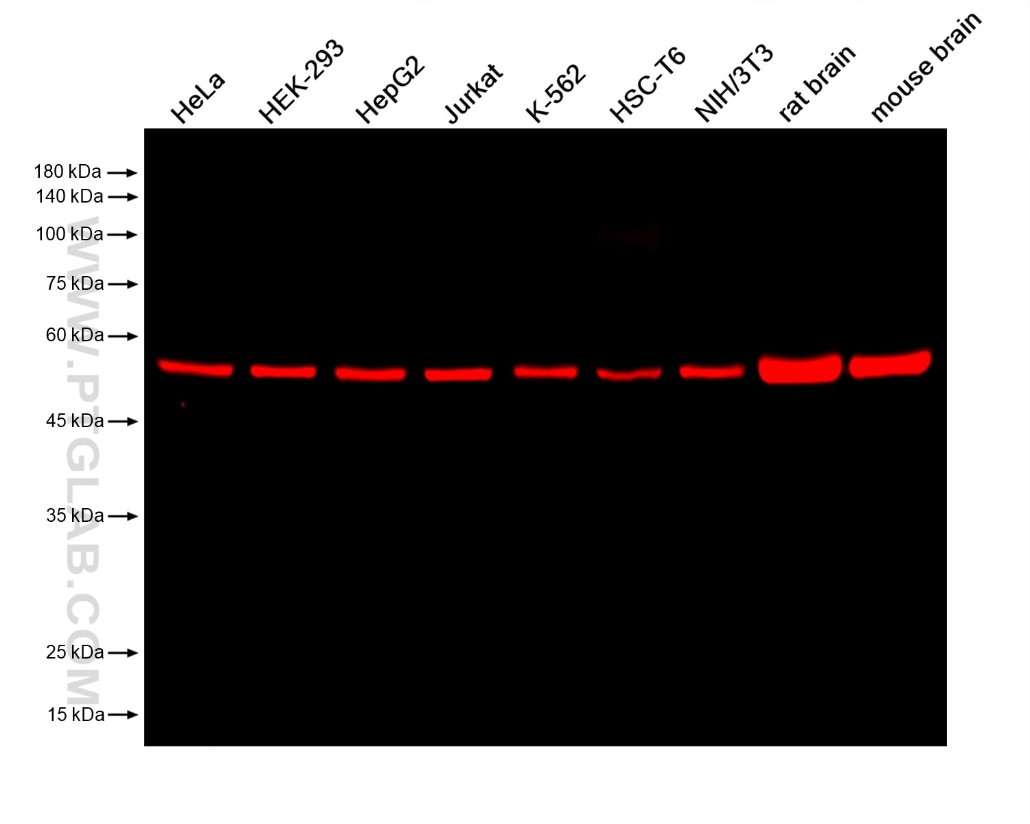
Various lysates were subjected to SDS-PAGE followed by western blot with anti-beta tubulin rabbit recombinant antibody (80713-1-RR) at a dilution of 1:20000. Multi-rAb CoraLite® Plus 750-Goat Anti-Rabbit Recombinant Secondary Antibody (H+L) (RGAR006) was used at a dilution of 1:10000 for detection.
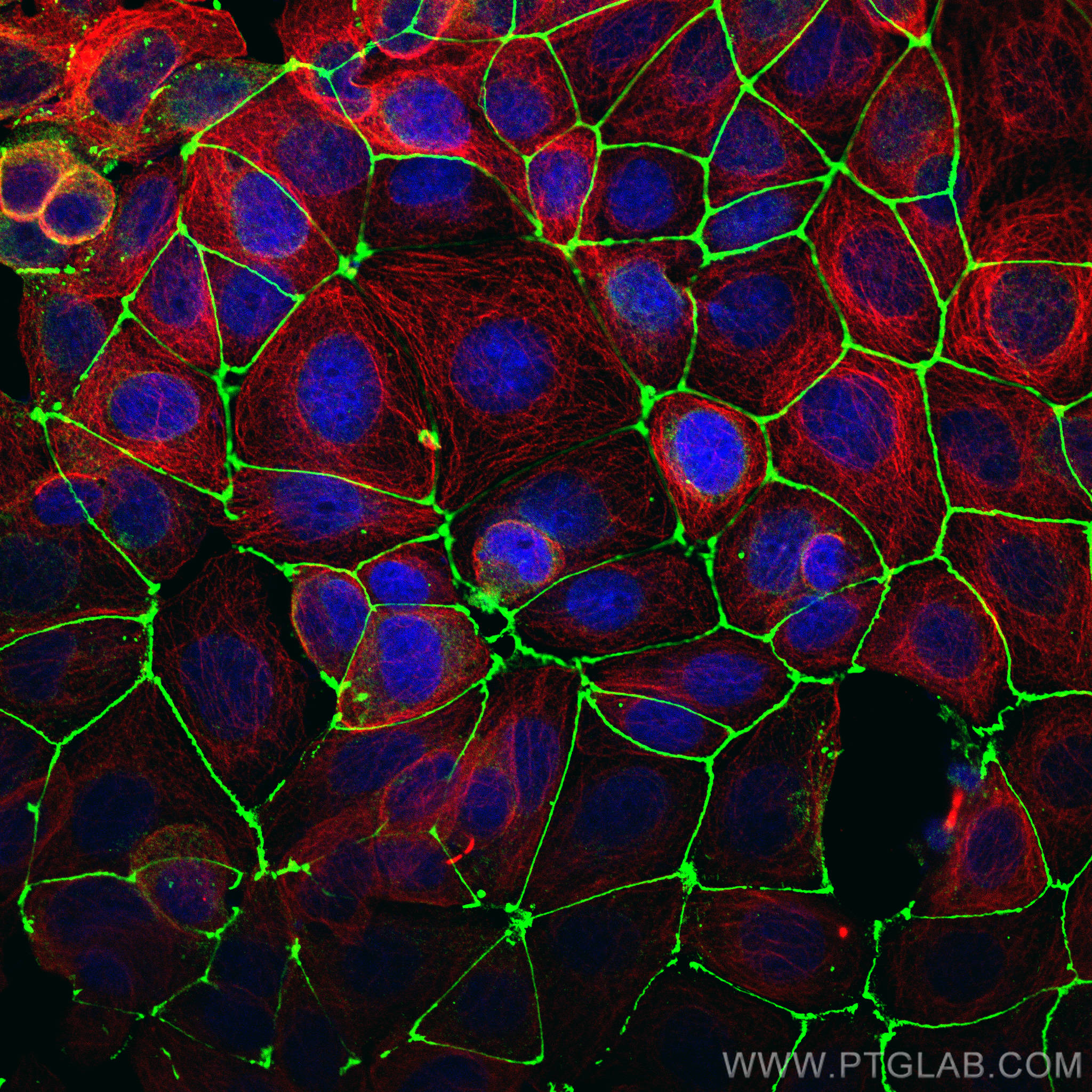
Immunofluorescence analysis of MCF-7 cells stained with rabbit anti-ZO1 polyclonal antibody (21773-1-AP, green) and mouse anti-Alpha Tubulin monoclonal antibody (66031-1-Ig, red). Multi-rAb CoraLite® Plus 488-Goat Anti-Rabbit Recombinant Secondary Antibody (H+L) (RGAR002, 1:500) and Multi-rAb CoraLite® Plus 594-Goat Anti-Mouse Recombinant Secondary Antibody (H+L) (RGAM004, 1:500) were used for detection.
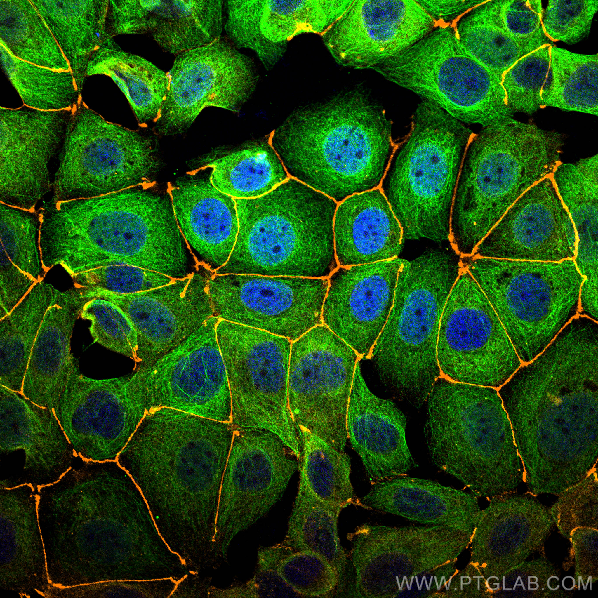
Immunofluorescence analysis of MCF-7 cells stained with rabbit anti-ZO1 polyclonal antibody (21773-1-AP, orange) and mouse anti-Alpha Tubulin monoclonal antibody (66031-1-Ig, green). Multi-rAb CoraLite® Plus 555-Goat Anti-Rabbit Recombinant Secondary Antibody (H+L) (RGAR003, 1:500) and Multi-rAb CoraLite® Plus 488-Goat Anti-Mouse Recombinant Secondary Antibody (H+L) (RGAM002, 1:500) were used for detection.
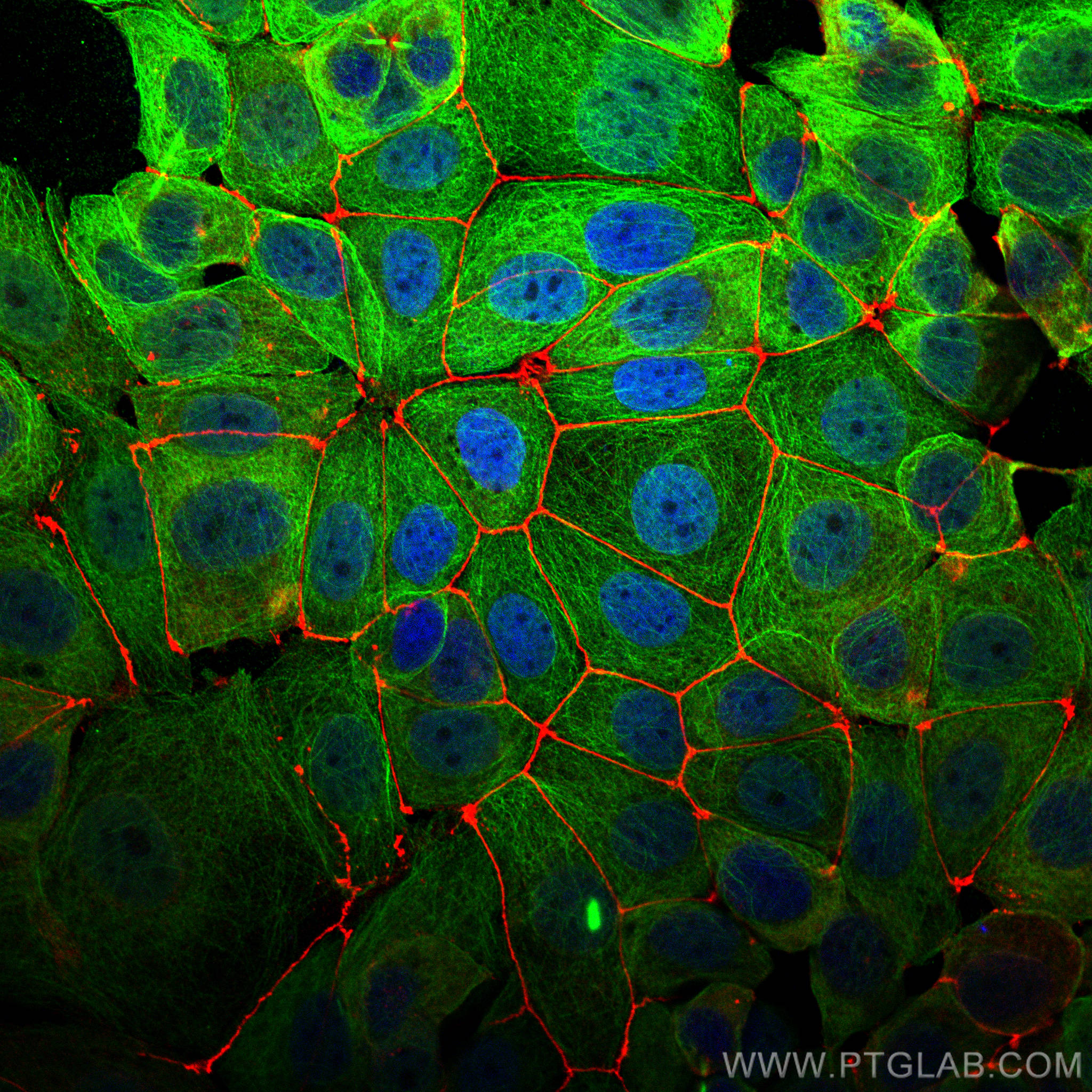
Immunofluorescence analysis of MCF-7 cells stained with rabbit anti-ZO1 polyclonal antibody (21773-1-AP, red) and mouse anti-Alpha Tubulin monoclonal antibody (66031-1-Ig, green). Multi-rAb CoraLite® Plus 594-Goat Anti-Rabbit Recombinant Secondary Antibody (H+L) (RGAR004, 1:500) and Multi-rAb CoraLite® Plus 488-Goat Anti-Mouse Recombinant Secondary Antibody (H+L) (RGAM002, 1:500) were used for detection.
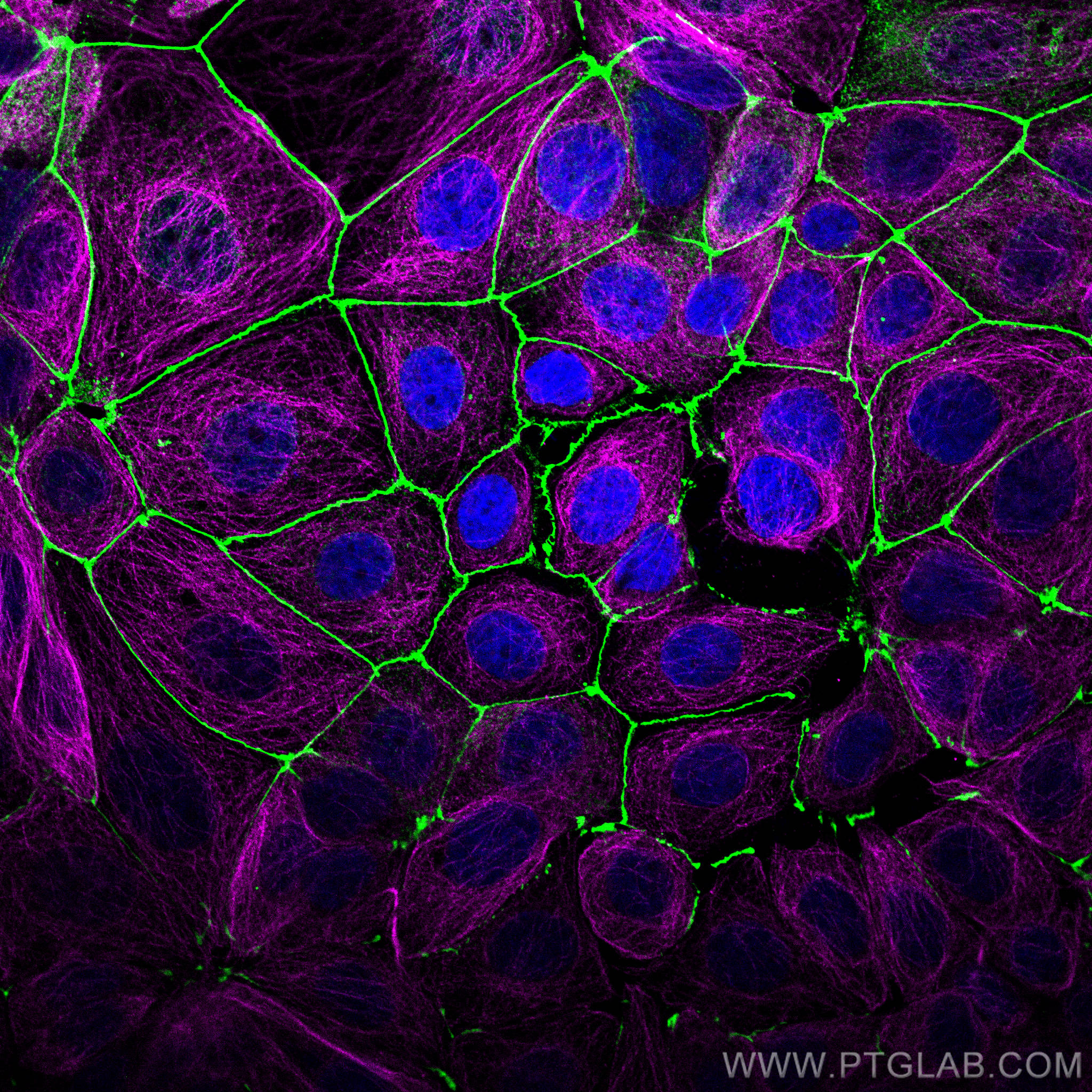
Immunofluorescence analysis of MCF-7 cells stained with rabbit anti-ZO1 polyclonal antibody (21773-1-AP, green) and mouse anti-Alpha Tubulin monoclonal antibody (66031-1-Ig, magenta). Multi-rAb CoraLite® Plus 488-Goat Anti-Rabbit Recombinant Secondary Antibody (H+L) (RGAR002, 1:500) and Multi-rAb CoraLite® Plus 647-Goat Anti-Mouse Recombinant Secondary Antibody (H+L) (RGAM005, 1:500) were used for detection.
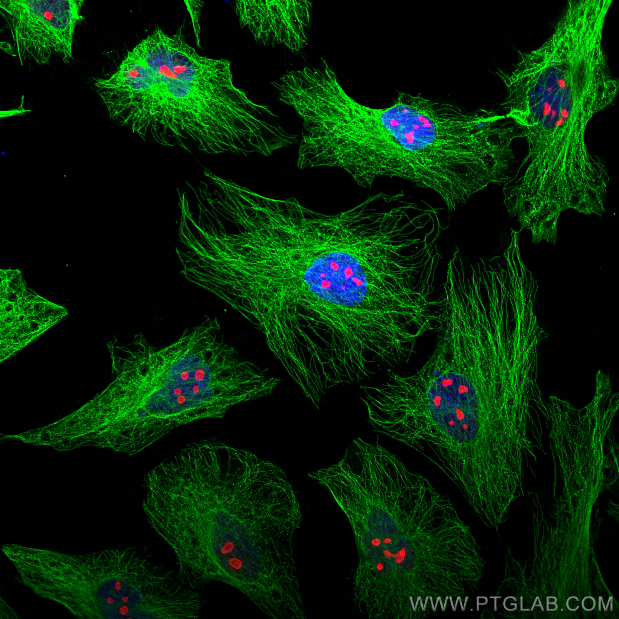
Immunofluorescence analysis of Hela cells stained with rabbit anti-Alpha Tubulin polyclonal antibody (11224-1-AP, green) and mouse anti-NPM1 monoclonal antibody (60096-1-Ig, red). Multi-rAb CoraLite® Plus 488-Goat Anti-Rabbit Recombinant Secondary Antibody (H+L) (RGAR002, 1:500) and Multi-rAb CoraLite® Plus 594-Goat Anti-Mouse Recombinant Secondary Antibody (H+L) (RGAM004, 1:500) were used for detection.
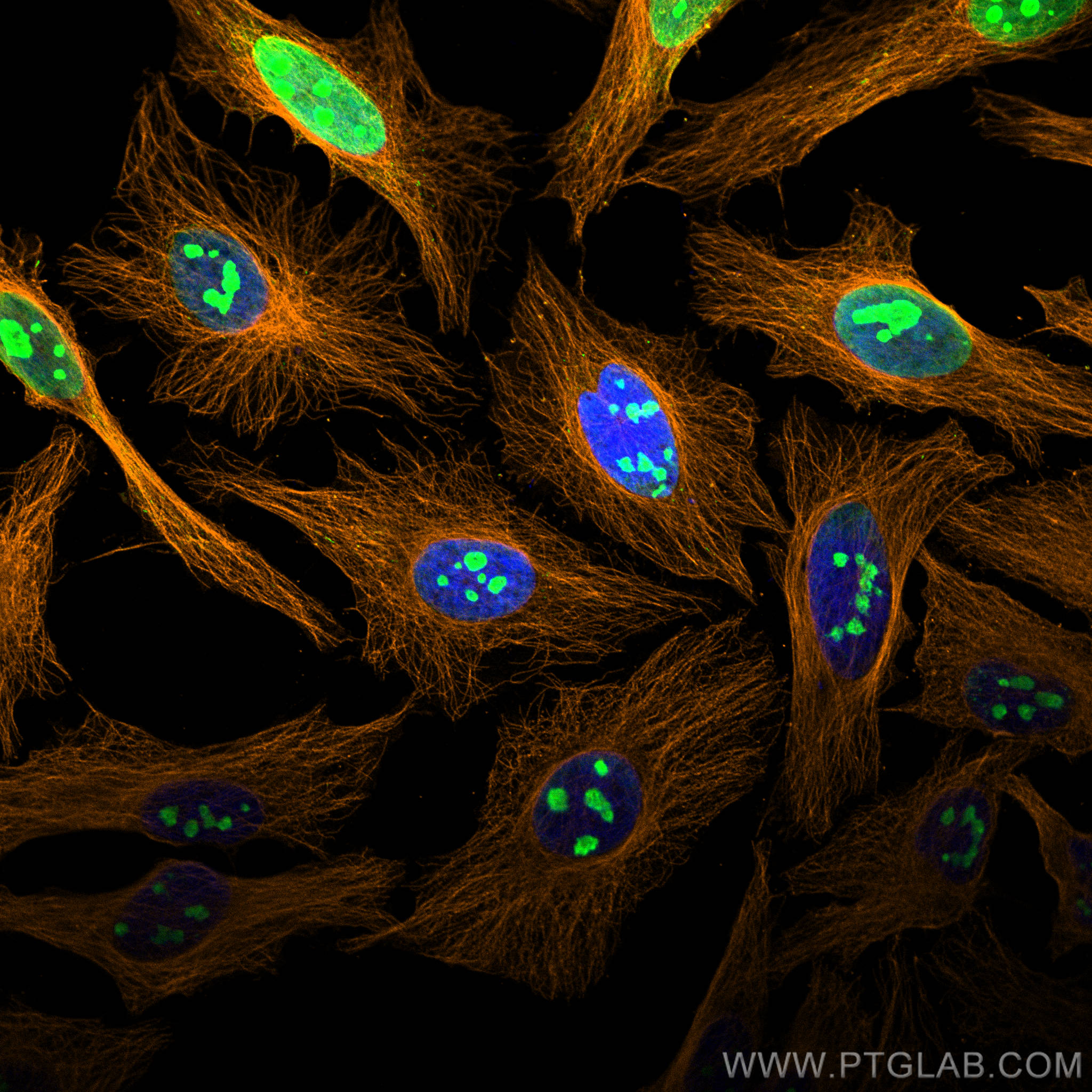
Immunofluorescence analysis of Hela cells stained with rabbit anti-Alpha Tubulin polyclonal antibody (11224-1-AP, orange) and mouse anti-NPM1 monoclonal antibody (60096-1-Ig, green). Multi-rAb CoraLite® Plus 555-Goat Anti-Rabbit Recombinant Secondary Antibody (H+L) (RGAR003, 1:500) and Multi-rAb CoraLite® Plus 488-Goat Anti-Mouse Recombinant Secondary Antibody (H+L) (RGAM002, 1:500) were used for detection.
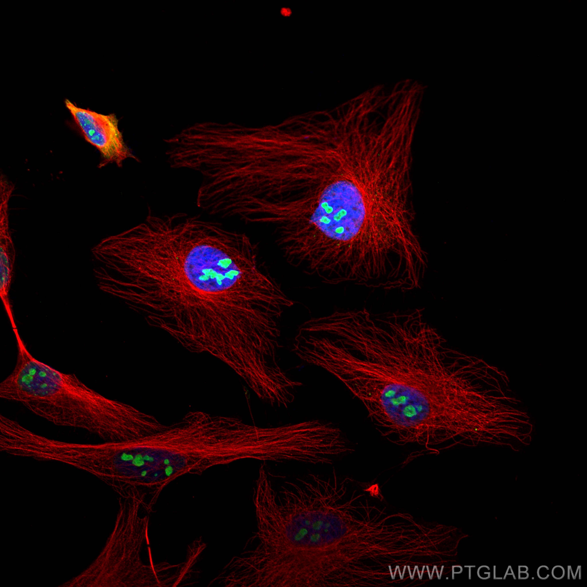
Immunofluorescence analysis of Hela cells stained with rabbit anti-Alpha Tubulin polyclonal antibody (11224-1-AP, red) and mouse anti-NPM1 monoclonal antibody (60096-1-Ig, green). Multi-rAb CoraLite® Plus 594-Goat Anti-Rabbit Recombinant Secondary Antibody (H+L) (RGAR004, 1:500) and Multi-rAb CoraLite® Plus 488-Goat Anti-Mouse Recombinant Secondary Antibody (H+L) (RGAM002, 1:500) were used for detection.
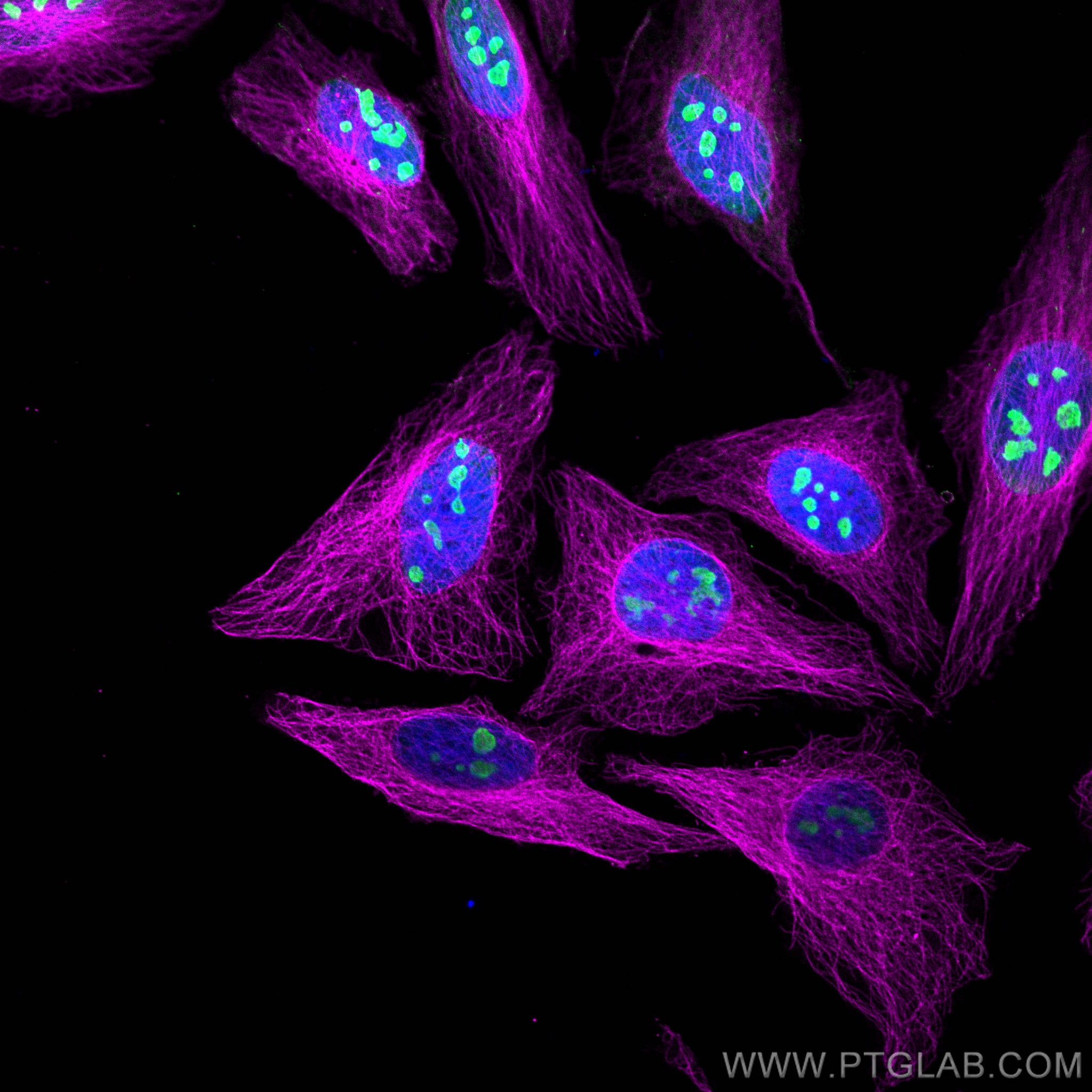
Immunofluorescence analysis of Hela cells stained with rabbit anti-Alpha Tubulin polyclonal antibody (11224-1-AP, magenta) and mouse anti-NPM1 monoclonal antibody (60096-1-Ig, green). Multi-rAb CoraLite® Plus 647-Goat Anti-Rabbit Recombinant Secondary Antibody (H+L) (RGAR005, 1:500) and Multi-rAb CoraLite® Plus 488-Goat Anti-Mouse Recombinant Secondary Antibody (H+L) (RGAM002, 1:500) were used for detection.
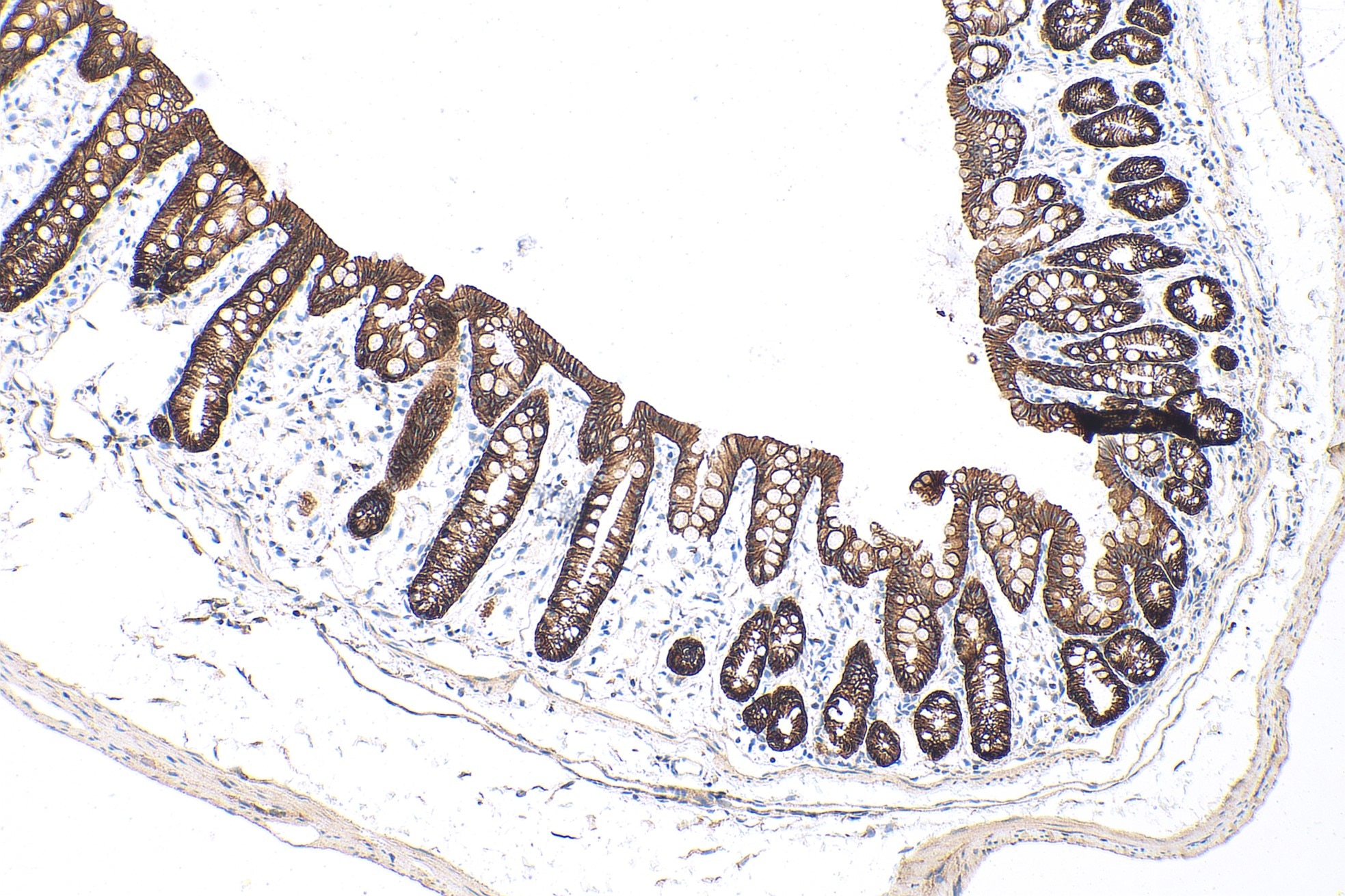
Immunohistochemical analysis of paraffin-embedded mouse colon tissue slide using 20874-1-AP (E-cadherin polyclonal antibody) at dilution of 1:5000 (under 10x lens). Heat mediated antigen retrieval with Tris-EDTA buffer (pH 9.0). Multi-rAb Polymer HRP-Goat anti-rabbit Recombinant secondary antibody (RGAR011) was used for detection..
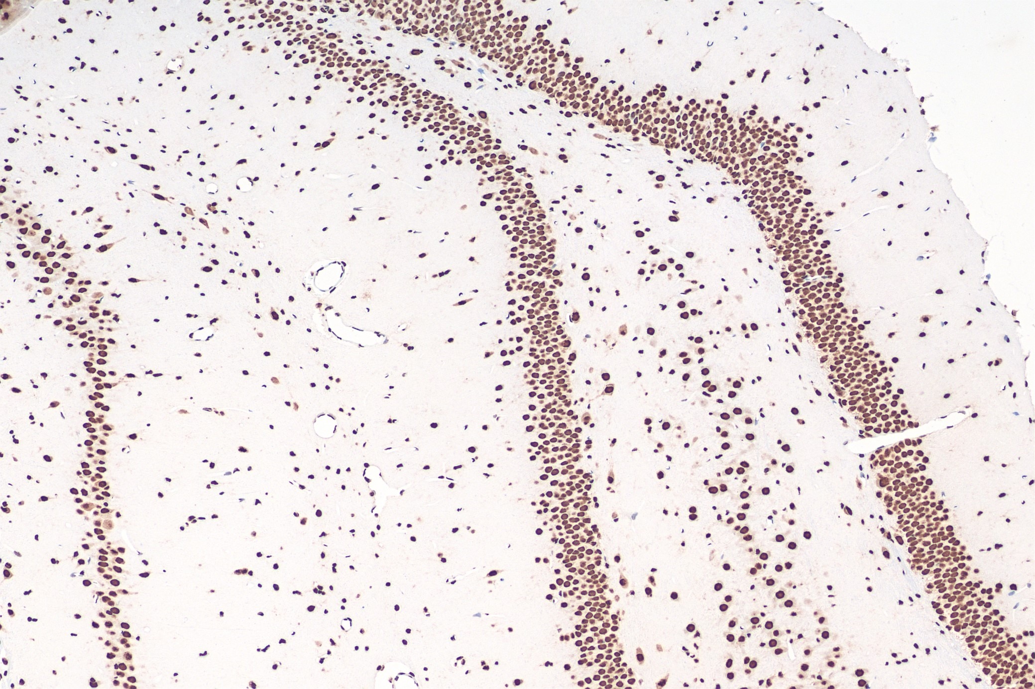
Immunohistochemical analysis of paraffin-embedded rat brain tissue slide using 80001-1-RR (TDP43 recombinant antibody) at dilution of 1:4000 (under 10x lens). Heat mediated antigen retrieval with Tris-EDTA buffer (pH 9.0). Multi-rAb Polymer HRP-Goat anti-rabbit Recombinant secondary antibody (RGAR011) was used for detection.
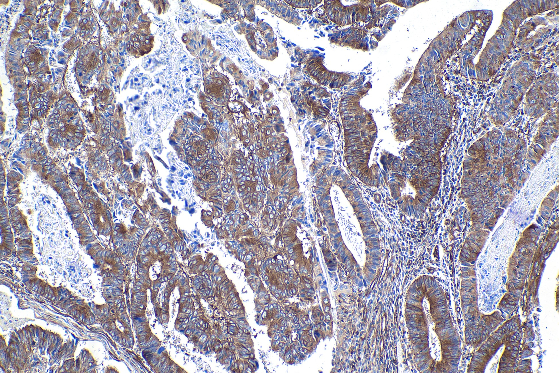
Immunohistochemical analysis of paraffin-embedded human colon cancer tissue slide using other vendor's NF-kappa B p65 rabbit recombinant antibody. Heat mediated antigen retrieval with Tris-EDTA buffer (pH 9.0). Multi-rAb Polymer HRP-Goat anti-rabbit Recombinant secondary antibody (RGAR011) was used for detection.
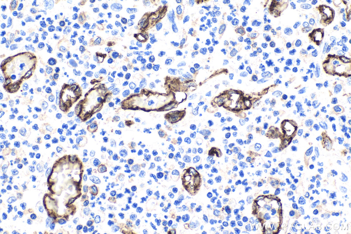
Immunohistochemical analysis of paraffin-embedded human colon cancer tissue slide using 66065-2-Ig (CD31 mouse monoclonal antibody) at dilution of 1:10000 . Heat mediated antigen retrieval with Tris-EDTA buffer (pH 9.0). Multi-rAb Polymer HRP-Goat anti-Mouse Recombinant secondary antibody (RGAM011) was used for detection.
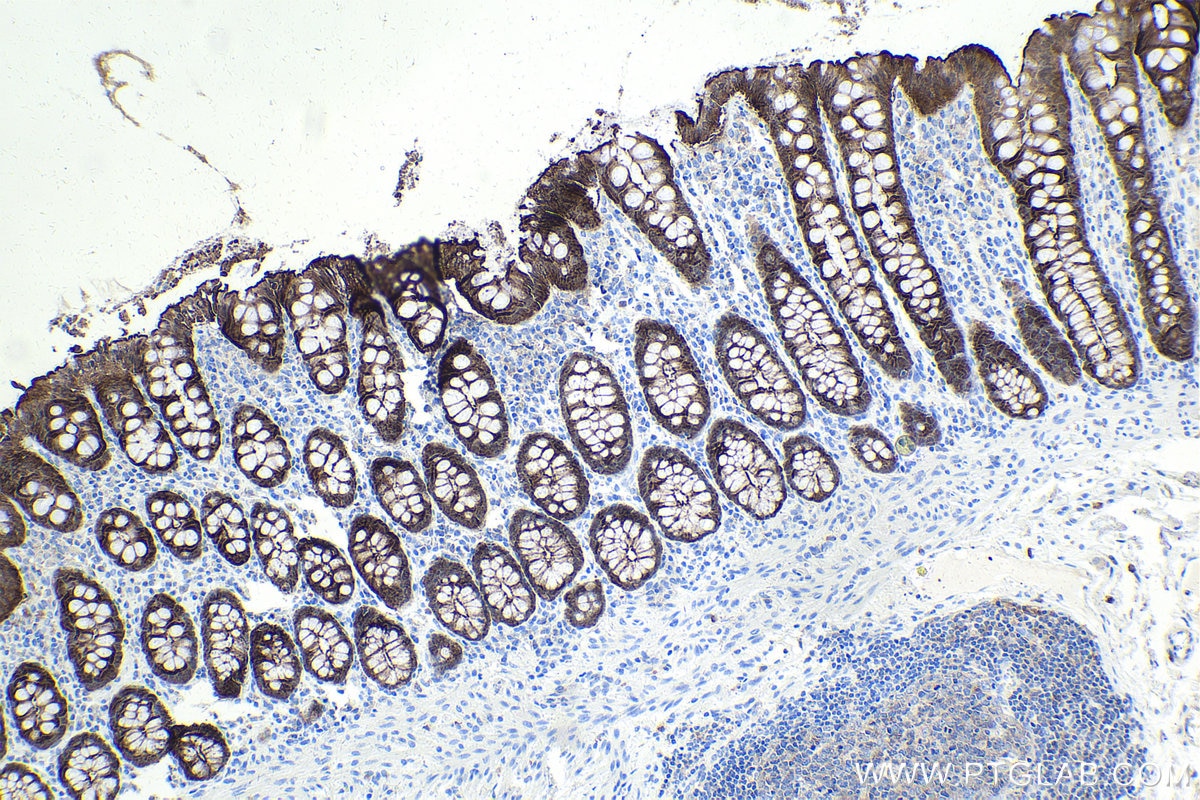
Immunohistochemical analysis of paraffin-embedded human colon tissue slide using 66096-1-Ig (Villin antibody) at dilution of 1:5000 (under 10x lens). Heat mediated antigen retrieval with Tris-EDTA buffer (pH 9.0). Multi-rAb Polymer HRP-Goat anti-Mouse Recombinant secondary antibod (RGAM011) was used for detection.
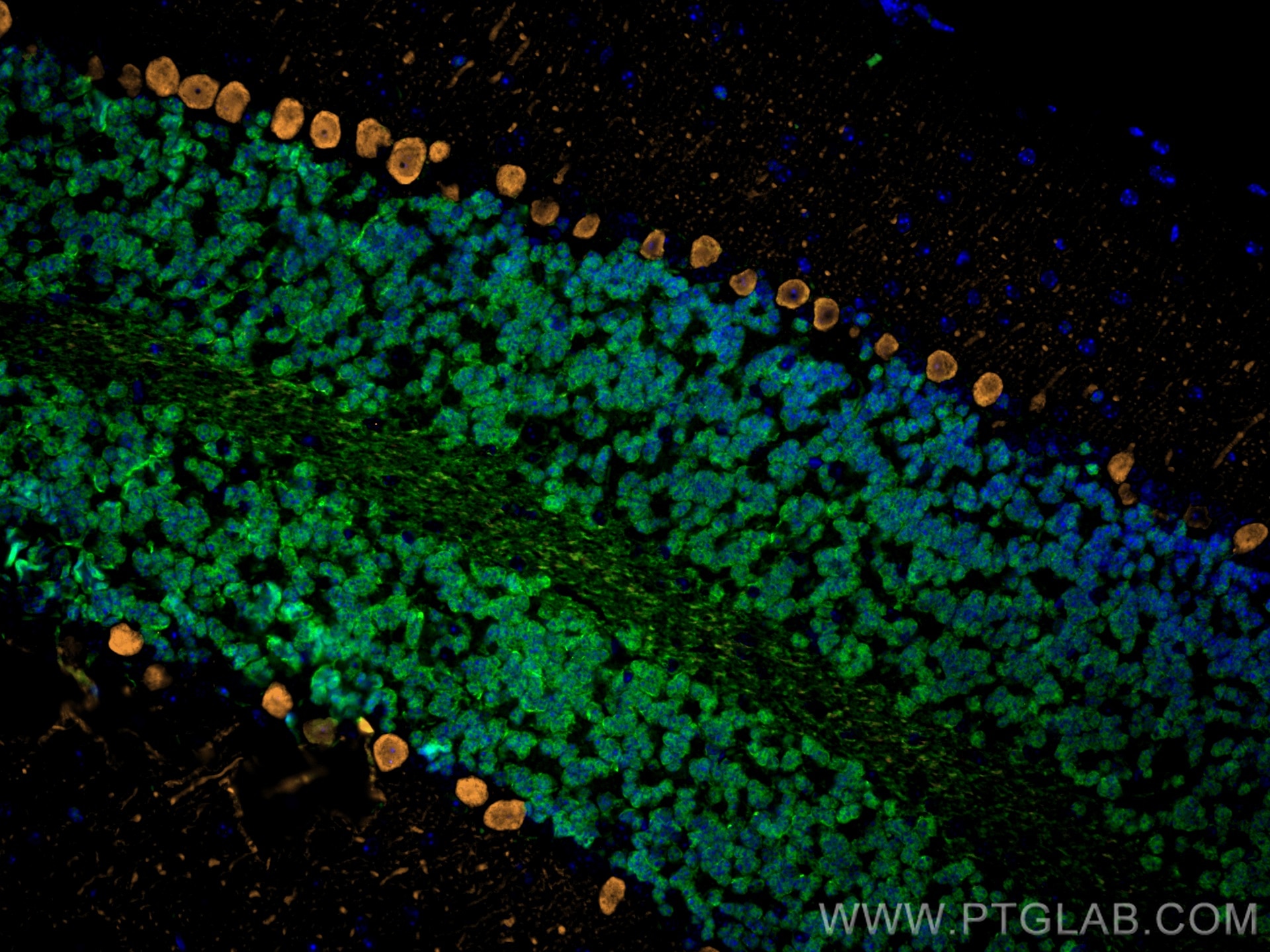
Immunofluorescence analysis of mouse cerebellum FFPE tissue stained with rabbit anti-NeuN polyclonal antibody (26975-1-AP, green) and mouse anti-Calbindin-D28k monoclonal antibody (66394-1-Ig, orange). Multi-rAb CoraLite® Plus 488-Goat Anti-Rabbit Recombinant Secondary Antibody (H+L) (RGAR002, 1:500) and Multi-rAb CoraLite® Plus 555-Goat Anti-Mouse Recombinant Secondary Antibody (H+L) (RGAM003, 1:500) were used for detection.
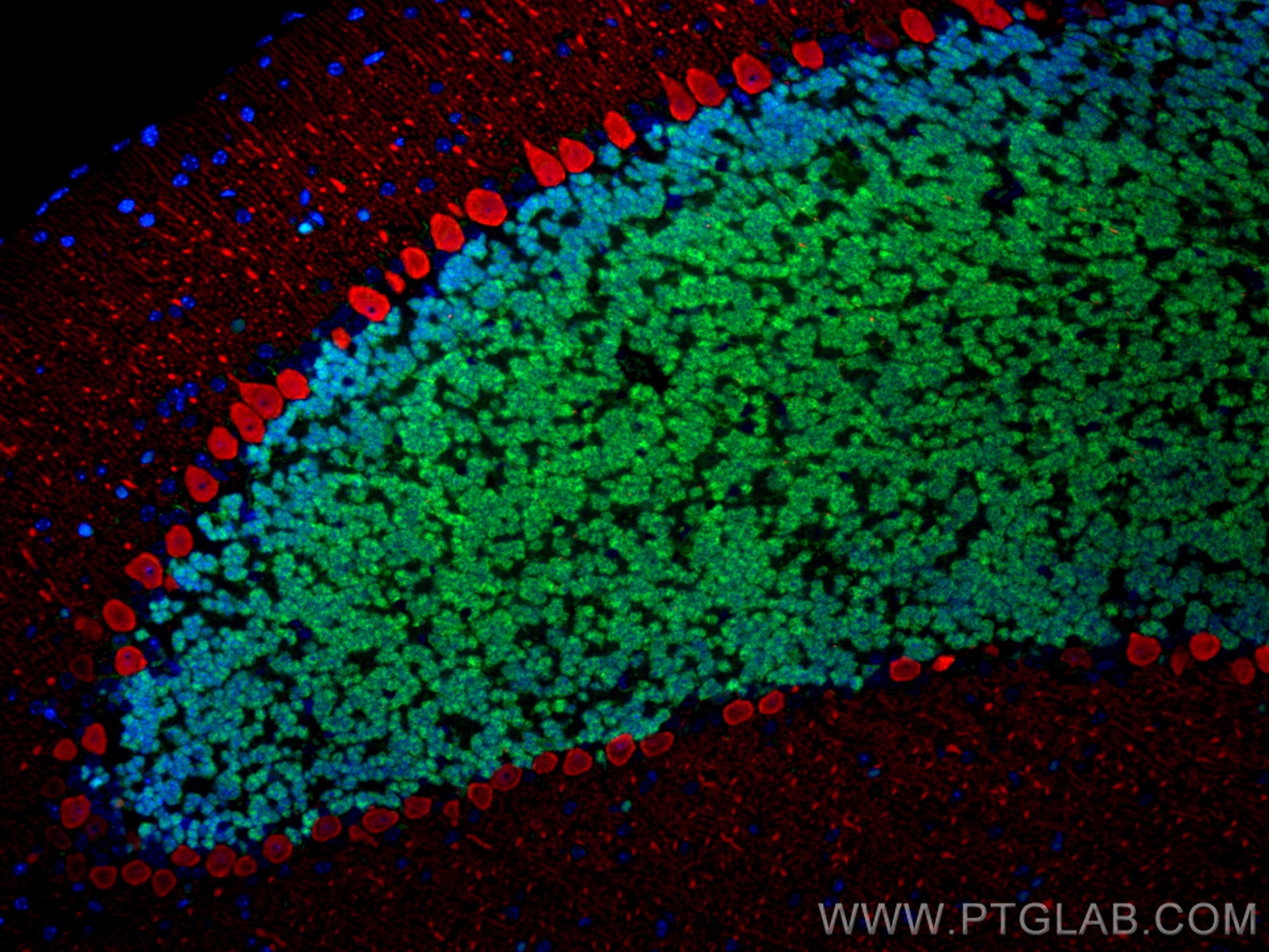
Immunofluorescence analysis of mouse cerebellum FFPE tissue stained with rabbit anti-NeuN polyclonal antibody (26975-1-AP, green) and mouse anti-Calbindin-D28k monoclonal antibody (66394-1-Ig, red). Multi-rAb CoraLite® Plus 488-Goat Anti-Rabbit Recombinant Secondary Antibody (H+L) (RGAR002, 1:500) and Multi-rAb CoraLite® Plus 594-Goat Anti-Mouse Recombinant Secondary Antibody (H+L) (RGAM004, 1:500) were used for detection.
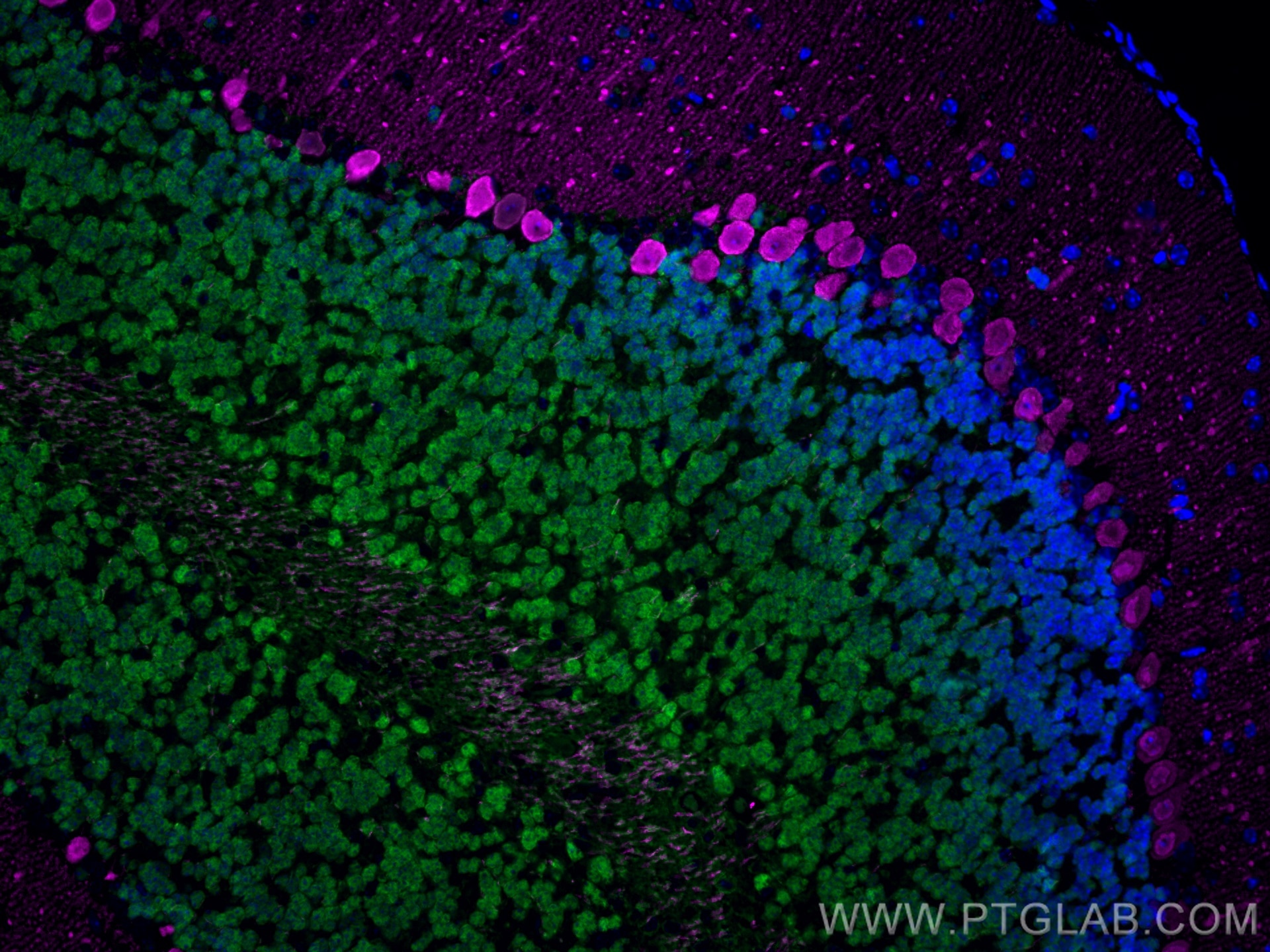
Immunofluorescence analysis of mouse cerebellum FFPE tissue stained with rabbit anti-NeuN polyclonal antibody (26975-1-AP, green) and mouse anti-Calbindin-D28k monoclonal antibody (66394-1-Ig, magenta). Multi-rAb CoraLite® Plus 488-Goat Anti-Rabbit Recombinant Secondary Antibody (H+L) (RGAR002, 1:500) and Multi-rAb CoraLite® Plus 647-Goat Anti-Mouse Recombinant Secondary Antibody (H+L) (RGAM005, 1:500) were used for detection.
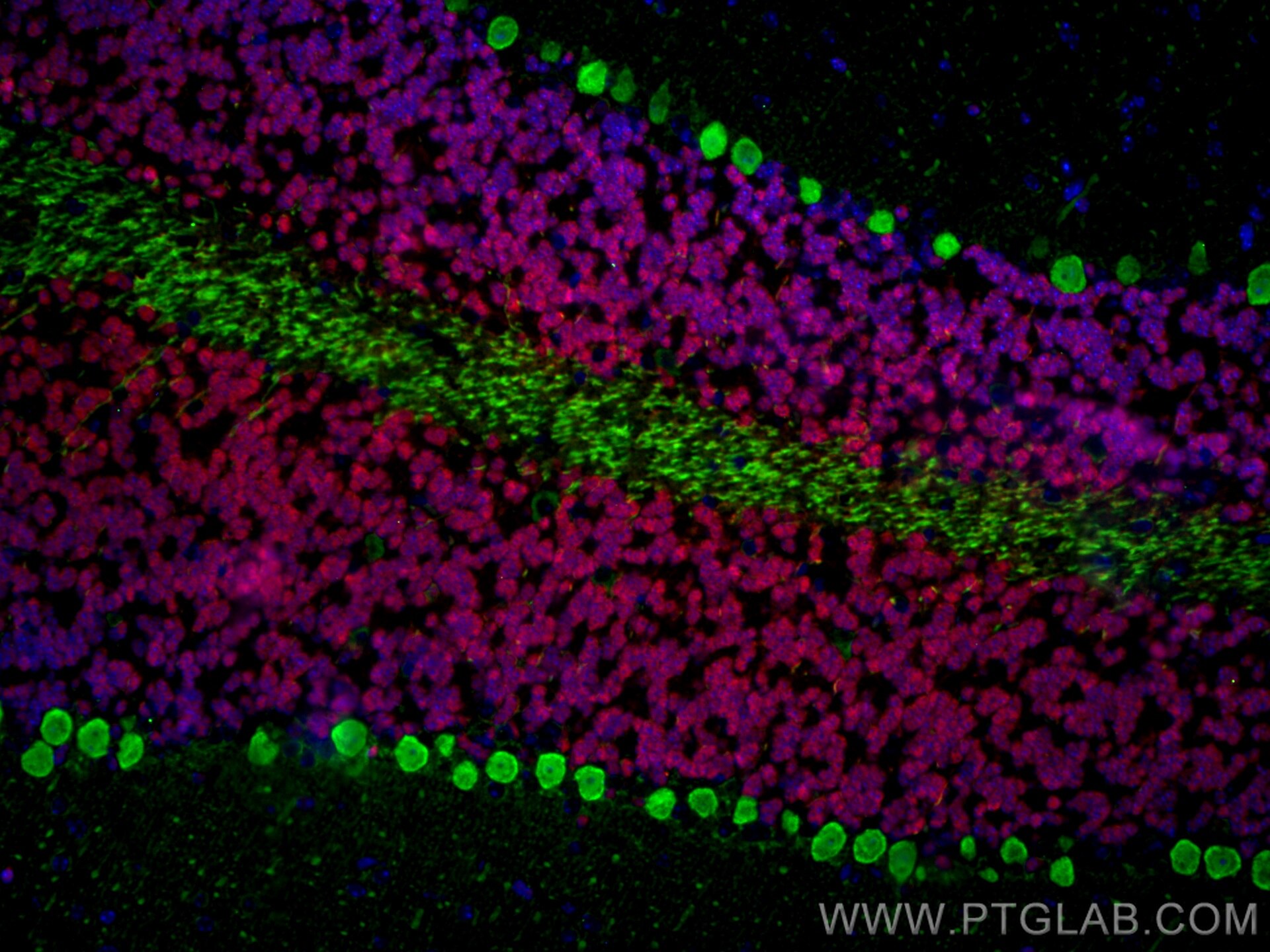
Immunofluorescence analysis of mouse cerebellum FFPE tissue stained with rabbit anti-NeuN polyclonal antibody (26975-1-AP, red) and mouse anti-Calbindin-D28k monoclonal antibody (66394-1-Ig, green). Multi-rAb CoraLite® Plus 594-Goat Anti-Rabbit Secondary Antibody (H+L) (RGAR004, 1:500) and Multi-rAb CoraLite® Plus 488-Goat Anti-Mouse Recombinant Secondary Antibody (H+L) (RGAM002, 1:500) were used for detection.
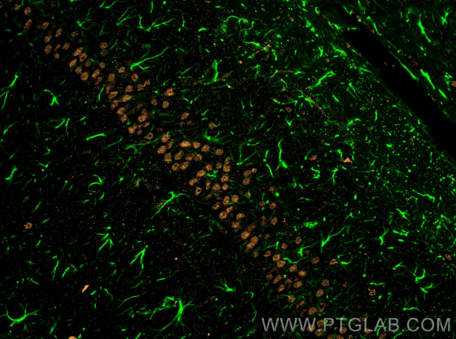
Immunofluorescence analysis of rat brain FFPE tissue stained with rabbit anti-GFAP polyclonal antibody (16825-1-AP, green) and mouse anti-NeuN monoclonal antibody (66836-1-Ig, orange). Multi-rAb CoraLite® Plus 488-Goat Anti-Rabbit Recombinant Secondary Antibody (H+L) (RGAR002, 1:500) and Multi-rAb CoraLite® Plus 555-Goat Anti-Mouse Recombinant Secondary Antibody (H+L) (RGAM003, 1:500) were used for detection.
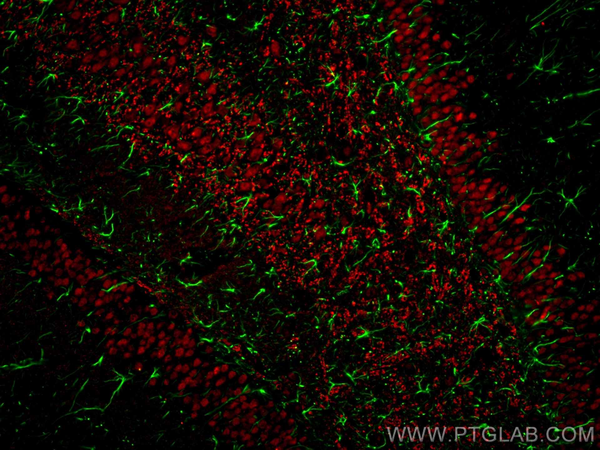
Immunofluorescence analysis of rat brain FFPE tissue stained with rabbit anti-GFAP polyclonal antibody (16825-1-AP, green) and mouse anti-NeuN monoclonal antibody (66836-1-Ig, red). Multi-rAb CoraLite® Plus 488-Goat Anti-Rabbit Recombinant Secondary Antibody (H+L) (RGAR002, 1:500) and Multi-rAb CoraLite® Plus 594-Goat Anti-Mouse Recombinant Secondary Antibody (H+L) were (RGAM004, 1:500) used for detection.
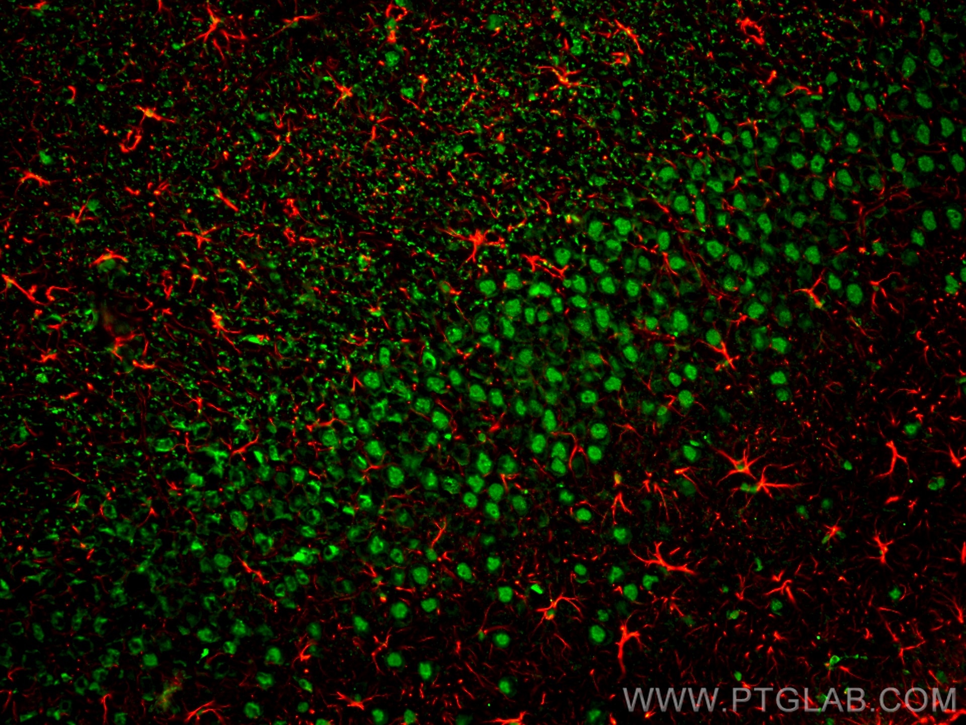
Immunofluorescence analysis of rat brain FFPE section stained with rabbit anti-GFAP polyclonal antibody (16825-1-AP, red) and mouse anti-NeuN monoclonal antibody (66836-1-Ig, green). Multi-rAb CoraLite® Plus 594-Goat Anti-Rabbit Recombinant Secondary Antibody (H+L) (RGAR004, 1:500) and Multi-rAb CoraLite® Plus 488-Goat Anti-Mouse Recombinant Secondary Antibody (H+L) (RGAM002, 1:500) were used for detection.
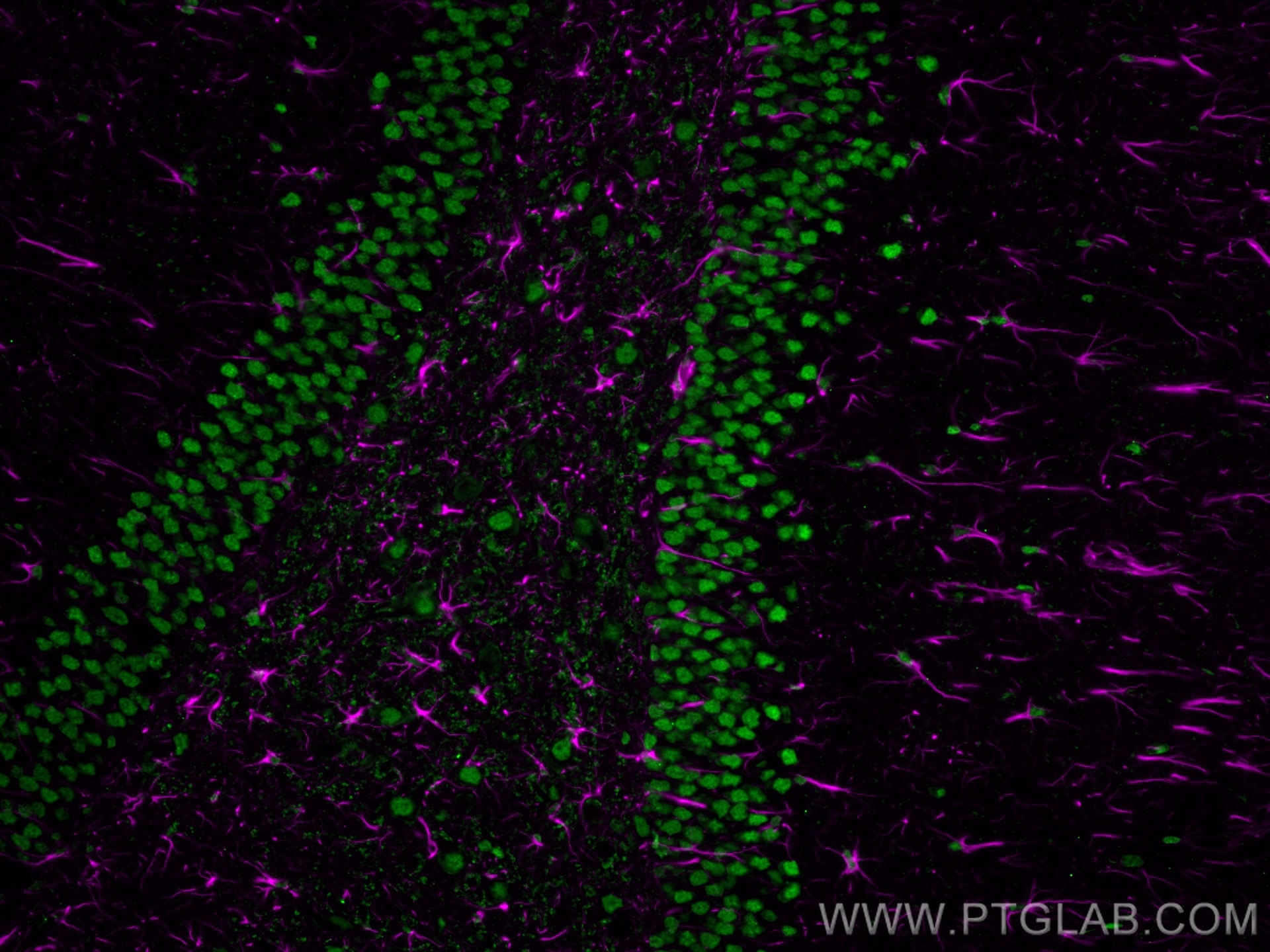
Immunofluorescence analysis of rat brain FFPE section stained with rabbit anti-GFAP polyclonal antibody (16825-1-AP, magenta) and mouse anti-NeuN monoclonal antibody (66836-1-Ig, green). Multi-rAb CoraLite® Plus 647-Goat Anti-Rabbit Secondary Antibody (H+L) (RGAR005, 1:500) and Multi-rAb CoraLite® Plus 488-Goat Anti-Mouse Recombinant Secondary Antibody (H+L) were (RGAM002, 1:500) used for detection.
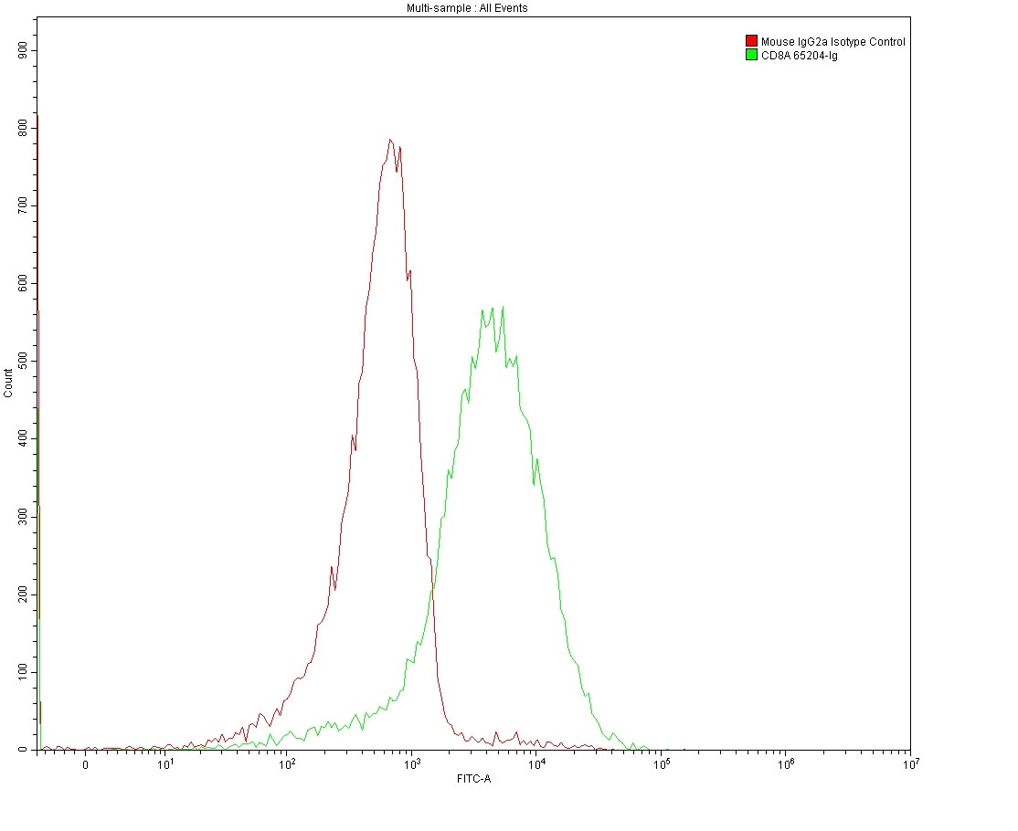
Flow cytometry analysis of 1X10^6 MOLT4 cells surface stained with 0.2 ug anti-Human CD8 antibody (65204-1-Ig, Clone: UCHT4) and mouse IgG2a isotype control antibody (66360-3-Ig). Multi-rAb CoraLite® Plus 488-Goat Anti-Mouse Recombinant Secondary Antibody (H+L) (RGAM002) was used for detection.
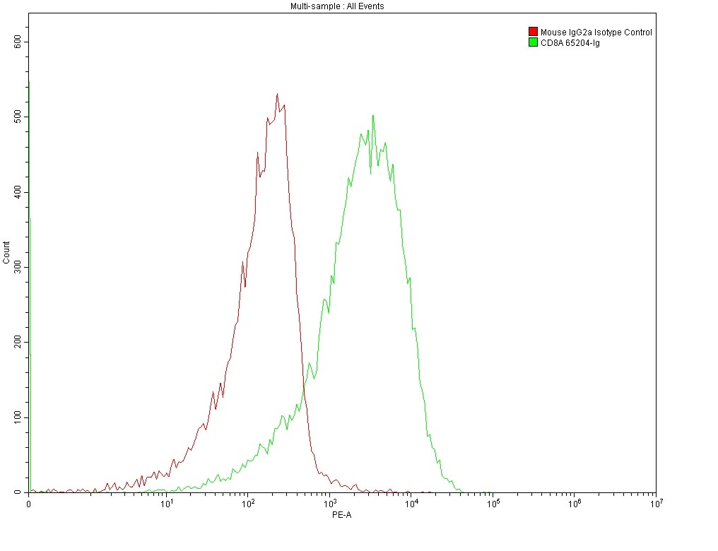
Flow cytometry analysis of 1X10^6 MOLT4 cells surface stained with 0.2 ug anti-Human CD8 antibody (65204-1-Ig, Clone: UCHT4) and mouse IgG2a isotype control antibody (66360-3-Ig). Multi-rAb CoraLite® Plus 555-Goat Anti-Mouse Recombinant Secondary Antibody (H+L) (RGAM003) was used for detection.
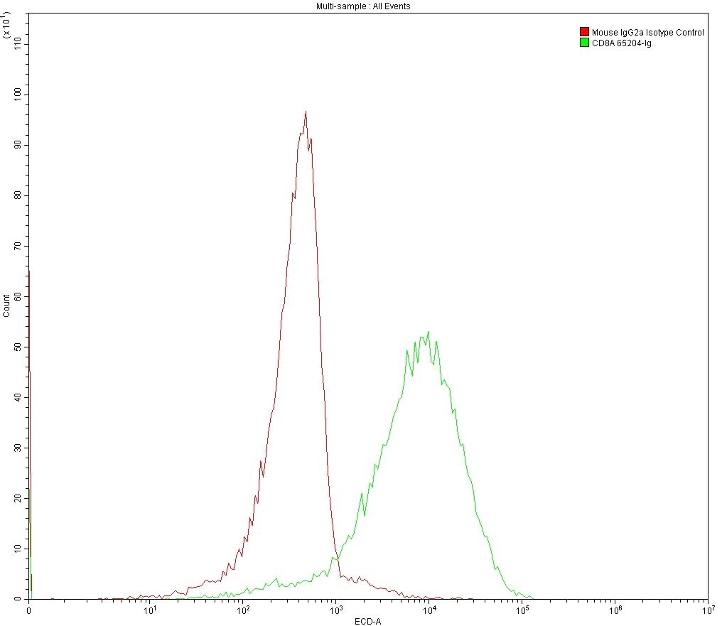
Flow cytometry analysis of 1X10^6 MOLT4 cells surface stained with 0.2 ug anti-Human CD8 antibody (65204-1-Ig, Clone: UCHT4) and mouse IgG2a isotype control antibody (66360-3-Ig). Multi-rAb CoraLite® Plus 594-Goat Anti-Mouse Recombinant Secondary Antibody (H+L) (RGAM004) was used for detection.
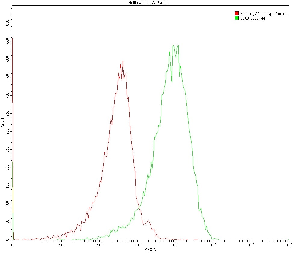
Flow cytometry analysis of 1X10^6 MOLT4 cells surface stained with 0.2 ug anti-Human CD8 antibody (65204-1-Ig, Clone: UCHT4) and mouse IgG2a isotype control antibody (66360-3-Ig). Multi-rAb CoraLite® Plus 647-Goat Anti-Mouse Recombinant Secondary Antibody (H+L) (RGAM005) was used for detection.
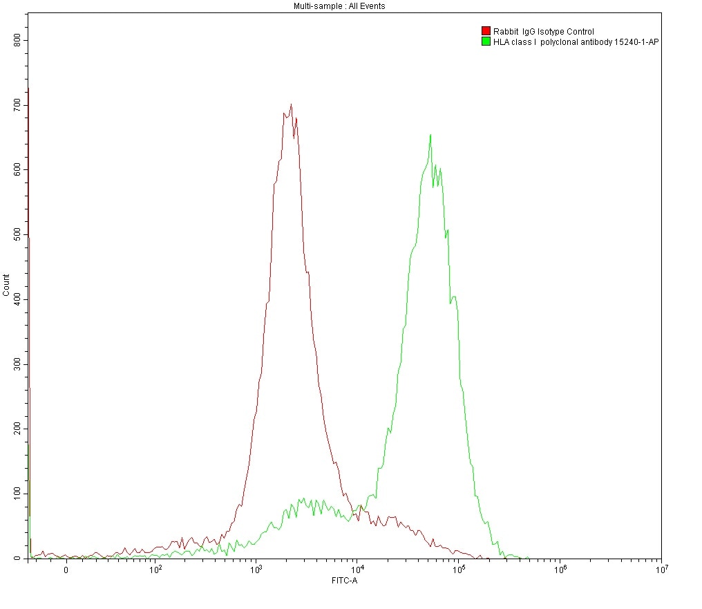
Flow cytometry analysis of 1X10^6 MOLT4 cells surface stained with 0.2 ug anti-HLA class I rabbit polyclonal antibody (15240-1-AP) and rabbit IgG isotype control antibody (30000-0-AP). Multi-rAb CoraLite® Plus 488-Goat Anti-Rabbit Recombinant Secondary Antibody (H+L) (RGAR002) was used for detection.
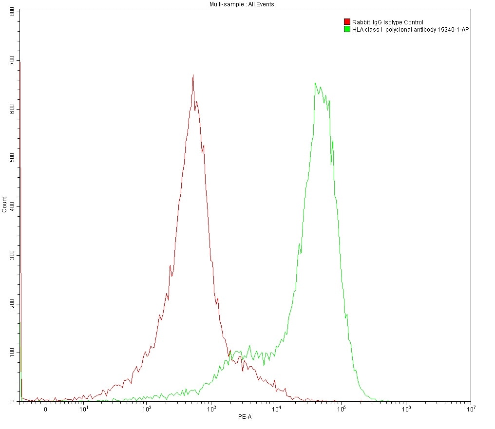
Flow cytometry analysis of 1X10^6 MOLT4 cells surface stained with 0.2 ug anti-HLA class I rabbit polyclonal antibody (15240-1-AP) and rabbit IgG isotype control antibody (30000-0-AP). Multi-rAb CoraLite® Plus 555-Goat Anti-Rabbit Recombinant Secondary Antibody (H+L) (RGAR003) was used for detection.
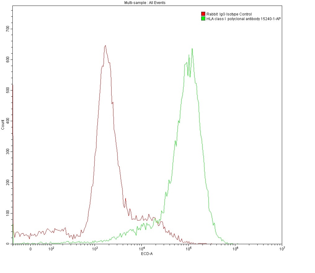
Flow cytometry analysis of 1X10^6 MOLT4 cells surface stained with 0.2 ug anti-HLA class I rabbit polyclonal antibody (15240-1-AP) and rabbit IgG isotype control antibody (30000-0-AP). Multi-rAb CoraLite® Plus 594-Goat Anti-Rabbit Recombinant Secondary Antibody (H+L) (RGAR004) was used for detection.
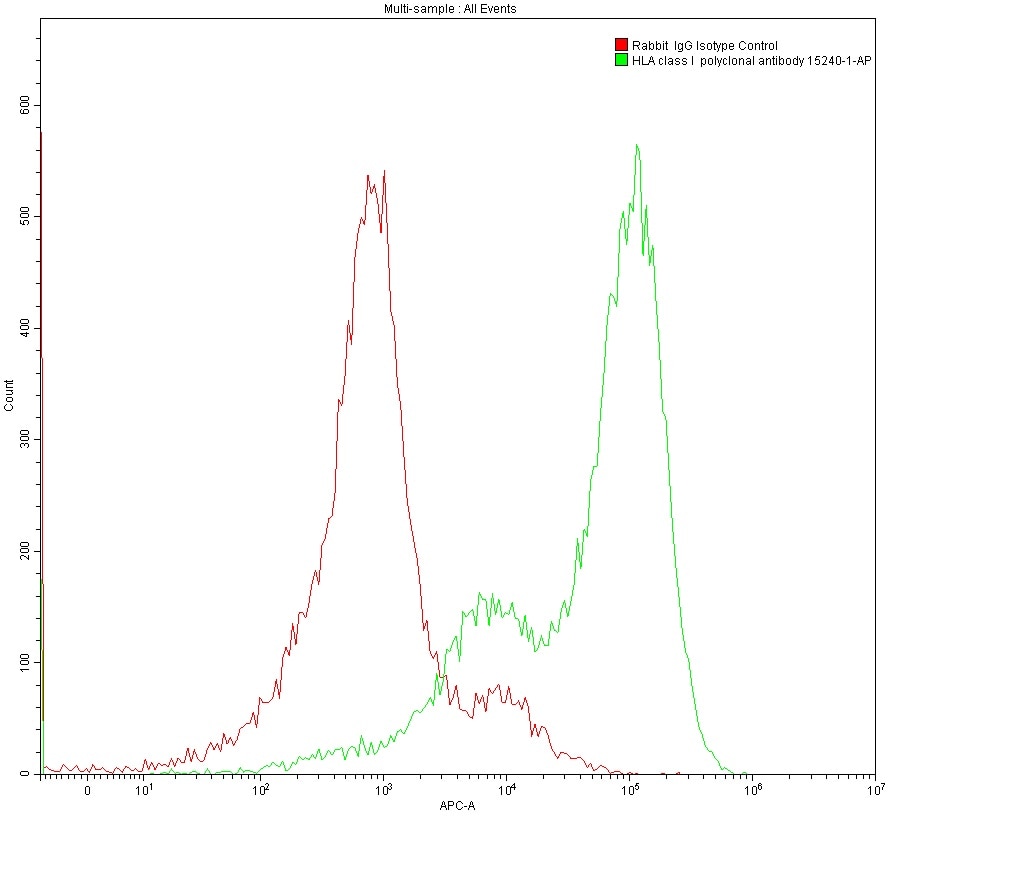
Flow cytometry analysis 1X10^6 MOLT4 cells surface stained with 0.2 ug anti-HLA class I rabbit polyclonal antibody (15240-1-AP) and rabbit IgG isotype control antibody (30000-0-AP). Multi-rAb CoraLite® Plus 647-Goat Anti-Rabbit Recombinant Secondary Antibody (H+L) (RGAR005) was used for detection.
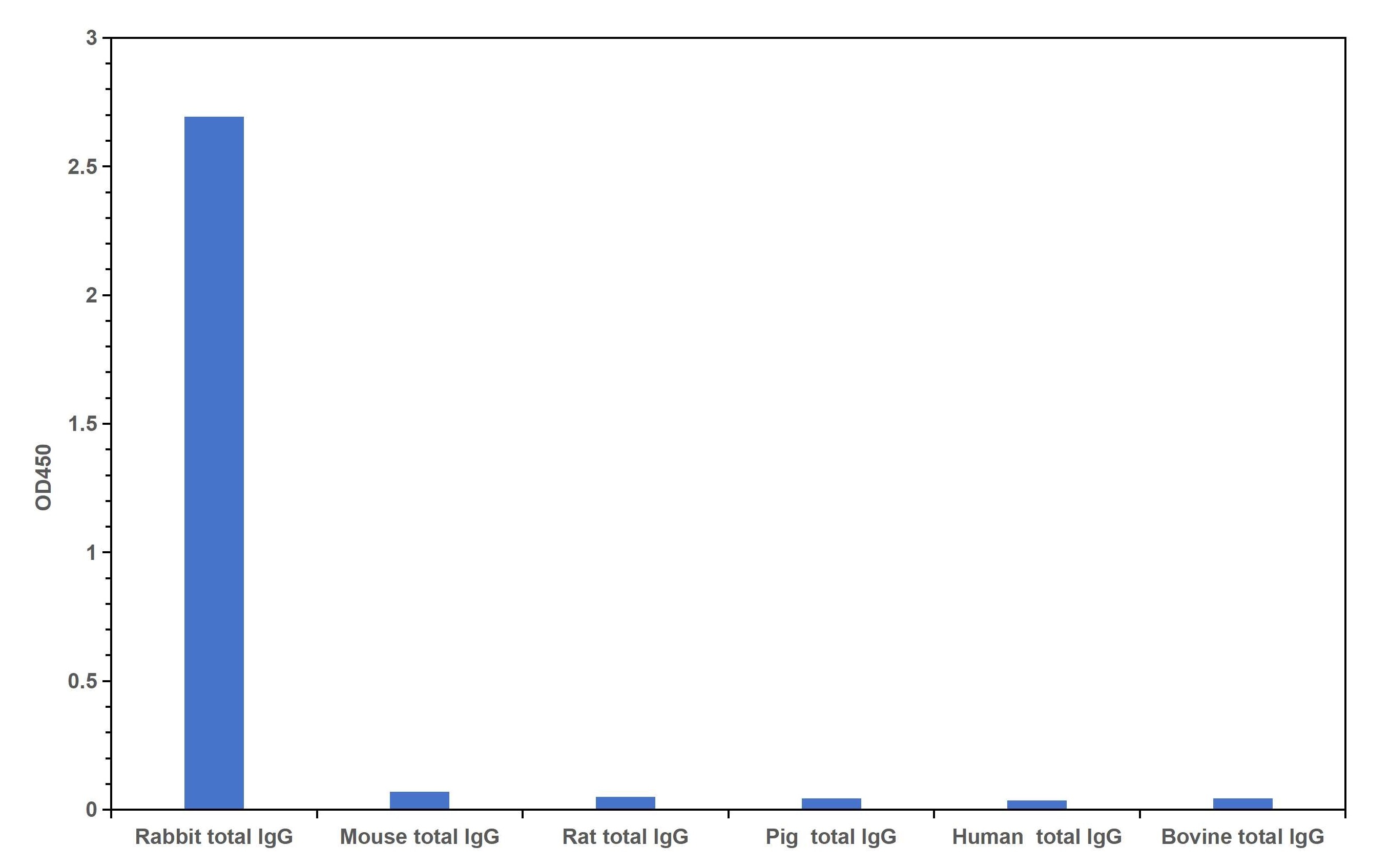
Cross reactivity test using Direct ELISA. Rabbit total IgG, Mouse total IgG, Rat total IgG, Pig total IgG, Human total IgG, Bovine total IgG were coated at 100 ng/well. 0.125 μg/mL of Multi-rAb HRP-Goat Anti-Rabbit Recombinant Secondary Antibody (H+L) (RGAR001) was used for detection. The result indicates that RGAM001 is highly specific for rabbit IgG and does not react with other species tested in the experiment.
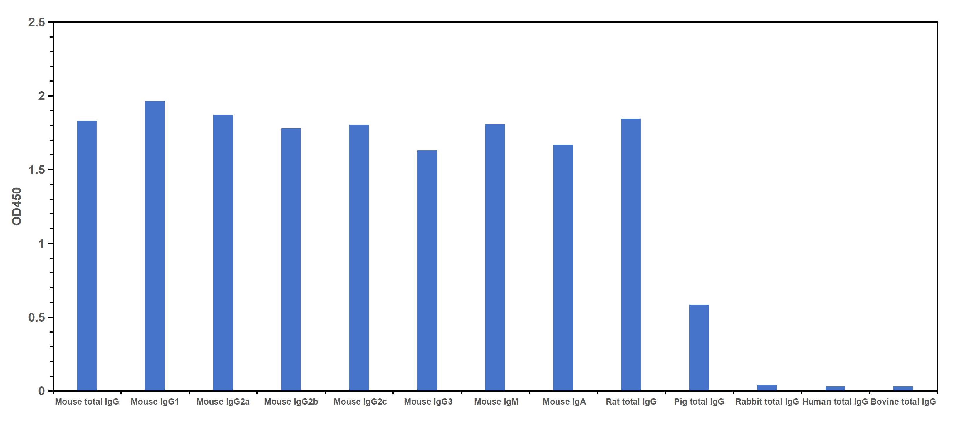
Cross reactivity test using Direct ELISA. Mouse total IgG, Mouse IgG1, IgG2a, IgG2b, IgG2c, IgG3, IgM, IgA monoclonal antibodies, Rat total IgG, Pig total IgG, Rabbit total IgG, Human total IgG, Bovine total IgG were coated at 100 ng/well. 0.125 μg/mL of Multi-rAb HRP-Goat Anti-Mouse Recombinant Secondary Antibody (H+L) (RGAM001) was used for detection. The result indicates that RGAM001 strongly binds to all Mouse IgGs, Mouse IgM and IgA as well as Rat IgG. It shows weak reactivity for pig IgG and does not react with other species tested in the experiment.
Secondary antibody cross-reactivity arises from the binding of a secondary antibody to an unintended IgG. This can lead to high background or non-specific signal when the secondary antibody binds to endogenous immunoglobulins in the sample or to off-target antibodies in multiplexing applications, respectively.
Multi-rAb Secondary Antibodies are a mixture of recombinant monoclonal antibodies that have been carefully selected for minimal cross-reactivity against IgGs from off-target species. Therefore, with Multi-rAb Secondaries you can achieve the same level of enhanced species specificity and reduced background as with highly cross-adsorbed traditional polyclonal secondary antibodies.
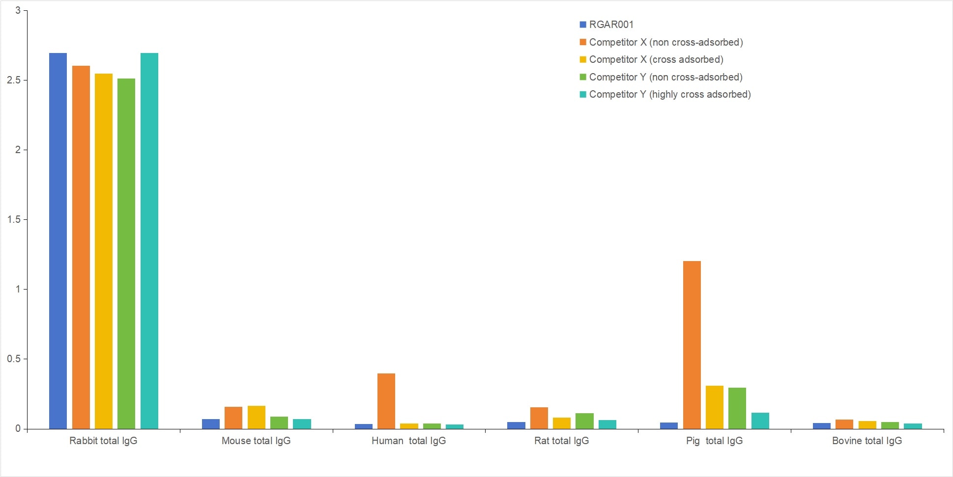
Cross reactivity comparison of RGAR001 with non-cross-adsorbed and cross-adsorbed secondary antibodies from leading competitors using Direct ELISA. Rabbit total IgG, Mouse total IgG, Human total IgG, Rat total IgG, Pig total IgG, and Bovine total IgG were coated at 100 ng/well. 0.125 μg/mL of Multi-rAb HRP-Goat Anti-Rabbit Recombinant Secondary Antibody (H+L) (RGAR001) and non-cross-adsorbed and cross-adsorbed secondary antibodies from two different competitors were used for detection.
Each lot of Multi-rAb Secondary Antibodies is produced by mixing the same high performing recombinant clones selected after several rounds of stringent screening and validation. This ensures that Multi-rAb Secondaries have high batch-to-batch consistency, which in turn can help you achieve reproducible results throughout your project.
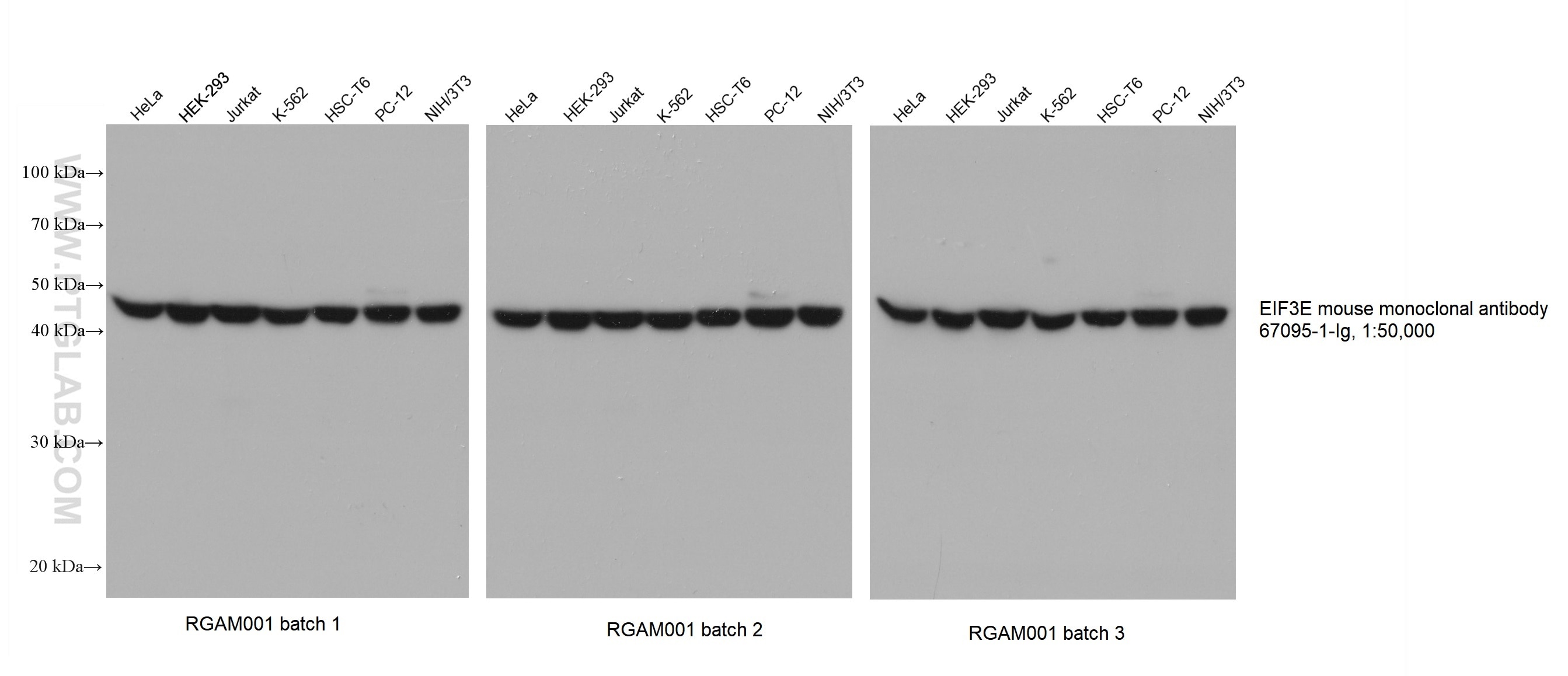
Various lysates were subjected to SDS-PAGE followed by western blot with EIF3E mouse monoclonal antibody (67095-1-Ig) at a dilution of 1:50000. Three separate batches of Multi-rAb HRP-Goat Anti-Mouse Recombinant Secondary Antibody (H+L) (RGAM001) were used at a dilution of 1:20000 for detection.
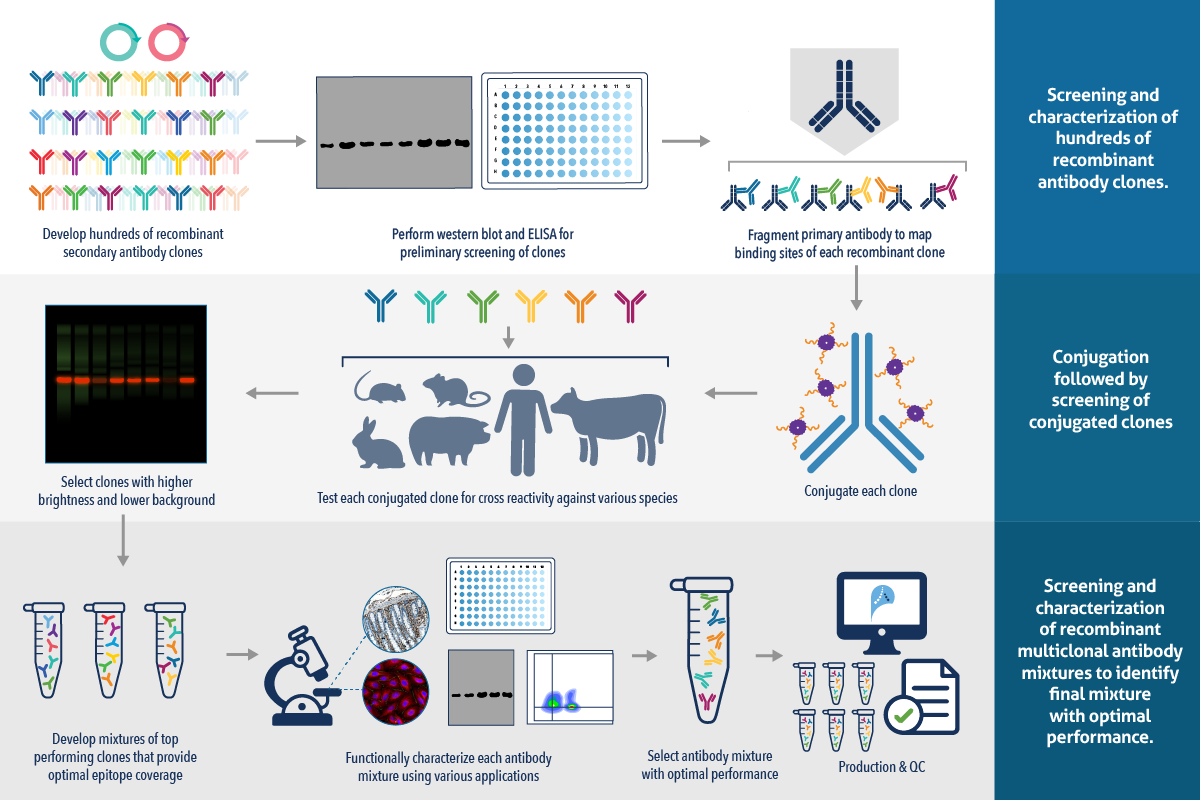
To ensure that you can achieve the highest level of performance with Multi-rAb Secondary Antibodies, several candidate clones are first conjugated to our advanced CoraLite Plus dyes and then individually characterized to select the clones with minimal cross-reactivity, low background and high brightness. The selected clones are mixed in various combinations, each of which is then functionally characterized in multiple applications to select the final multiclonal mixture with optimal performance.
Proteintech's CoraLite Plus dyes are developed with SuperHydrophilic dPEG® technology that facilitates unmatched improvements in brightness and photostability. Learn more about CoraLite Plus dyes.

Various lysates were subjected to SDS-PAGE followed by western blot with anti-beta tubulin rabbit recombinant antibody (80713-1-RR) at a dilution of 1:20000. Multi-rAb CoraLite® Plus 750-Goat Anti-Rabbit Recombinant Secondary Antibody (H+L) (RGAR006) was used at a dilution of 1:10000 for detection.
Multi-rAb Secondary Antibodies are highly suitable for multiplex applications requiring multiple primary –secondary antibody combinations. The composition of Multi-rAb Secondary Antibodies from top-performing recombinant clones that have been individually characterized for minimal cross-reactivity with IgGs from off-target species ensures that they do not bind to any unintended primary antibodies used in previous steps.
Therefore, Multi-rAb Secondary Antibodies can be used for multiplex experiments in combination with:
- Commercially available unconjugated primary antibodies and other Multi-rAb Secondary Antibodies
- Commercially available unconjugated primary antibodies and traditional polyclonal secondary antibodies

Immunofluorescence analysis of mouse cerebellum FFPE tissue stained with anti-NeuN rabbit polyclonal antibody (26975-1-AP, green) and anti-Calbindin-D28k mouse monoclonal antibody (66394-1-Ig, red). Multi-rAb CoraLite® Plus 488-Goat Anti-Rabbit Recombinant Secondary Antibody (H+L) (RGAR002, 1:500) and Multi-rAb CoraLite® Plus 594-Goat Anti-Mouse Recombinant Secondary Antibody (H+L) (RGAM004, 1:500) were used for detection.