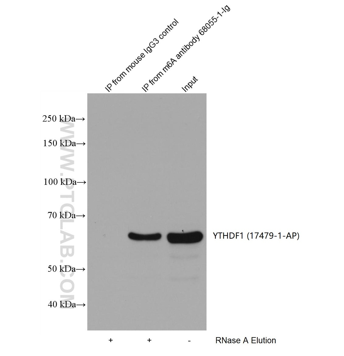
RIP experiment of RNA using 68055-1-Ig (same clone as 68055-1-PBS)
HEK-293 cells were lysised and immunoprecipitated with Protein A-m6A antibody and Protein A-mouse IgG3 control antibody respectively in the presence of RNAase inhibotor cocktail. The immunoprecipitated complex was washed diggested by RNAse A followed by western blot with YTHDF1(m6A reader) antibody 17479-1-AP (1:2000). (Lysate: 3.6mg per IP; IP: 15µg antibody and 50µL beads, 4 hours at 4℃; Diggestion: 50µg/mL * 80µL RNAse A for 1 hour at 37℃; Loading: 20% of elution; Input: 10µg.) This data was developed using the same antibody clone with 68055-1-PBS in a different storage buffer formulation.
× HEK-293 cells were lysised and immunoprecipitated with Protein A-m6A antibody and Protein A-mouse IgG3 control antibody respectively in the presence of RNAase inhibotor cocktail. The immunoprecipitated complex was washed diggested by RNAse A followed by western blot with YTHDF1(m6A reader) antibody 17479-1-AP (1:2000). (Lysate: 3.6mg per IP; IP: 15µg antibody and 50µL beads, 4 hours at 4℃; Diggestion: 50µg/mL * 80µL RNAse A for 1 hour at 37℃; Loading: 20% of elution; Input: 10µg.) This data was developed using the same antibody clone with 68055-1-PBS in a different storage buffer formulation.
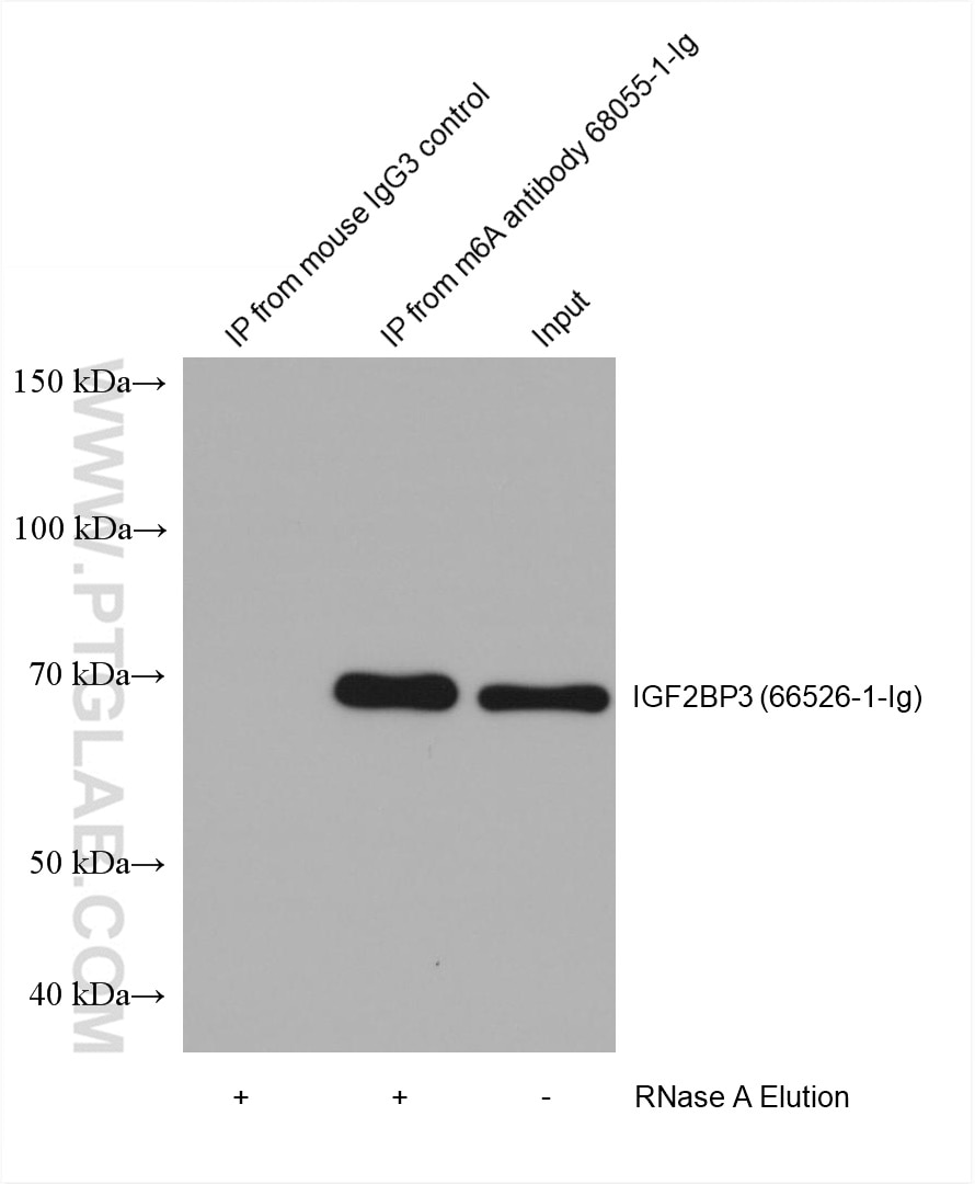
RIP experiment of RNA using 68055-1-Ig (same clone as 68055-1-PBS)
HEK-293 cells were lysised and immunoprecipitated with Protein A-m6A antibody and Protein A-mouse IgG3 control antibody respectively in the presence of RNAase inhibotor cocktail. The immunoprecipitated complex was washed diggested by RNAse A followed by western blot with IGF2BP3 (m6A reader) antibody 66526-1-Ig (1:2000). (Lysate: 4.0 mg per IP; IP: 30µg antibody and 50µL beads, 4 hours at 4℃; Diggestion: 50µg/mL * 80µL RNAse A for 1 hour at 37℃; Loading: 20% of elution; Input: 10µg.) This data was developed using the same antibody clone with 68055-1-PBS in a different storage buffer formulation.
× HEK-293 cells were lysised and immunoprecipitated with Protein A-m6A antibody and Protein A-mouse IgG3 control antibody respectively in the presence of RNAase inhibotor cocktail. The immunoprecipitated complex was washed diggested by RNAse A followed by western blot with IGF2BP3 (m6A reader) antibody 66526-1-Ig (1:2000). (Lysate: 4.0 mg per IP; IP: 30µg antibody and 50µL beads, 4 hours at 4℃; Diggestion: 50µg/mL * 80µL RNAse A for 1 hour at 37℃; Loading: 20% of elution; Input: 10µg.) This data was developed using the same antibody clone with 68055-1-PBS in a different storage buffer formulation.
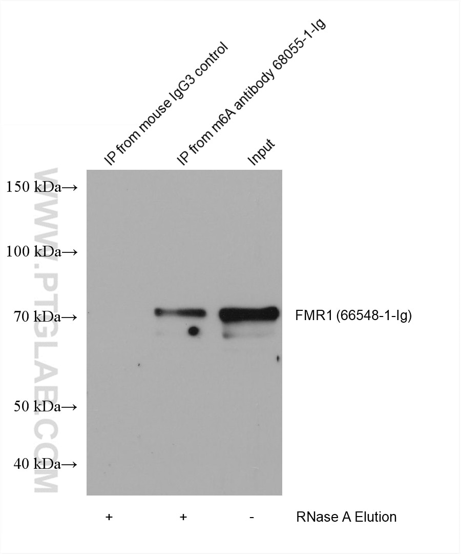
RIP experiment of RNA using 68055-1-Ig (same clone as 68055-1-PBS)
HEK-293 cells were lysised and immunoprecipitated with Protein A-m6A antibody and Protein A-mouse IgG3 control antibody respectively in the presence of RNAase inhibotor cocktail. The immunoprecipitated complex was washed diggested by RNAse A followed by western blot with FMR1 (m6A reader) antibody 66548-1-Ig (1:5000). (Lysate: 4.0 mg per IP; IP: 30µg antibody and 50µL beads, 4 hours at 4℃; Diggestion: 50µg/mL * 80µL RNAse A for 1 hour at 37℃; Loading: 20% of elution; Input: 10µg.) This data was developed using the same antibody clone with 68055-1-PBS in a different storage buffer formulation.
× HEK-293 cells were lysised and immunoprecipitated with Protein A-m6A antibody and Protein A-mouse IgG3 control antibody respectively in the presence of RNAase inhibotor cocktail. The immunoprecipitated complex was washed diggested by RNAse A followed by western blot with FMR1 (m6A reader) antibody 66548-1-Ig (1:5000). (Lysate: 4.0 mg per IP; IP: 30µg antibody and 50µL beads, 4 hours at 4℃; Diggestion: 50µg/mL * 80µL RNAse A for 1 hour at 37℃; Loading: 20% of elution; Input: 10µg.) This data was developed using the same antibody clone with 68055-1-PBS in a different storage buffer formulation.
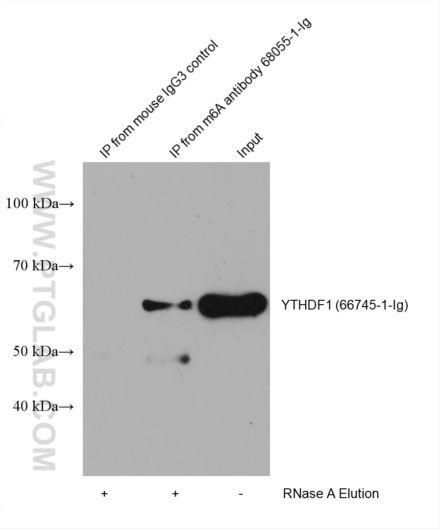
RIP experiment of RNA using 68055-1-Ig (same clone as 68055-1-PBS)
HEK-293 cells were lysised and immunoprecipitated with Protein A-m6A antibody and Protein A-mouse IgG3 control antibody respectively in the presence of RNAase inhibotor cocktail. The immunoprecipitated complex was washed diggested by RNAse A followed by western blot with YTHDF1 (m6A reader) antibody 66745-1-Ig (1:2000). (Lysate: 4.0 mg per IP; IP: 30µg antibody and 50µL beads, 4 hours at 4℃; Diggestion: 50µg/mL * 80µL RNAse A for 1 hour at 37℃; Loading: 20% of elution; Input: 10µg.) This data was developed using the same antibody clone with 68055-1-PBS in a different storage buffer formulation.
× HEK-293 cells were lysised and immunoprecipitated with Protein A-m6A antibody and Protein A-mouse IgG3 control antibody respectively in the presence of RNAase inhibotor cocktail. The immunoprecipitated complex was washed diggested by RNAse A followed by western blot with YTHDF1 (m6A reader) antibody 66745-1-Ig (1:2000). (Lysate: 4.0 mg per IP; IP: 30µg antibody and 50µL beads, 4 hours at 4℃; Diggestion: 50µg/mL * 80µL RNAse A for 1 hour at 37℃; Loading: 20% of elution; Input: 10µg.) This data was developed using the same antibody clone with 68055-1-PBS in a different storage buffer formulation.
RIP experiment of RNA using 68055-1-Ig (same clone as 68055-1-PBS)
HEK-293 cells were lysised and immunoprecipitated with Protein A-m6A antibody and Protein A-mouse IgG3 control antibody respectively in the presence of RNAase inhibotor cocktail. The immunoprecipitated complex was washed diggested by RNAse A followed by western blot with FMR1 (m6A reader) antibody 13755-1-AP (1:2000). (Lysate: 4.0 mg per IP; IP: 30µg antibody and 50µL beads, 4 hours at 4℃; Diggestion: 50µg/mL * 80µL RNAse A for 1 hour at 37℃; Loading: 20% of elution; Input: 10µg.) This data was developed using the same antibody clone with 68055-1-PBS in a different storage buffer formulation.
× HEK-293 cells were lysised and immunoprecipitated with Protein A-m6A antibody and Protein A-mouse IgG3 control antibody respectively in the presence of RNAase inhibotor cocktail. The immunoprecipitated complex was washed diggested by RNAse A followed by western blot with FMR1 (m6A reader) antibody 13755-1-AP (1:2000). (Lysate: 4.0 mg per IP; IP: 30µg antibody and 50µL beads, 4 hours at 4℃; Diggestion: 50µg/mL * 80µL RNAse A for 1 hour at 37℃; Loading: 20% of elution; Input: 10µg.) This data was developed using the same antibody clone with 68055-1-PBS in a different storage buffer formulation.
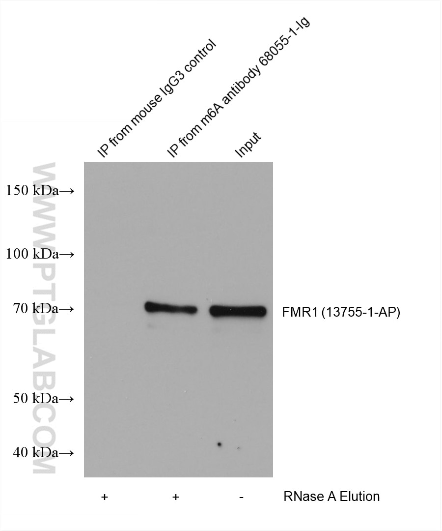
RIP experiment of RNA using 68055-1-Ig (same clone as 68055-1-PBS)
HEK-293 cells were lysised and immunoprecipitated with Protein A-m6A antibody and Protein A-mouse IgG3 control antibody respectively in the presence of RNAase inhibotor cocktail. The immunoprecipitated complex was washed diggested by RNAse A followed by western blot with YTHDF2 (m6A reader) antibody 24744-1-Ig (1:2000). (Lysate: 4.0 mg per IP; IP: 30µg antibody and 50µL beads, 4 hours at 4℃; Diggestion: 50µg/mL * 80µL RNAse A for 1 hour at 37℃; Loading: 20% of elution; Input: 10µg.) This data was developed using the same antibody clone with 68055-1-PBS in a different storage buffer formulation.
× HEK-293 cells were lysised and immunoprecipitated with Protein A-m6A antibody and Protein A-mouse IgG3 control antibody respectively in the presence of RNAase inhibotor cocktail. The immunoprecipitated complex was washed diggested by RNAse A followed by western blot with YTHDF2 (m6A reader) antibody 24744-1-Ig (1:2000). (Lysate: 4.0 mg per IP; IP: 30µg antibody and 50µL beads, 4 hours at 4℃; Diggestion: 50µg/mL * 80µL RNAse A for 1 hour at 37℃; Loading: 20% of elution; Input: 10µg.) This data was developed using the same antibody clone with 68055-1-PBS in a different storage buffer formulation.
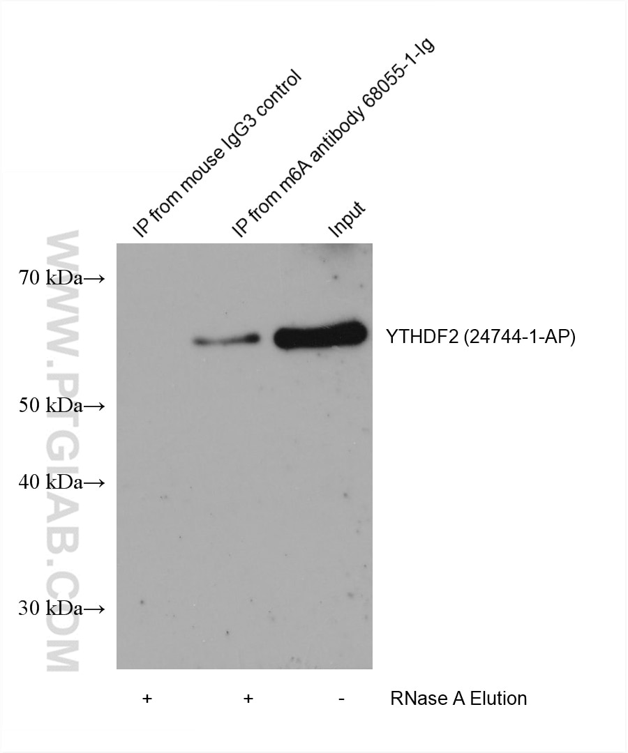
RIP experiment of RNA using 68055-1-Ig (same clone as 68055-1-PBS)
HEK-293 cells were lysised and immunoprecipitated with Protein A-m6A antibody and Protein A-mouse IgG3 control antibody respectively in the presence of RNAase inhibotor cocktail. The immunoprecipitated complex was washed diggested by RNAse A followed by western blot with YTHDC2 (m6A reader) antibody 27779-1-Ig (1:4000). (Lysate: 4.0 mg per IP; IP: 30µg antibody and 50µL beads, 4 hours at 4℃; Diggestion: 50µg/mL * 80µL RNAse A for 1 hour at 37℃; Loading: 20% of elution; Input: 10µg.) This data was developed using the same antibody clone with 68055-1-PBS in a different storage buffer formulation.
× HEK-293 cells were lysised and immunoprecipitated with Protein A-m6A antibody and Protein A-mouse IgG3 control antibody respectively in the presence of RNAase inhibotor cocktail. The immunoprecipitated complex was washed diggested by RNAse A followed by western blot with YTHDC2 (m6A reader) antibody 27779-1-Ig (1:4000). (Lysate: 4.0 mg per IP; IP: 30µg antibody and 50µL beads, 4 hours at 4℃; Diggestion: 50µg/mL * 80µL RNAse A for 1 hour at 37℃; Loading: 20% of elution; Input: 10µg.) This data was developed using the same antibody clone with 68055-1-PBS in a different storage buffer formulation.
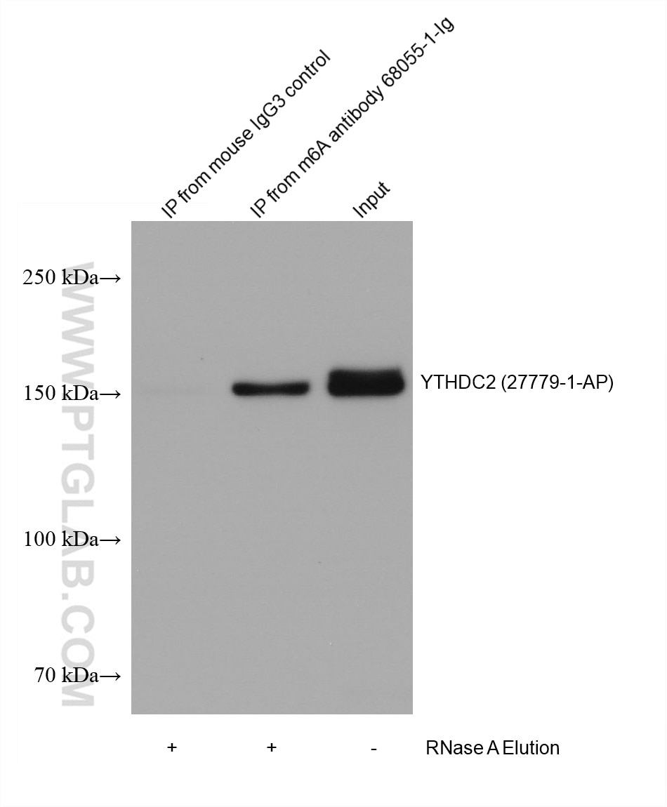
RIP experiment of RNA using 68055-1-Ig (same clone as 68055-1-PBS)
HEK-293 cells were lysised and immunoprecipitated with Protein A-m6A antibody and Protein A-mouse IgG3 control antibody respectively in the presence of RNAase inhibotor cocktail. The immunoprecipitated complex was washed diggested by RNAse A followed by western blot with LRPPRC (m6A reader) antibody 21175-1-AP (1:5000). (Lysate: 4.0 mg per IP; IP: 30µg antibody and 50µL beads, 4 hours at 4℃; Diggestion: 50µg/mL * 80µL RNAse A for 1 hour at 37℃; Loading: 20% of elution; Input: 10µg.) This data was developed using the same antibody clone with 68055-1-PBS in a different storage buffer formulation.
× HEK-293 cells were lysised and immunoprecipitated with Protein A-m6A antibody and Protein A-mouse IgG3 control antibody respectively in the presence of RNAase inhibotor cocktail. The immunoprecipitated complex was washed diggested by RNAse A followed by western blot with LRPPRC (m6A reader) antibody 21175-1-AP (1:5000). (Lysate: 4.0 mg per IP; IP: 30µg antibody and 50µL beads, 4 hours at 4℃; Diggestion: 50µg/mL * 80µL RNAse A for 1 hour at 37℃; Loading: 20% of elution; Input: 10µg.) This data was developed using the same antibody clone with 68055-1-PBS in a different storage buffer formulation.
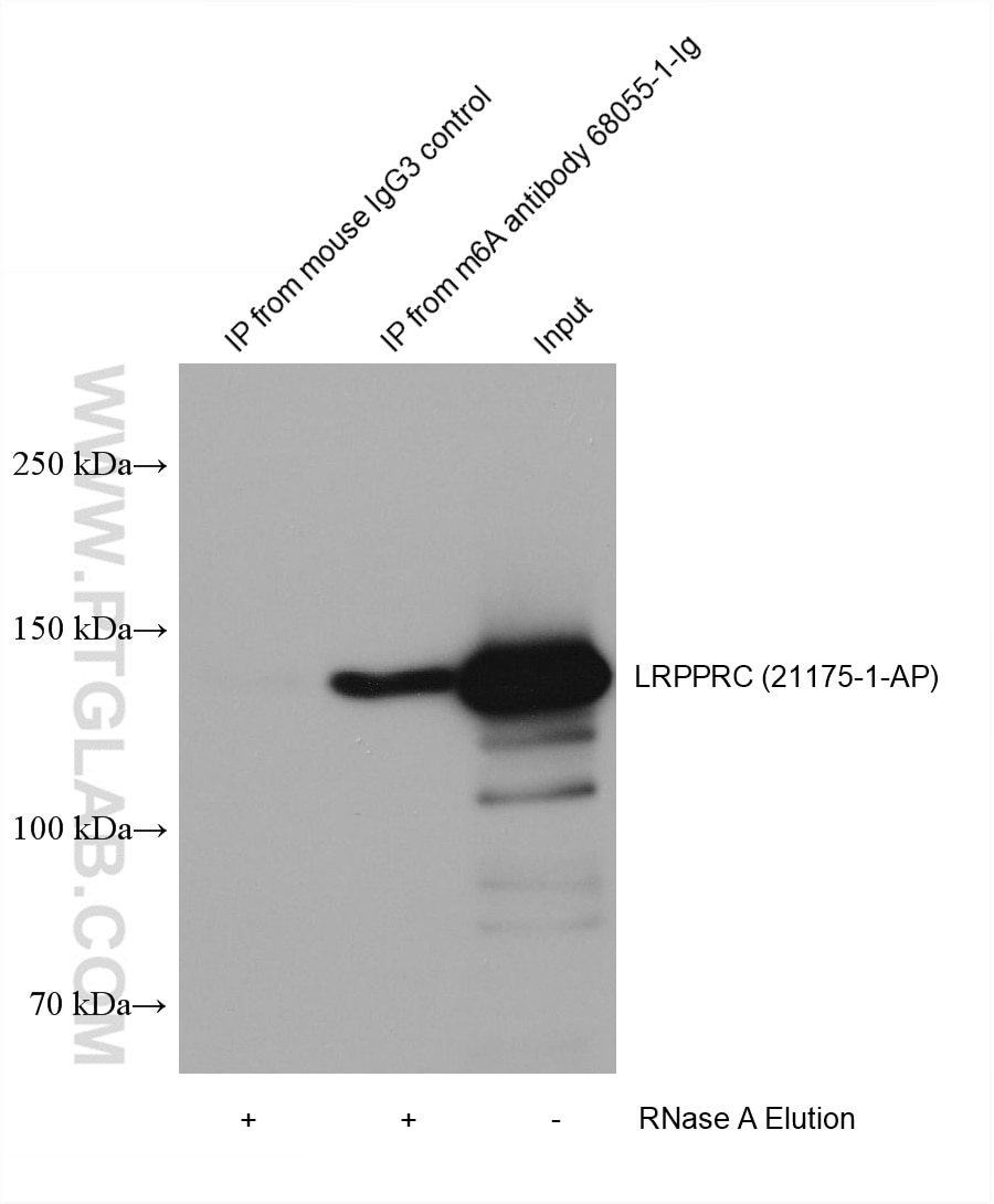
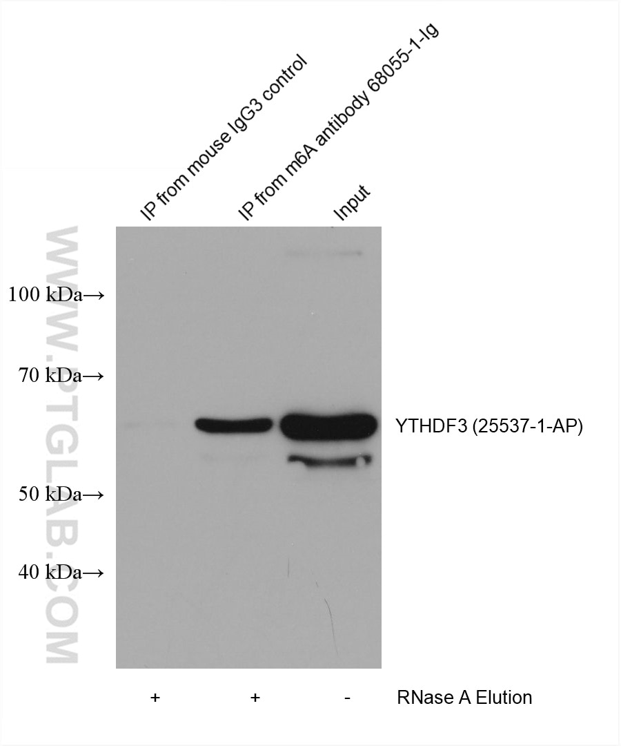
RIP experiment of RNA using 68055-1-Ig (same clone as 68055-1-PBS)
HEK-293 cells were lysised and immunoprecipitated with Protein A-m6A antibody and Protein A-mouse IgG3 control antibody respectively in the presence of RNAase inhibotor cocktail. The immunoprecipitated complex was washed diggested by RNAse A followed by western blot with YTHDF3 (m6A reader) antibody 25537-1-Ig (1:5000). (Lysate: 4.0 mg per IP; IP: 30µg antibody and 50µL beads, 4 hours at 4℃; Diggestion: 50µg/mL * 80µL RNAse A for 1 hour at 37℃; Loading: 20% of elution; Input: 10µg.) This data was developed using the same antibody clone with 68055-1-PBS in a different storage buffer formulation.
× HEK-293 cells were lysised and immunoprecipitated with Protein A-m6A antibody and Protein A-mouse IgG3 control antibody respectively in the presence of RNAase inhibotor cocktail. The immunoprecipitated complex was washed diggested by RNAse A followed by western blot with YTHDF3 (m6A reader) antibody 25537-1-Ig (1:5000). (Lysate: 4.0 mg per IP; IP: 30µg antibody and 50µL beads, 4 hours at 4℃; Diggestion: 50µg/mL * 80µL RNAse A for 1 hour at 37℃; Loading: 20% of elution; Input: 10µg.) This data was developed using the same antibody clone with 68055-1-PBS in a different storage buffer formulation.
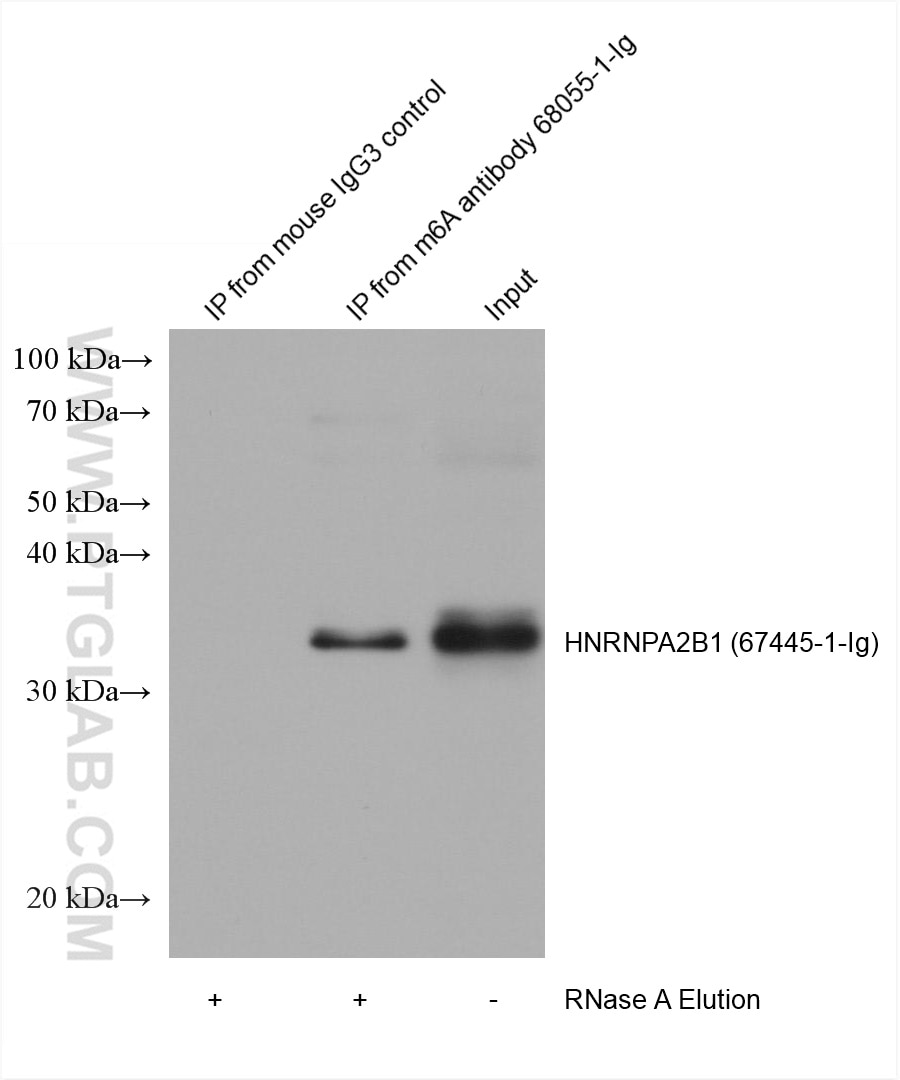
RIP experiment of RNA using 68055-1-Ig (same clone as 68055-1-PBS)
HEK-293 cells were lysised and immunoprecipitated with Protein A-m6A antibody and Protein A-mouse IgG3 control antibody respectively in the presence of RNAase inhibotor cocktail. The immunoprecipitated complex was washed diggested by RNAse A followed by western blot with HNRNPA2B1 (m6A reader) antibody 67445-1-Ig (1:2000). (Lysate: 4.0 mg per IP; IP: 30µg antibody and 50µL beads, 4 hours at 4℃; Diggestion: 50µg/mL * 80µL RNAse A for 1 hour at 37℃; Loading: 20% of elution; Input: 10µg.) This data was developed using the same antibody clone with 68055-1-PBS in a different storage buffer formulation.
× HEK-293 cells were lysised and immunoprecipitated with Protein A-m6A antibody and Protein A-mouse IgG3 control antibody respectively in the presence of RNAase inhibotor cocktail. The immunoprecipitated complex was washed diggested by RNAse A followed by western blot with HNRNPA2B1 (m6A reader) antibody 67445-1-Ig (1:2000). (Lysate: 4.0 mg per IP; IP: 30µg antibody and 50µL beads, 4 hours at 4℃; Diggestion: 50µg/mL * 80µL RNAse A for 1 hour at 37℃; Loading: 20% of elution; Input: 10µg.) This data was developed using the same antibody clone with 68055-1-PBS in a different storage buffer formulation.
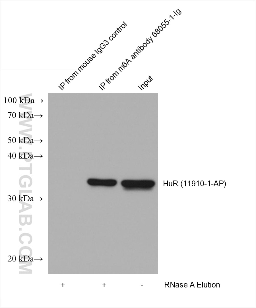
RIP experiment of RNA using 68055-1-Ig (same clone as 68055-1-PBS)
HEK-293 cells were lysised and immunoprecipitated with Protein A-m6A antibody and Protein A-mouse IgG3 control antibody respectively in the presence of RNAase inhibotor cocktail. The immunoprecipitated complex was washed diggested by RNAse A followed by western blot with HuR (m6A reader) antibody 11910-1-AP (1:5000). (Lysate: 4.0 mg per IP; IP: 30µg antibody and 50µL beads, 4 hours at 4℃; Diggestion: 50µg/mL * 80µL RNAse A for 1 hour at 37℃; Loading: 20% of elution; Input: 10µg.) This data was developed using the same antibody clone with 68055-1-PBS in a different storage buffer formulation.
× HEK-293 cells were lysised and immunoprecipitated with Protein A-m6A antibody and Protein A-mouse IgG3 control antibody respectively in the presence of RNAase inhibotor cocktail. The immunoprecipitated complex was washed diggested by RNAse A followed by western blot with HuR (m6A reader) antibody 11910-1-AP (1:5000). (Lysate: 4.0 mg per IP; IP: 30µg antibody and 50µL beads, 4 hours at 4℃; Diggestion: 50µg/mL * 80µL RNAse A for 1 hour at 37℃; Loading: 20% of elution; Input: 10µg.) This data was developed using the same antibody clone with 68055-1-PBS in a different storage buffer formulation.
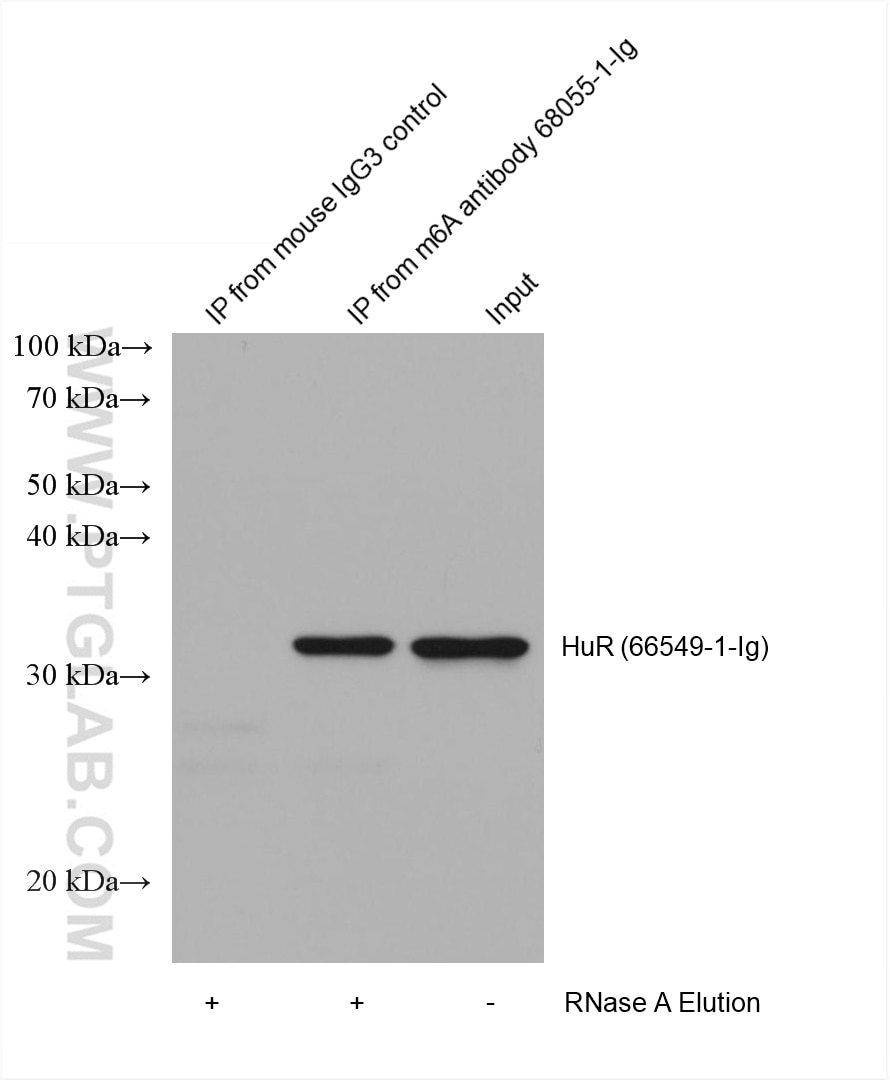
RIP experiment of RNA using 68055-1-Ig (same clone as 68055-1-PBS)
HEK-293 cells were lysised and immunoprecipitated with Protein A-m6A antibody and Protein A-mouse IgG3 control antibody respectively in the presence of RNAase inhibotor cocktail. The immunoprecipitated complex was washed diggested by RNAse A followed by western blot with HuR (m6A reader) antibody 66549-1-Ig (1:2000). (Lysate: 4.0 mg per IP; IP: 30µg antibody and 50µL beads, 4 hours at 4℃; Diggestion: 50µg/mL * 80µL RNAse A for 1 hour at 37℃; Loading: 20% of elution; Input: 10µg.) This data was developed using the same antibody clone with 68055-1-PBS in a different storage buffer formulation.
× HEK-293 cells were lysised and immunoprecipitated with Protein A-m6A antibody and Protein A-mouse IgG3 control antibody respectively in the presence of RNAase inhibotor cocktail. The immunoprecipitated complex was washed diggested by RNAse A followed by western blot with HuR (m6A reader) antibody 66549-1-Ig (1:2000). (Lysate: 4.0 mg per IP; IP: 30µg antibody and 50µL beads, 4 hours at 4℃; Diggestion: 50µg/mL * 80µL RNAse A for 1 hour at 37℃; Loading: 20% of elution; Input: 10µg.) This data was developed using the same antibody clone with 68055-1-PBS in a different storage buffer formulation.
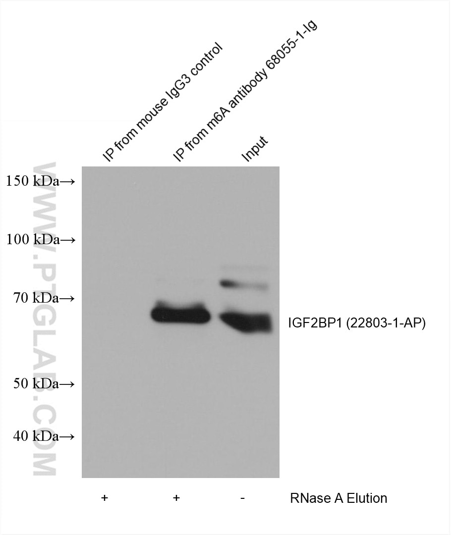
RIP experiment of RNA using 68055-1-Ig (same clone as 68055-1-PBS)
HEK-293 cells were lysised and immunoprecipitated with Protein A-m6A antibody and Protein A-mouse IgG3 control antibody respectively in the presence of RNAase inhibotor cocktail. The immunoprecipitated complex was washed diggested by RNAse A followed by western blot with IGF2BP1 (m6A reader) antibody 22803-1-AP (1:5000). (Lysate: 4.0 mg per IP; IP: 30µg antibody and 50µL beads, 4 hours at 4℃; Diggestion: 50µg/mL * 80µL RNAse A for 1 hour at 37℃; Loading: 20% of elution; Input: 10µg.) This data was developed using the same antibody clone with 68055-1-PBS in a different storage buffer formulation.
× HEK-293 cells were lysised and immunoprecipitated with Protein A-m6A antibody and Protein A-mouse IgG3 control antibody respectively in the presence of RNAase inhibotor cocktail. The immunoprecipitated complex was washed diggested by RNAse A followed by western blot with IGF2BP1 (m6A reader) antibody 22803-1-AP (1:5000). (Lysate: 4.0 mg per IP; IP: 30µg antibody and 50µL beads, 4 hours at 4℃; Diggestion: 50µg/mL * 80µL RNAse A for 1 hour at 37℃; Loading: 20% of elution; Input: 10µg.) This data was developed using the same antibody clone with 68055-1-PBS in a different storage buffer formulation.
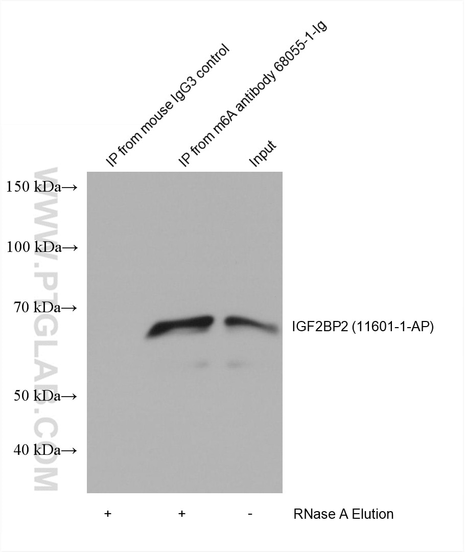
RIP experiment of RNA using 68055-1-Ig (same clone as 68055-1-PBS)
HEK-293 cells were lysised and immunoprecipitated with Protein A-m6A antibody and Protein A-mouse IgG3 control antibody respectively in the presence of RNAase inhibotor cocktail. The immunoprecipitated complex was washed diggested by RNAse A followed by western blot with IGF2BP2 (m6A reader) antibody 11601-1-AP (1:2000). (Lysate: 4.0 mg per IP; IP: 30µg antibody and 50µL beads, 4 hours at 4℃; Diggestion: 50µg/mL * 80µL RNAse A for 1 hour at 37℃; Loading: 20% of elution; Input: 10µg.) This data was developed using the same antibody clone with 68055-1-PBS in a different storage buffer formulation.
× HEK-293 cells were lysised and immunoprecipitated with Protein A-m6A antibody and Protein A-mouse IgG3 control antibody respectively in the presence of RNAase inhibotor cocktail. The immunoprecipitated complex was washed diggested by RNAse A followed by western blot with IGF2BP2 (m6A reader) antibody 11601-1-AP (1:2000). (Lysate: 4.0 mg per IP; IP: 30µg antibody and 50µL beads, 4 hours at 4℃; Diggestion: 50µg/mL * 80µL RNAse A for 1 hour at 37℃; Loading: 20% of elution; Input: 10µg.) This data was developed using the same antibody clone with 68055-1-PBS in a different storage buffer formulation.
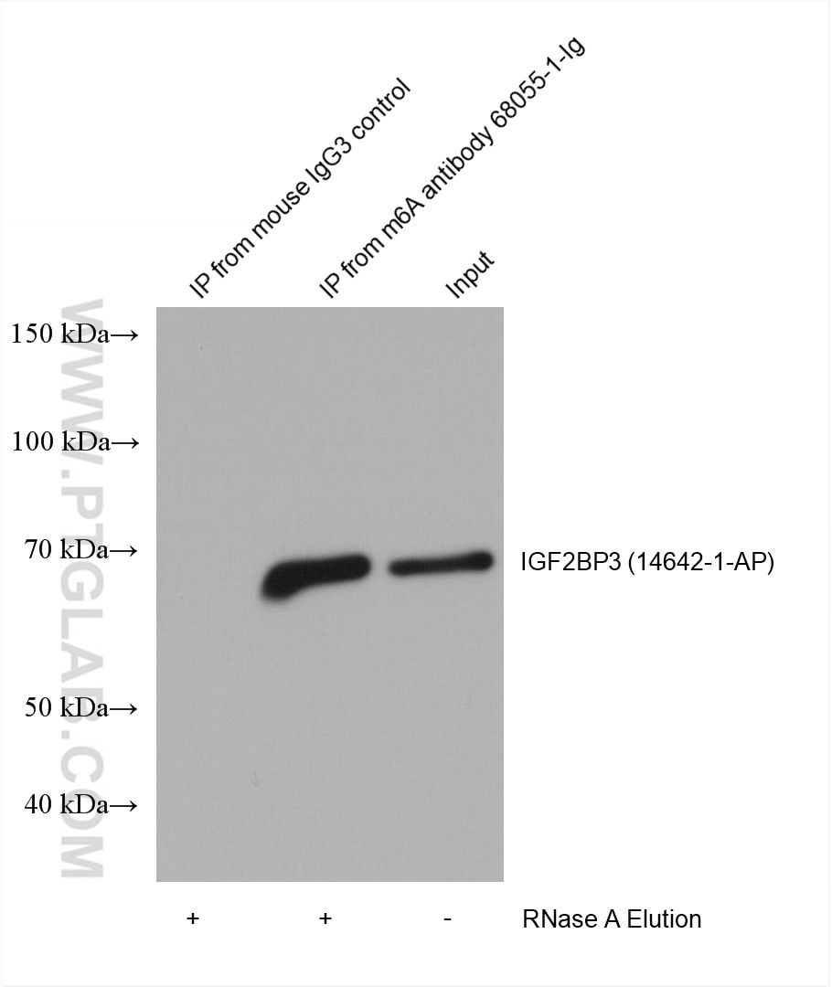
RIP experiment of RNA using 68055-1-Ig (same clone as 68055-1-PBS)
HEK-293 cells were lysised and immunoprecipitated with Protein A-m6A antibody and Protein A-mouse IgG3 control antibody respectively in the presence of RNAase inhibotor cocktail. The immunoprecipitated complex was washed diggested by RNAse A followed by western blot with IGF2BP3 (m6A reader) antibody 14642-1-AP (1:5000). (Lysate: 4.0 mg per IP; IP: 30µg antibody and 50µL beads, 4 hours at 4℃; Diggestion: 50µg/mL * 80µL RNAse A for 1 hour at 37℃; Loading: 20% of elution; Input: 10µg.) This data was developed using the same antibody clone with 68055-1-PBS in a different storage buffer formulation.
× HEK-293 cells were lysised and immunoprecipitated with Protein A-m6A antibody and Protein A-mouse IgG3 control antibody respectively in the presence of RNAase inhibotor cocktail. The immunoprecipitated complex was washed diggested by RNAse A followed by western blot with IGF2BP3 (m6A reader) antibody 14642-1-AP (1:5000). (Lysate: 4.0 mg per IP; IP: 30µg antibody and 50µL beads, 4 hours at 4℃; Diggestion: 50µg/mL * 80µL RNAse A for 1 hour at 37℃; Loading: 20% of elution; Input: 10µg.) This data was developed using the same antibody clone with 68055-1-PBS in a different storage buffer formulation.
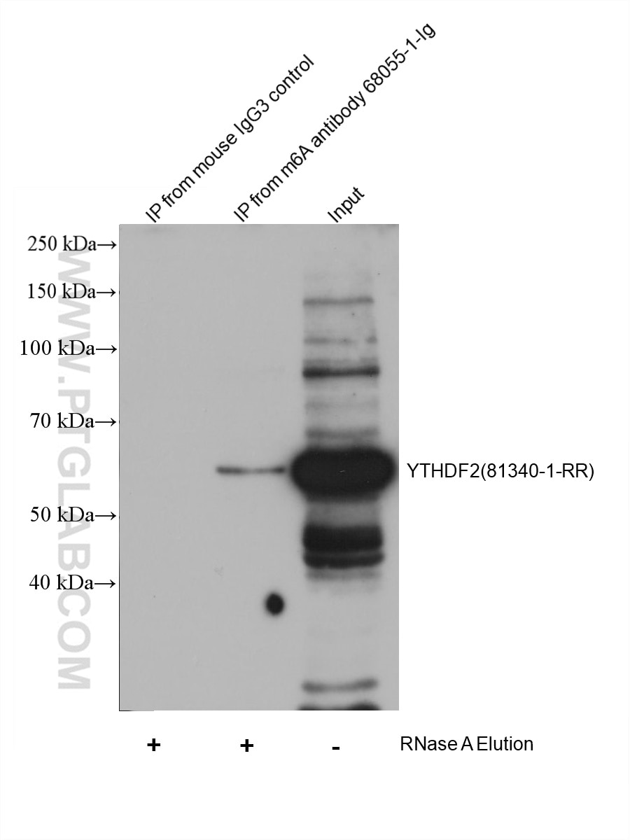
IP experiment of HEK-293 using 68055-1-Ig (same clone as 68055-1-PBS)
HEK-293 cells were lysised and immunoprecipitated with Protein A-m6A antibody and Protein A-mouse IgG3 control antibody respectively in the presence of RNAase inhibotor cocktail. The immunoprecipitated complex was washed diggested by RNAse A followed by western blot with YTHDF2 (m6A reader) antibody 81340-1-RR (1:5000). (Lysate: 4.0 mg per IP; IP: 30µg antibody and 50µL beads, 4 hours at 4℃; Diggestion: 50µg/mL * 80µL RNAse A for 1 hour at 37℃; Loading: 20% of elution; Input: 10µg.) This data was developed using the same antibody clone with 68055-1-PBS in a different storage buffer formulation.
× HEK-293 cells were lysised and immunoprecipitated with Protein A-m6A antibody and Protein A-mouse IgG3 control antibody respectively in the presence of RNAase inhibotor cocktail. The immunoprecipitated complex was washed diggested by RNAse A followed by western blot with YTHDF2 (m6A reader) antibody 81340-1-RR (1:5000). (Lysate: 4.0 mg per IP; IP: 30µg antibody and 50µL beads, 4 hours at 4℃; Diggestion: 50µg/mL * 80µL RNAse A for 1 hour at 37℃; Loading: 20% of elution; Input: 10µg.) This data was developed using the same antibody clone with 68055-1-PBS in a different storage buffer formulation.
IHC staining of mouse testis using 68055-1-Ig (same clone as 68055-1-PBS)
Immunohistochemical analysis of paraffin-embedded mouse testis tissue slide using 68055-1-Ig (m6A antibody) at dilution of 1:4000 (under 40x lens). Heat mediated antigen retrieval with Tris-EDTA buffer (pH 9.0). This data was developed using the same antibody clone with 68055-1-PBS in a different storage buffer formulation.
× Immunohistochemical analysis of paraffin-embedded mouse testis tissue slide using 68055-1-Ig (m6A antibody) at dilution of 1:4000 (under 40x lens). Heat mediated antigen retrieval with Tris-EDTA buffer (pH 9.0). This data was developed using the same antibody clone with 68055-1-PBS in a different storage buffer formulation.
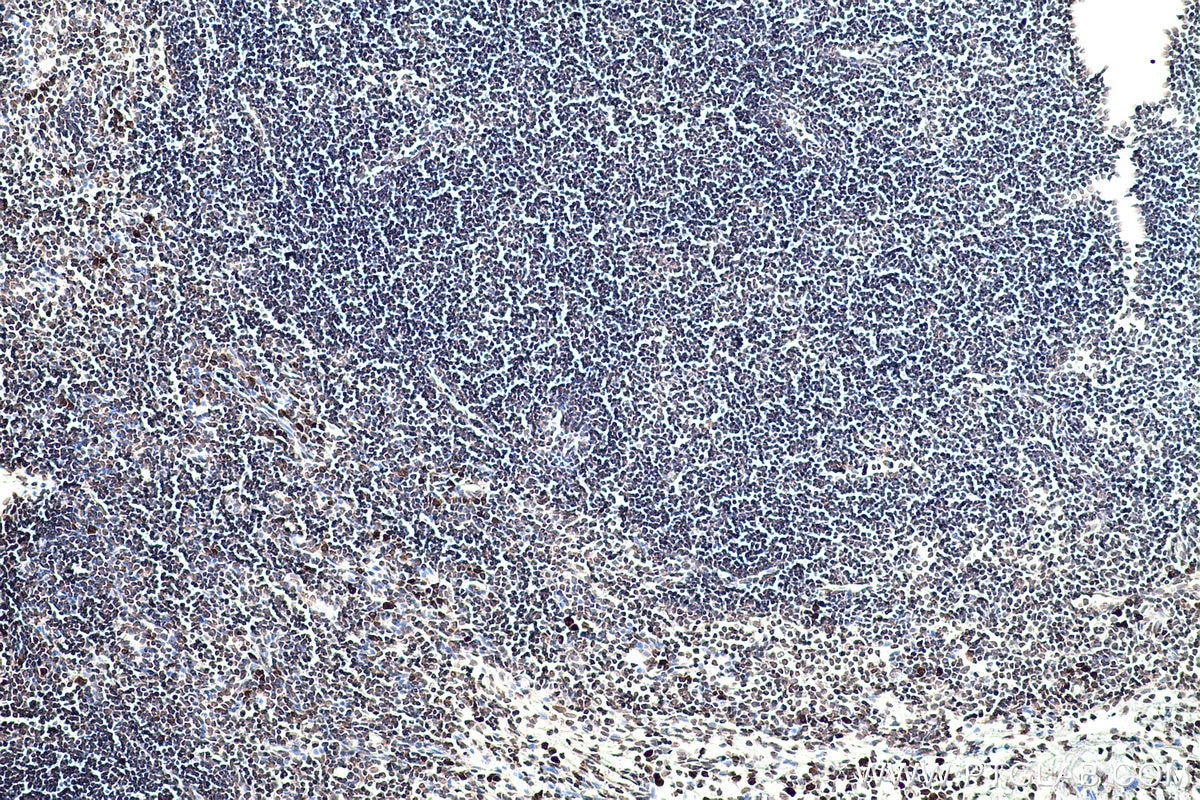
IHC staining of rat lymph node using 68055-1-Ig (same clone as 68055-1-PBS)
Immunohistochemical analysis of paraffin-embedded rat lymph node slide using 68055-1-Ig (m6A antibody) at dilution of 1:4000 (under 10x lens). Heat mediated antigen retrieval with Tris-EDTA buffer (pH 9.0). This data was developed using the same antibody clone with 68055-1-PBS in a different storage buffer formulation.
× Immunohistochemical analysis of paraffin-embedded rat lymph node slide using 68055-1-Ig (m6A antibody) at dilution of 1:4000 (under 10x lens). Heat mediated antigen retrieval with Tris-EDTA buffer (pH 9.0). This data was developed using the same antibody clone with 68055-1-PBS in a different storage buffer formulation.
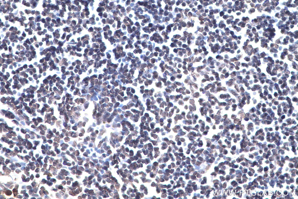
IHC staining of rat lymph node using 68055-1-Ig (same clone as 68055-1-PBS)
Immunohistochemical analysis of paraffin-embedded rat lymph node slide using 68055-1-Ig (m6A antibody) at dilution of 1:4000 (under 40x lens). Heat mediated antigen retrieval with Tris-EDTA buffer (pH 9.0). This data was developed using the same antibody clone with 68055-1-PBS in a different storage buffer formulation.
× Immunohistochemical analysis of paraffin-embedded rat lymph node slide using 68055-1-Ig (m6A antibody) at dilution of 1:4000 (under 40x lens). Heat mediated antigen retrieval with Tris-EDTA buffer (pH 9.0). This data was developed using the same antibody clone with 68055-1-PBS in a different storage buffer formulation.
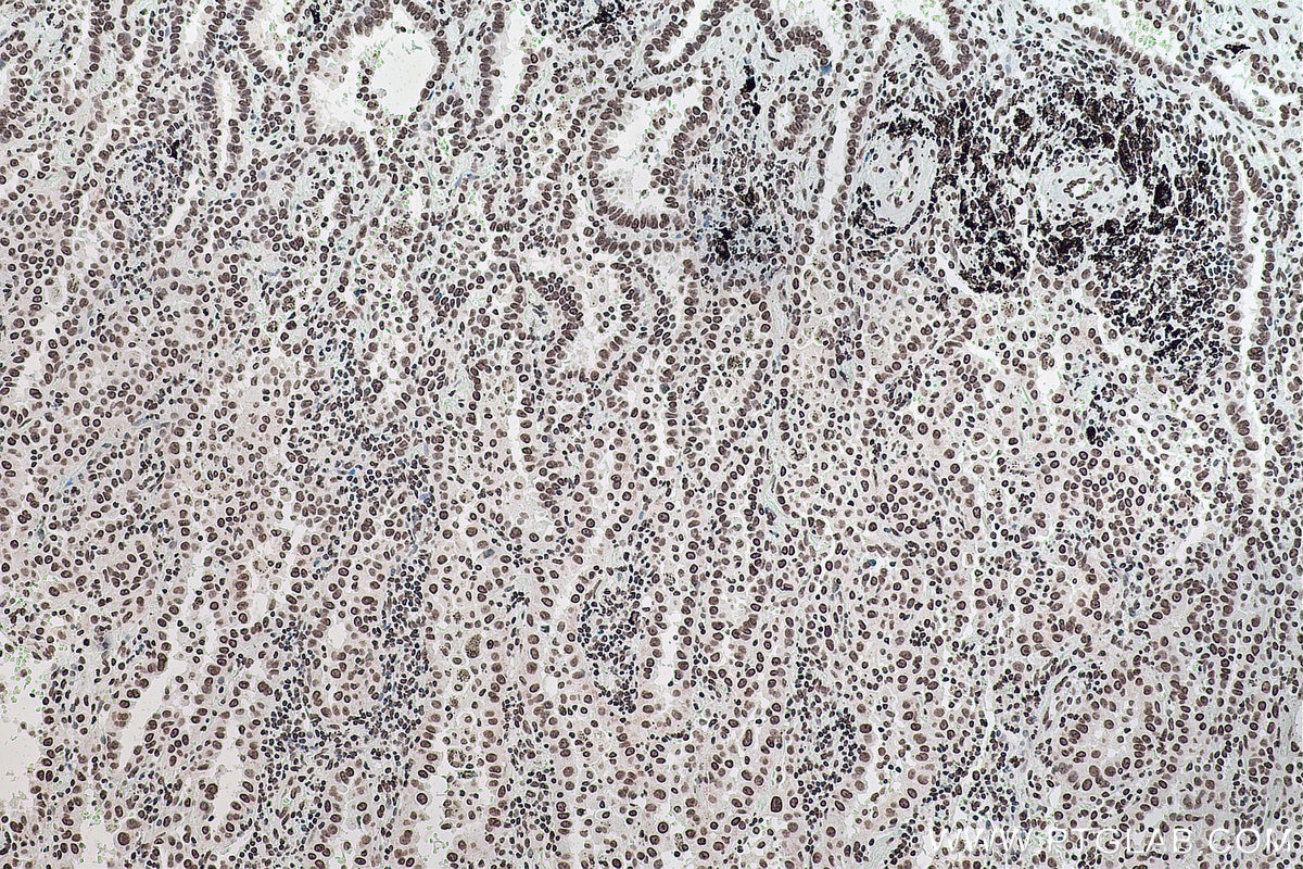
IHC staining of human lung cancer using 68055-1-Ig (same clone as 68055-1-PBS)
Immunohistochemical analysis of paraffin-embedded human lung cancer tissue slide using 68055-1-Ig (m6A antibody) at dilution of 1:8000 (under 10x lens). Heat mediated antigen retrieval with Tris-EDTA buffer (pH 9.0). This data was developed using the same antibody clone with 68055-1-PBS in a different storage buffer formulation.
× Immunohistochemical analysis of paraffin-embedded human lung cancer tissue slide using 68055-1-Ig (m6A antibody) at dilution of 1:8000 (under 10x lens). Heat mediated antigen retrieval with Tris-EDTA buffer (pH 9.0). This data was developed using the same antibody clone with 68055-1-PBS in a different storage buffer formulation.
IHC staining of human lung cancer using 68055-1-Ig (same clone as 68055-1-PBS)
Immunohistochemical analysis of paraffin-embedded human lung cancer tissue slide using 68055-1-Ig (m6A antibody) at dilution of 1:8000 (under 40x lens). Heat mediated antigen retrieval with Tris-EDTA buffer (pH 9.0). This data was developed using the same antibody clone with 68055-1-PBS in a different storage buffer formulation.
× Immunohistochemical analysis of paraffin-embedded human lung cancer tissue slide using 68055-1-Ig (m6A antibody) at dilution of 1:8000 (under 40x lens). Heat mediated antigen retrieval with Tris-EDTA buffer (pH 9.0). This data was developed using the same antibody clone with 68055-1-PBS in a different storage buffer formulation.
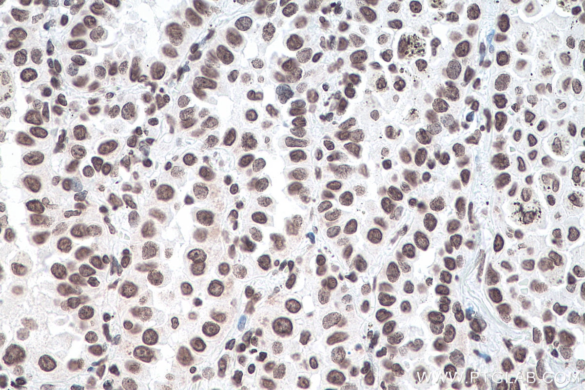
IHC staining of human breast cancer using 68055-1-Ig (same clone as 68055-1-PBS)
Immunohistochemical analysis of paraffin-embedded human breast cancer tissue slide using 68055-1-Ig (m6A antibody) at dilution of 1:4000 (under 10x lens). Heat mediated antigen retrieval with Tris-EDTA buffer (pH 9.0). This data was developed using the same antibody clone with 68055-1-PBS in a different storage buffer formulation.
× Immunohistochemical analysis of paraffin-embedded human breast cancer tissue slide using 68055-1-Ig (m6A antibody) at dilution of 1:4000 (under 10x lens). Heat mediated antigen retrieval with Tris-EDTA buffer (pH 9.0). This data was developed using the same antibody clone with 68055-1-PBS in a different storage buffer formulation.
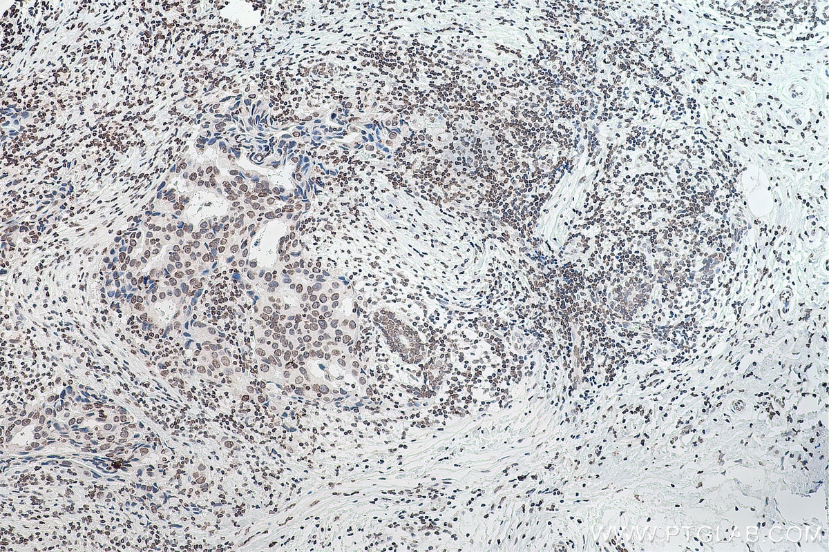
IHC staining of human breast cancer using 68055-1-Ig (same clone as 68055-1-PBS)
Immunohistochemical analysis of paraffin-embedded human breast cancer tissue slide using 68055-1-Ig (m6A antibody) at dilution of 1:4000 (under 40x lens). Heat mediated antigen retrieval with Tris-EDTA buffer (pH 9.0). This data was developed using the same antibody clone with 68055-1-PBS in a different storage buffer formulation.
× Immunohistochemical analysis of paraffin-embedded human breast cancer tissue slide using 68055-1-Ig (m6A antibody) at dilution of 1:4000 (under 40x lens). Heat mediated antigen retrieval with Tris-EDTA buffer (pH 9.0). This data was developed using the same antibody clone with 68055-1-PBS in a different storage buffer formulation.
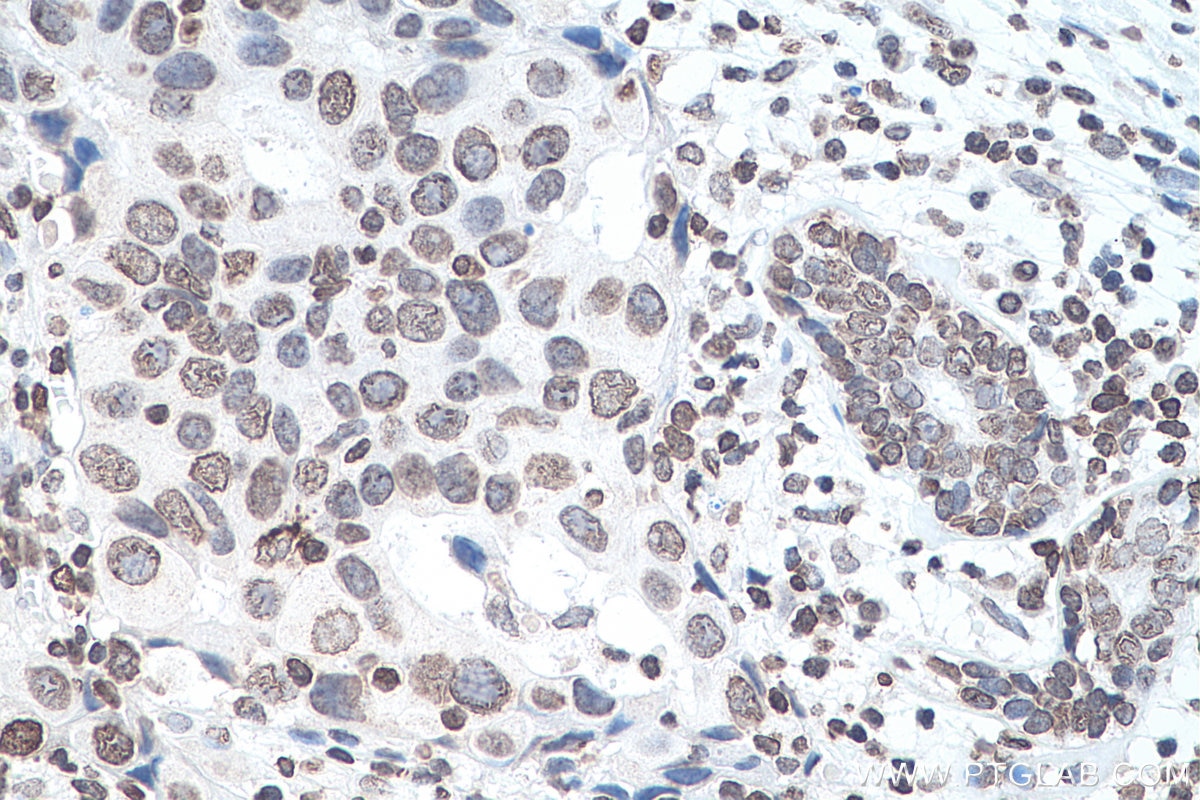
IHC staining of human colon cancer using 68055-1-Ig (same clone as 68055-1-PBS)
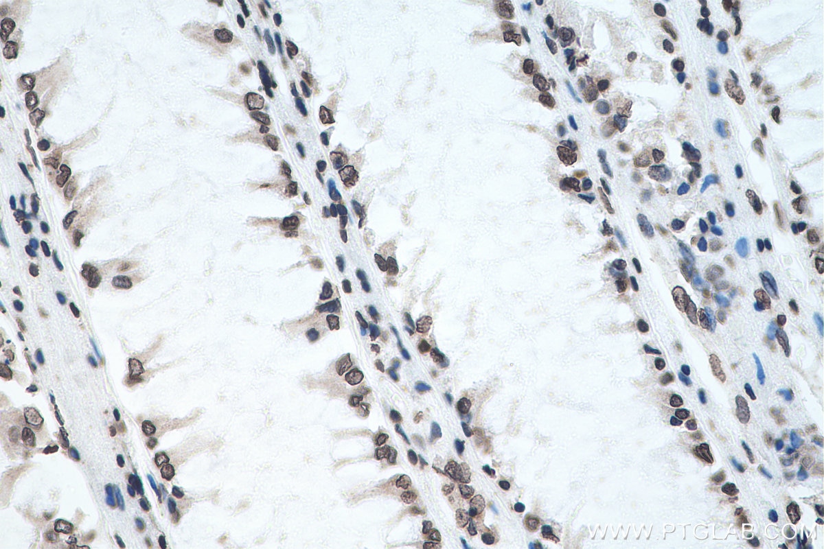
Immunohistochemical analysis of paraffin-embedded human colon cancer tissue slide using 68055-1-Ig (m6A antibody) at dilution of 1:4000 (under 10x lens). Heat mediated antigen retrieval with Tris-EDTA buffer (pH 9.0). This data was developed using the same antibody clone with 68055-1-PBS in a different storage buffer formulation.
× Immunohistochemical analysis of paraffin-embedded human colon cancer tissue slide using 68055-1-Ig (m6A antibody) at dilution of 1:4000 (under 10x lens). Heat mediated antigen retrieval with Tris-EDTA buffer (pH 9.0). This data was developed using the same antibody clone with 68055-1-PBS in a different storage buffer formulation.
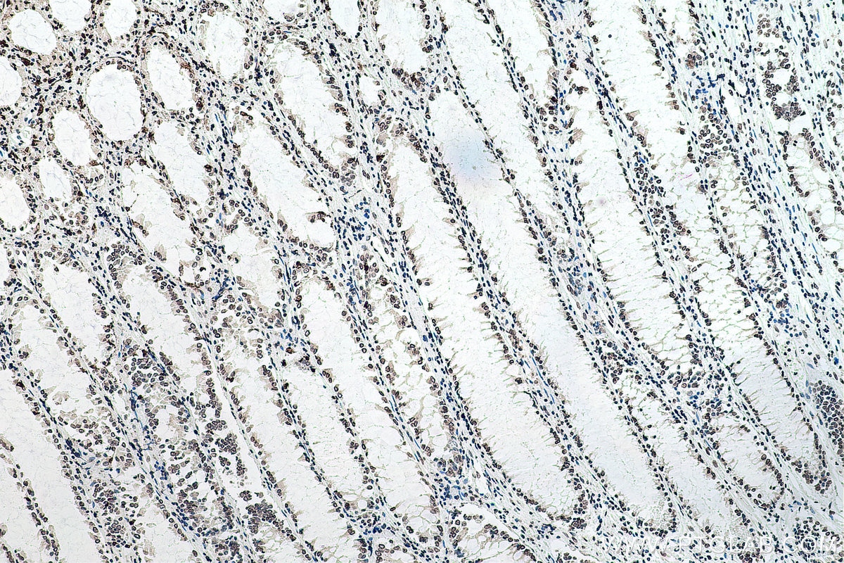
IHC staining of human colon cancer using 68055-1-Ig (same clone as 68055-1-PBS)
Immunohistochemical analysis of paraffin-embedded human colon cancer tissue slide using 68055-1-Ig (m6A antibody) at dilution of 1:4000 (under 40x lens). Heat mediated antigen retrieval with Tris-EDTA buffer (pH 9.0). This data was developed using the same antibody clone with 68055-1-PBS in a different storage buffer formulation.
× Immunohistochemical analysis of paraffin-embedded human colon cancer tissue slide using 68055-1-Ig (m6A antibody) at dilution of 1:4000 (under 40x lens). Heat mediated antigen retrieval with Tris-EDTA buffer (pH 9.0). This data was developed using the same antibody clone with 68055-1-PBS in a different storage buffer formulation.
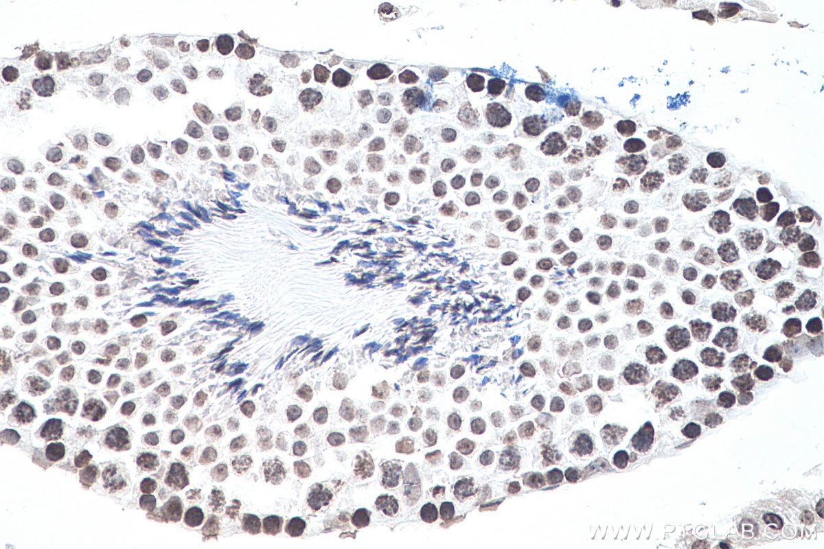
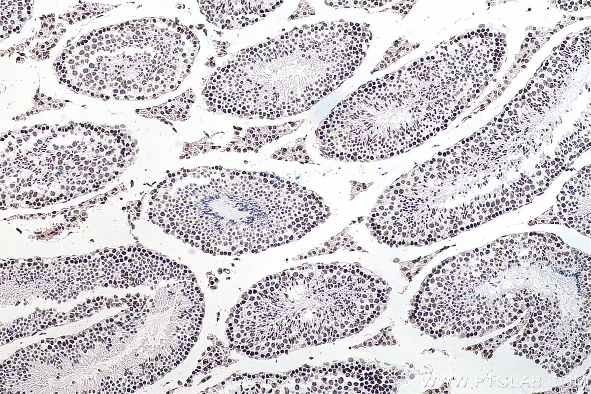
IHC staining of mouse testis using 68055-1-Ig (same clone as 68055-1-PBS)
Immunohistochemical analysis of paraffin-embedded mouse testis tissue slide using 68055-1-Ig (m6A antibody) at dilution of 1:4000 (under 10x lens). Heat mediated antigen retrieval with Tris-EDTA buffer (pH 9.0). This data was developed using the same antibody clone with 68055-1-PBS in a different storage buffer formulation.
× Immunohistochemical analysis of paraffin-embedded mouse testis tissue slide using 68055-1-Ig (m6A antibody) at dilution of 1:4000 (under 10x lens). Heat mediated antigen retrieval with Tris-EDTA buffer (pH 9.0). This data was developed using the same antibody clone with 68055-1-PBS in a different storage buffer formulation.
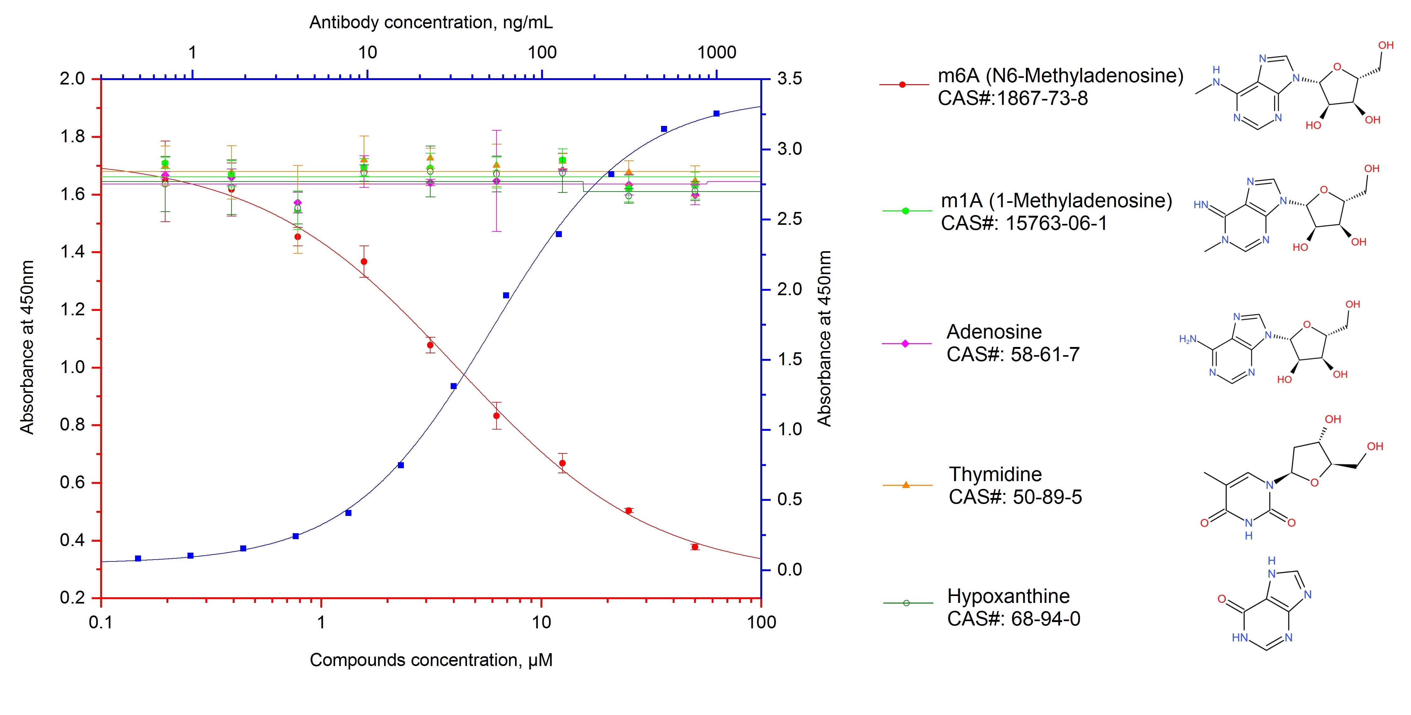
ELISA experiment of m6A using 68055-1-Ig (same clone as 68055-1-PBS)
Indirect ELISA and competitive ELISA results show that this antibody is specific to m6A. Indirect ELISA was performed by coating BSA conjugated m6A at 20ng/well followed by blocking with 1% BSA. Serial diluted primary antibody was added to the plates and incubated at 37℃. HRP-goat anti-mouse was used for detection. Competitive ELISA was performed similarly except that different concentration of m6A or its structure analogue compounds are mixed in primary antibody (fixed at 0.05μg/mL). This data was developed using the same antibody clone with 68055-1-PBS in a different storage buffer formulation.
× Indirect ELISA and competitive ELISA results show that this antibody is specific to m6A. Indirect ELISA was performed by coating BSA conjugated m6A at 20ng/well followed by blocking with 1% BSA. Serial diluted primary antibody was added to the plates and incubated at 37℃. HRP-goat anti-mouse was used for detection. Competitive ELISA was performed similarly except that different concentration of m6A or its structure analogue compounds are mixed in primary antibody (fixed at 0.05μg/mL). This data was developed using the same antibody clone with 68055-1-PBS in a different storage buffer formulation.
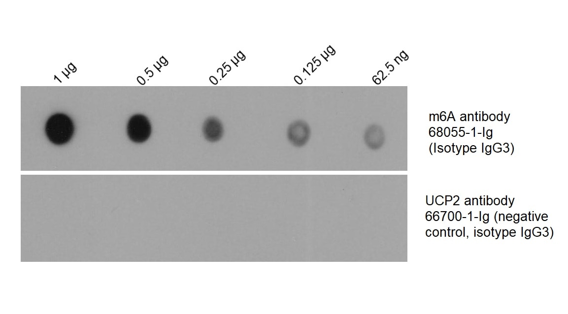
Dot Blot experiment of RNA using 68055-1-Ig (same clone as 68055-1-PBS)
Total RNA was isolated from HEK-293 cell line and was dotted to NC membrane at different amount as indicated above the dots. The membrane was blocked with BSA and blotted with m6A antibody 68055-1-Ig at 1:2000 followed by incubation of HRP-goat anti-mouse secondary antibody. Signal was developed by ECL substrate. A parallel dot blot was performed using unrelated antibody with the same isotype (UCP2 antibody 66700-1-Ig) at the same dose. This data was developed using the same antibody clone with 68055-1-PBS in a different storage buffer formulation.
× Total RNA was isolated from HEK-293 cell line and was dotted to NC membrane at different amount as indicated above the dots. The membrane was blocked with BSA and blotted with m6A antibody 68055-1-Ig at 1:2000 followed by incubation of HRP-goat anti-mouse secondary antibody. Signal was developed by ECL substrate. A parallel dot blot was performed using unrelated antibody with the same isotype (UCP2 antibody 66700-1-Ig) at the same dose. This data was developed using the same antibody clone with 68055-1-PBS in a different storage buffer formulation.
Proteintech Guarantee
The Proteintech guarantee covers Proteintech antibodies in any species and any application, including those not listed on the datasheet. If the antibody doesn’t perform, you can receive a hassle-free refund or credit note.
Product Information
68055-1-PBS targets m6A in IHC, RIP, Dot Blot, ELISA, Indirect ELISA applications and shows reactivity with chemical compound, m6a samples.
| Tested Reactivity | chemical compound, m6a |
| Host / Isotype | Mouse / IgG3 |
| Class | Monoclonal |
| Type | Antibody |
| Immunogen |
Peptide Predict reactive species |
| Full Name | m6A |
| GenBank Accession Number | m6A |
| Gene Symbol | |
| Gene ID (NCBI) | |
| RRID | AB_2918796 |
| Conjugate | Unconjugated |
| Form | Liquid |
| Purification Method | Protein A purification |
| Storage Buffer | PBS only, pH 7.3. |
| Storage Conditions | Store at -80°C. |
Background Information
m6A (N6-methyladenosine) is the most abundant internal modification in mammalian mRNA. This modification is installed by the m6A methyltransferases or termed "writers"such as METTL3 and METTL14, and can be reversed by demethylases that serve as "erasers" such as FTO and ALKBH5. The stability of m6A-modified mRNA is regulated by m6A reader protein YTHDFs, which recognizes m6A and reduces the stability of target transcripts. m6A modification and its regulatory proteins play critical roles in cancer pathogenesis and progression. m6A modification is also invovled in viruses life cycles, suggesting that drugs targets to m6A pathway could be used for antiviral thereapy.
Protocol for Dot Blot:
https://www.ptglab.com/protocol/68055-1-IgDotBlot.pdf


