Filter:
 with sh-Control and sh-TOMM40 transfected HeLa cells. This data was developed using the same antibody clone with 66658-1-PBS in a different storage buffer formulation.")
 at dilution of 1:20000 incubated at room temperature for 1.5 hours. The membrane was stripped and reblotted with HRP-conjugated Lamin B1 Monoclonal antibody (HRP-66095) as loading control. This data was developed using the same antibody clone with 66658-1-PBS in a different storage buffer formulation.")
 at dilution of 1:20000 incubated at room temperature for 1.5 hours. This data was developed using the same antibody clone with 66658-1-PBS in a different storage buffer formulation.")
 at dilution of 1:20000 incubated at room temperature for 1.5 hours. This data was developed using the same antibody clone with 66658-1-PBS in a different storage buffer formulation.")
 at dilution of 1:20000 incubated at room temperature for 1.5 hours. This data was developed using the same antibody clone with 66658-1-PBS in a different storage buffer formulation.")
 at dilution of 1:20000 incubated at room temperature for 1.5 hours. This data was developed using the same antibody clone with 66658-1-PBS in a different storage buffer formulation.")
 at dilution of 1:20000 incubated at room temperature for 1.5 hours. This data was developed using the same antibody clone with 66658-1-PBS in a different storage buffer formulation.")
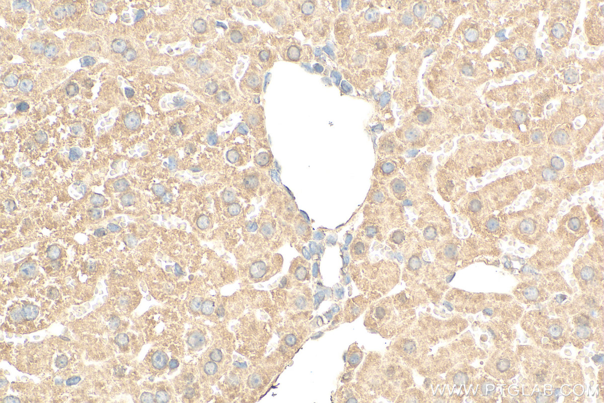
 at dilution of 1:200 (under 40x lens). Heat mediated antigen retrieval with Tris-EDTA buffer (pH 9.0). This data was developed using the same antibody clone with 66658-1-PBS in a different storage buffer formulation.")
 fixed HepG2 cells using 66658-1-Ig (TOMM40 antibody) at dilution of 1:100 and CoraLite488-Conjugated AffiniPure Goat Anti-Mouse IgG(H+L). This data was developed using the same antibody clone with 66658-1-PBS in a different storage buffer formulation.")
 fixed HeLa cells using 66658-1-Ig (TOMM40 antibody) at dilution of 1:100 and CoraLite488-Conjugated AffiniPure Goat Anti-Mouse IgG(H+L). This data was developed using the same antibody clone with 66658-1-PBS in a different storage buffer formulation.")
 fixed HepG2 cells using 66658-1-Ig (TOMM40 antibody) at dilution of 1:100 and CoraLite488-Conjugated AffiniPure Goat Anti-Mouse IgG(H+L). This data was developed using the same antibody clone with 66658-1-PBS in a different storage buffer formulation.")
Product Information
66658-1-PBS targets TOMM40 in WB, IHC, IF/ICC, Indirect ELISA applications and shows reactivity with human, mouse, rat, pig samples.
| Tested Reactivity | human, mouse, rat, pig |
| Host / Isotype | Mouse / IgG1 |
| Class | Monoclonal |
| Type | Antibody |
| Immunogen |
CatNo: Ag13065 Product name: Recombinant human TOMM40 protein Source: e coli.-derived, PET28a Tag: 6*His Domain: 1-361 aa of BC017224 Sequence: MGNVLAASSPPAGPPPPPAPALVGLPPPPPSPPGFTLPPLGGSLGAGTSTSRSSERTPGAATASASGAAEDGACGCLPNPGTFEECHRKCKELFPIQMEGVKLTVNKGLSNHFQVNHTVALSTIGESNYHFGVTYVGTKQLSPTEAFPVLVGDMDNSGSLNAQVIHQLGPGLRSKMAIQTQQSKFVNWQVDGEYRGSDFTAAVTLGNPDVLVGSGILVAHYLQSITPCLALGGELVYHRRPGEEGTVMSLAGKYTLNNWLATVTLGQAGMHATYYHKASDQLQVGVEFEASTRMQDTSVSFGYQLDLPKANLLFKGSVDSNWIVGATLEKKLPPLPLTLALGAFLNHRKNKFQCGFGLTIG Predict reactive species |
| Full Name | translocase of outer mitochondrial membrane 40 homolog (yeast) |
| Calculated Molecular Weight | 38 kDa |
| Observed Molecular Weight | 38 kDa |
| GenBank Accession Number | BC017224 |
| Gene Symbol | TOMM40 |
| Gene ID (NCBI) | 10452 |
| RRID | AB_2882015 |
| Conjugate | Unconjugated |
| Form | Liquid |
| Purification Method | Protein A purification |
| UNIPROT ID | O96008 |
| Storage Buffer | PBS only, pH 7.3. |
| Storage Conditions | Store at -80°C. |