Filter:
 with sh-Control and sh-TOMM40 transfected HeLa cells.")
 at dilution of 1:20000 incubated at room temperature for 1.5 hours. The membrane was stripped and reblotted with HRP-conjugated Lamin B1 Monoclonal antibody (HRP-66095) as loading control.")
 at dilution of 1:20000 incubated at room temperature for 1.5 hours.")
 at dilution of 1:20000 incubated at room temperature for 1.5 hours.")
 at dilution of 1:20000 incubated at room temperature for 1.5 hours.")
 at dilution of 1:20000 incubated at room temperature for 1.5 hours.")
 at dilution of 1:20000 incubated at room temperature for 1.5 hours.")
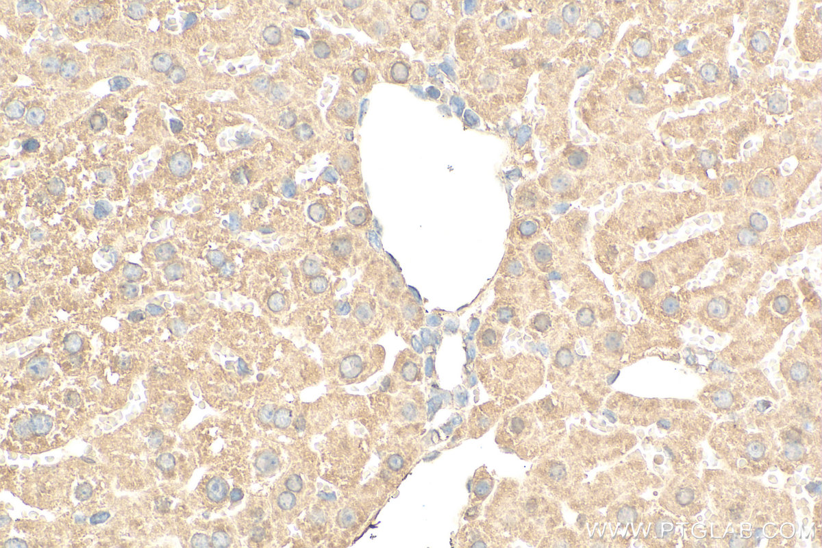
 at dilution of 1:200 (under 40x lens). Heat mediated antigen retrieval with Tris-EDTA buffer (pH 9.0).")
 fixed HepG2 cells using 66658-1-Ig (TOMM40 antibody) at dilution of 1:100 and CoraLite488-Conjugated AffiniPure Goat Anti-Mouse IgG(H+L).")
 fixed HeLa cells using 66658-1-Ig (TOMM40 antibody) at dilution of 1:100 and CoraLite488-Conjugated AffiniPure Goat Anti-Mouse IgG(H+L).")
 fixed HepG2 cells using 66658-1-Ig (TOMM40 antibody) at dilution of 1:100 and CoraLite488-Conjugated AffiniPure Goat Anti-Mouse IgG(H+L).")
Tested Applications
| Positive WB detected in | LNCaP cells, HeLa cells, HEK-293 cells, HepG2 cells, human brain tissue, pig brain tissue, Jurkat cells, HSC-T6 cells, PC-12 cells, NIH/3T3 cells, RAW264.7 cells, K-562 cells, rat brain tissue, mouse brain tissue |
| Positive IHC detected in | mouse liver tissue Note: suggested antigen retrieval with TE buffer pH 9.0; (*) Alternatively, antigen retrieval may be performed with citrate buffer pH 6.0 |
| Positive IF/ICC detected in | HepG2 cells, HeLa cells |
Recommended dilution
| Application | Dilution |
|---|---|
| Western Blot (WB) | WB : 1:5000-1:50000 |
| Immunohistochemistry (IHC) | IHC : 1:50-1:500 |
| Immunofluorescence (IF)/ICC | IF/ICC : 1:50-1:500 |
| It is recommended that this reagent should be titrated in each testing system to obtain optimal results. | |
| Sample-dependent, Check data in validation data gallery. | |
Published Applications
| WB | See 10 publications below |
| IHC | See 1 publications below |
| IF | See 2 publications below |
Product Information
66658-1-Ig targets TOMM40 in WB, IHC, IF/ICC, ELISA applications and shows reactivity with human, mouse, rat, pig samples.
| Tested Reactivity | human, mouse, rat, pig |
| Cited Reactivity | human, mouse, rat |
| Host / Isotype | Mouse / IgG1 |
| Class | Monoclonal |
| Type | Antibody |
| Immunogen |
CatNo: Ag13065 Product name: Recombinant human TOMM40 protein Source: e coli.-derived, PET28a Tag: 6*His Domain: 1-361 aa of BC017224 Sequence: MGNVLAASSPPAGPPPPPAPALVGLPPPPPSPPGFTLPPLGGSLGAGTSTSRSSERTPGAATASASGAAEDGACGCLPNPGTFEECHRKCKELFPIQMEGVKLTVNKGLSNHFQVNHTVALSTIGESNYHFGVTYVGTKQLSPTEAFPVLVGDMDNSGSLNAQVIHQLGPGLRSKMAIQTQQSKFVNWQVDGEYRGSDFTAAVTLGNPDVLVGSGILVAHYLQSITPCLALGGELVYHRRPGEEGTVMSLAGKYTLNNWLATVTLGQAGMHATYYHKASDQLQVGVEFEASTRMQDTSVSFGYQLDLPKANLLFKGSVDSNWIVGATLEKKLPPLPLTLALGAFLNHRKNKFQCGFGLTIG Predict reactive species |
| Full Name | translocase of outer mitochondrial membrane 40 homolog (yeast) |
| Calculated Molecular Weight | 38 kDa |
| Observed Molecular Weight | 38 kDa |
| GenBank Accession Number | BC017224 |
| Gene Symbol | TOMM40 |
| Gene ID (NCBI) | 10452 |
| RRID | AB_2882015 |
| Conjugate | Unconjugated |
| Form | Liquid |
| Purification Method | Protein G purification |
| UNIPROT ID | O96008 |
| Storage Buffer | PBS with 0.02% sodium azide and 50% glycerol, pH 7.3. |
| Storage Conditions | Store at -20°C. Stable for one year after shipment. Aliquoting is unnecessary for -20oC storage. 20ul sizes contain 0.1% BSA. |
Protocols
| Product Specific Protocols | |
|---|---|
| IF protocol for TOMM40 antibody 66658-1-Ig | Download protocol |
| IHC protocol for TOMM40 antibody 66658-1-Ig | Download protocol |
| WB protocol for TOMM40 antibody 66658-1-Ig | Download protocol |
| Standard Protocols | |
|---|---|
| Click here to view our Standard Protocols |
Publications
| Species | Application | Title |
|---|---|---|
Science Structural insight into the SAM-mediated assembly of the mitochondrial TOM core complex. | ||
Cell Death Differ Tufm lactylation regulates neuronal apoptosis by modulating mitophagy in traumatic brain injury | ||
Sci Signal A genome-wide screen uncovers multiple roles for mitochondrial nucleoside diphosphate kinase D in inflammasome activation. | ||
Apoptosis Lactate accelerates vascular calcification through NR4A1-regulated mitochondrial fission and BNIP3-related mitophagy. |