PC-12 cells were subjected to SDS PAGE followed by western blot with 67204-1-Ig (STMN2 antibody) at dilution of 1:5000 incubated at room temperature for 1.5 hours.
PC-12 cells were subjected to SDS PAGE followed by western blot with 67204-1-Ig (STMN2 antibody) at dilution of 1:5000 incubated at room temperature for 1.5 hours.
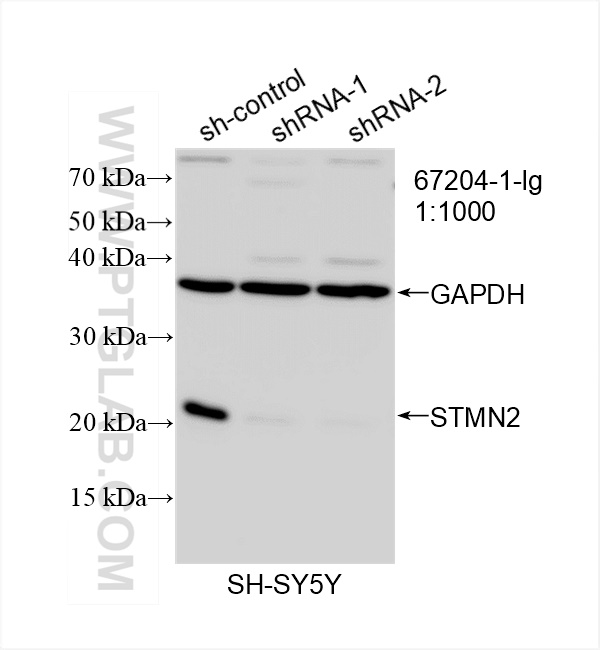
WB analysis of SH-SY5Y using 67204-1-Ig
WB result of STMN2 antibody (67204-1-Ig; 1:1000; incubated at room temperature for 1.5 hours) with sh-Control and sh-STMN2 transfected SH-SY5Y cells.
WB result of STMN2 antibody (67204-1-Ig; 1:1000; incubated at room temperature for 1.5 hours) with sh-Control and sh-STMN2 transfected SH-SY5Y cells.
WB analysis of mouse brain using 67204-1-Ig
mouse brain tissue were subjected to SDS PAGE followed by western blot with 67204-1-Ig (STMN2 antibody) at dilution of 1:5000 incubated at room temperature for 1.5 hours.
mouse brain tissue were subjected to SDS PAGE followed by western blot with 67204-1-Ig (STMN2 antibody) at dilution of 1:5000 incubated at room temperature for 1.5 hours.
WB analysis of fetal human brain using 67204-1-Ig
fetal human brain tissue were subjected to SDS PAGE followed by western blot with 67204-1-Ig (STMN2 antibody) at dilution of 1:5000 incubated at room temperature for 1.5 hours.
fetal human brain tissue were subjected to SDS PAGE followed by western blot with 67204-1-Ig (STMN2 antibody) at dilution of 1:5000 incubated at room temperature for 1.5 hours.
WB analysis of pig brain using 67204-1-Ig
pig brain tissue were subjected to SDS PAGE followed by western blot with 67204-1-Ig (STMN2 antibody) at dilution of 1:5000 incubated at room temperature for 1.5 hours.
pig brain tissue were subjected to SDS PAGE followed by western blot with 67204-1-Ig (STMN2 antibody) at dilution of 1:5000 incubated at room temperature for 1.5 hours.
WB analysis of rat brain using 67204-1-Ig
rat brain tissue were subjected to SDS PAGE followed by western blot with 67204-1-Ig (STMN2 antibody) at dilution of 1:5000 incubated at room temperature for 1.5 hours.
rat brain tissue were subjected to SDS PAGE followed by western blot with 67204-1-Ig (STMN2 antibody) at dilution of 1:5000 incubated at room temperature for 1.5 hours.
IHC staining of mouse brain using 67204-1-Ig
Immunohistochemical analysis of paraffin-embedded mouse brain tissue slide using 67204-1-Ig (STMN2 antibody) at dilution of 1:200 (under 10x lens). Heat mediated antigen retrieval with Tris-EDTA buffer (pH 9.0).
Immunohistochemical analysis of paraffin-embedded mouse brain tissue slide using 67204-1-Ig (STMN2 antibody) at dilution of 1:200 (under 40x lens). Heat mediated antigen retrieval with Tris-EDTA buffer (pH 9.0).
IF Staining of SH-SY5Y using 67204-1-Ig
Immunofluorescent analysis of (-20°C Ethanol) fixed SH-SY5Y cells using STMN2 antibody (67204-1-Ig, Clone: 1F6C4 ) at dilution of 1:1000 and CoraLite®488-Conjugated AffiniPure Goat Anti-Mouse IgG(H+L).
Immunofluorescent analysis of (-20°C Ethanol) fixed SH-SY5Y cells using STMN2 antibody (67204-1-Ig, Clone: 1F6C4 ) at dilution of 1:1000 and CoraLite®488-Conjugated AffiniPure Goat Anti-Mouse IgG(H+L).
The Proteintech guarantee covers Proteintech antibodies in any species and any application, including those not listed on the datasheet. If the antibody doesn’t perform, you can receive a hassle-free refund or credit note.
PC-12 cells, fetal human brain tissue, mouse brain tissue, pig brain tissue, rat brain tissue, SH-SY5Y cells
Positive IHC detected in
mouse brain tissue Note: suggested antigen retrieval with TE buffer pH 9.0; (*) Alternatively, antigen retrieval may be performed with citrate buffer pH 6.0
Positive IF/ICC detected in
SH-SY5Y cells
Recommended dilution
Application
Dilution
Western Blot (WB)
WB : 1:2000-1:10000
Immunohistochemistry (IHC)
IHC : 1:50-1:500
Immunofluorescence (IF)/ICC
IF/ICC : 1:500-1:2000
It is recommended that this reagent should be titrated in each testing system to obtain optimal results.
Sample-dependent, Check data in validation data gallery.
PBS with 0.02% sodium azide and 50% glycerol , pH 7.3
Storage Conditions
Store at -20°C. Stable for one year after shipment. Aliquoting is unnecessary for -20oC storage. 20ul sizes contain 0.1% BSA.
Background Information
STMN2 gene encodes a SCG10 (superior cervical ganglion-10) protein, a regulator of microtubule stability. When phosphorylated by MAPK8, SCG10 stabilizes microtubules and consequently controls neurite length in cortical neurons. SCG10 was early considered a neuron-specific growth-associated protein in Alzheimer's disease. In the developing brain, SCG10 negatively regulates the rate of exit from multipolar stage and retards radial migration from the ventricular zone.
PC-12 cells were subjected to SDS PAGE followed by western blot with 67204-1-Ig (STMN2 antibody) at dilution of 1:5000 incubated at room temperature for 1.5 hours.
WB analysis of SH-SY5Y using 67204-1-Ig
WB result of STMN2 antibody (67204-1-Ig; 1:1000; incubated at room temperature for 1.5 hours) with sh-Control and sh-STMN2 transfected SH-SY5Y cells.
WB analysis of mouse brain using 67204-1-Ig
mouse brain tissue were subjected to SDS PAGE followed by western blot with 67204-1-Ig (STMN2 antibody) at dilution of 1:5000 incubated at room temperature for 1.5 hours.
WB analysis of fetal human brain using 67204-1-Ig
fetal human brain tissue were subjected to SDS PAGE followed by western blot with 67204-1-Ig (STMN2 antibody) at dilution of 1:5000 incubated at room temperature for 1.5 hours.
WB analysis of pig brain using 67204-1-Ig
pig brain tissue were subjected to SDS PAGE followed by western blot with 67204-1-Ig (STMN2 antibody) at dilution of 1:5000 incubated at room temperature for 1.5 hours.
WB analysis of rat brain using 67204-1-Ig
rat brain tissue were subjected to SDS PAGE followed by western blot with 67204-1-Ig (STMN2 antibody) at dilution of 1:5000 incubated at room temperature for 1.5 hours.
IHC Figures
IHC staining of mouse brain using 67204-1-Ig
Immunohistochemical analysis of paraffin-embedded mouse brain tissue slide using 67204-1-Ig (STMN2 antibody) at dilution of 1:200 (under 10x lens). Heat mediated antigen retrieval with Tris-EDTA buffer (pH 9.0).
IHC staining of mouse brain using 67204-1-Ig
Immunohistochemical analysis of paraffin-embedded mouse brain tissue slide using 67204-1-Ig (STMN2 antibody) at dilution of 1:200 (under 40x lens). Heat mediated antigen retrieval with Tris-EDTA buffer (pH 9.0).
IF/ICC Figures
IF Staining of SH-SY5Y using 67204-1-Ig
Immunofluorescent analysis of (-20°C Ethanol) fixed SH-SY5Y cells using STMN2 antibody (67204-1-Ig, Clone: 1F6C4 ) at dilution of 1:1000 and CoraLite®488-Conjugated AffiniPure Goat Anti-Mouse IgG(H+L).
The species listed in Tested Reactivity are in-house verified and applicable species. For unlisted species, please refer to the homology analysis of the immunogen sequence and related species. For rabbit polyclonal antibodies, homology >70% is recommended. For mouse monoclonal antibodies and rabbit recombinant antibodies, homology >90% is recommended. Generally, the higher the homology, the greater the applicability. However, there will be certain differences in protein expression in different species, tissues or cells. Therefore, the homology analysis results are for reference only and do not serve as a guarantee.
At Proteintech, we pride ourselves on our antibody quality, customer service and transparency. As such, we are comparing our antibodies with other vendors, enabling easy identification and comparisons of key data to help you choose the suitable antibody for your needs.
We have selected the top cited antibodies from these vendors for you to compare.
 at dilution of 1:5000 incubated at room temperature for 1.5 hours.")
 with sh-Control and sh-STMN2 transfected SH-SY5Y cells.")
 at dilution of 1:5000 incubated at room temperature for 1.5 hours.")
 at dilution of 1:5000 incubated at room temperature for 1.5 hours.")
 at dilution of 1:5000 incubated at room temperature for 1.5 hours.")
 at dilution of 1:5000 incubated at room temperature for 1.5 hours.")
 at dilution of 1:200 (under 10x lens). Heat mediated antigen retrieval with Tris-EDTA buffer (pH 9.0).")
 at dilution of 1:200 (under 40x lens). Heat mediated antigen retrieval with Tris-EDTA buffer (pH 9.0).")
 fixed SH-SY5Y cells using STMN2 antibody (67204-1-Ig, Clone: 1F6C4 ) at dilution of 1:1000 and CoraLite®488-Conjugated AffiniPure Goat Anti-Mouse IgG(H+L).")