Various lysates were subjected to SDS PAGE followed by western blot with 82016-1-RR (STAT1 antibody) at dilution of 1:20000 incubated at room temperature for 1.5 hours.
Various lysates were subjected to SDS PAGE followed by western blot with 82016-1-RR (STAT1 antibody) at dilution of 1:20000 incubated at room temperature for 1.5 hours.
WB analysis of HEK-293 using 82016-1-RR
WB result of STAT1 antibody (82016-1-RR; 1:40000; incubated at room temperature for 1.5 hours) with sh-Control and sh-STAT1 transfected HEK-293 cells.
IP result of anti-STAT1 (IP:82016-1-RR, 4ug; Detection:82016-1-RR 1:10000) with PC-3 cells lysate 2000 ug.
IHC staining of human ovary tumor using 82016-1-RR
Immunohistochemical analysis of paraffin-embedded human ovary tumor tissue slide using 82016-1-RR (STAT1 antibody) at dilution of 1:400 (under 10x lens). Heat mediated antigen retrieval with Tris-EDTA buffer (pH 9.0).
Immunohistochemical analysis of paraffin-embedded human ovary tumor tissue slide using 82016-1-RR (STAT1 antibody) at dilution of 1:400 (under 10x lens). Heat mediated antigen retrieval with Tris-EDTA buffer (pH 9.0).
IHC staining of human ovary tumor using 82016-1-RR
Immunohistochemical analysis of paraffin-embedded human ovary tumor tissue slide using 82016-1-RR (STAT1 antibody) at dilution of 1:400 (under 40x lens). Heat mediated antigen retrieval with Tris-EDTA buffer (pH 9.0).
Immunohistochemical analysis of paraffin-embedded human ovary tumor tissue slide using 82016-1-RR (STAT1 antibody) at dilution of 1:400 (under 40x lens). Heat mediated antigen retrieval with Tris-EDTA buffer (pH 9.0).
IHC staining of mouse spleen using 82016-1-RR
Immunohistochemical analysis of paraffin-embedded mouse spleen tissue slide using 82016-1-RR (STAT1 antibody) at dilution of 1:1000 (under 10x lens). Heat mediated antigen retrieval with Tris-EDTA buffer (pH 9.0).
Immunohistochemical analysis of paraffin-embedded mouse colon tissue slide using 82016-1-RR (STAT1 antibody) at dilution of 1:4000 (under 10x lens). Heat mediated antigen retrieval with Tris-EDTA buffer (pH 9.0).
IHC staining of rat colon using 82016-1-RR
Immunohistochemical analysis of paraffin-embedded rat colon tissue slide using 82016-1-RR (STAT1 antibody) at dilution of 1:4000 (under 10x lens). Heat mediated antigen retrieval with Tris-EDTA buffer (pH 9.0).
Immunohistochemical analysis of paraffin-embedded rat colon tissue slide using 82016-1-RR (STAT1 antibody) at dilution of 1:4000 (under 10x lens). Heat mediated antigen retrieval with Tris-EDTA buffer (pH 9.0).
IHC staining of rat spleen using 82016-1-RR
Immunohistochemical analysis of paraffin-embedded rat spleen tissue slide using 82016-1-RR (STAT1 antibody) at dilution of 1:4000 (under 10x lens). Heat mediated antigen retrieval with Tris-EDTA buffer (pH 9.0).
Immunohistochemical analysis of paraffin-embedded rat spleen tissue slide using 82016-1-RR (STAT1 antibody) at dilution of 1:4000 (under 10x lens). Heat mediated antigen retrieval with Tris-EDTA buffer (pH 9.0).
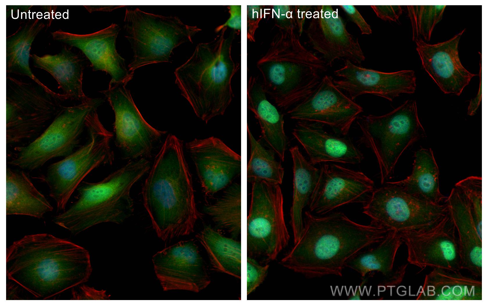
IF Staining of HeLa using 82016-1-RR
Immunofluorescent analysis of (4% PFA) fixed IFN alpha treated HeLa cells using STAT1 antibody (82016-1-RR, Clone: 2F16 ) at dilution of 1:1000 and CoraLite®488-Conjugated Goat Anti-Rabbit IgG(H+L) (SA00013-2), CL594-phalloidin (red).
Immunofluorescent analysis of (4% PFA) fixed IFN alpha treated HeLa cells using STAT1 antibody (82016-1-RR, Clone: 2F16 ) at dilution of 1:1000 and CoraLite®488-Conjugated Goat Anti-Rabbit IgG(H+L) (SA00013-2), CL594-phalloidin (red).
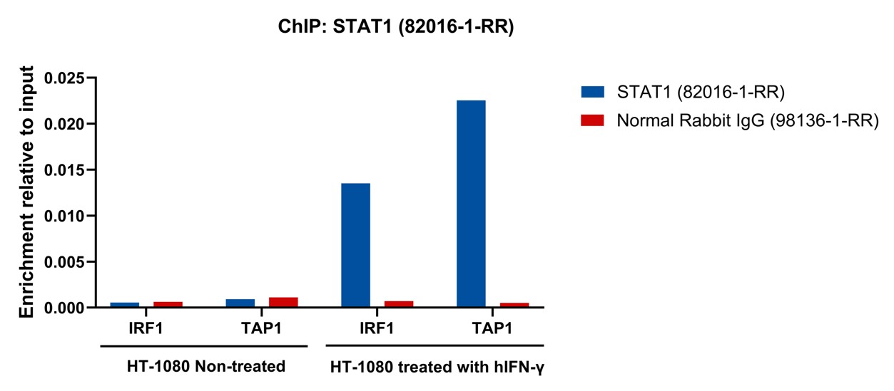
ChIP experiment of HT-1080 using 82016-1-RR
Chromatin was prepared from HT-1080 cells either non-treated or treated with hIFN-γ (50 ng/ml, 30min). Cells were fixed with formaldehyde for 10 minutes. The ChIP was performed with 15 µg of cross-linked chromatin, 5 µg of STAT1 (82016-1-RR) or 5 ug of Normal Rabbit IgG (98136-1-RR), and 20 µl of Protein A Magarose Beads. The immunoprecipitated DNA was quantified by real-time PCR.
Chromatin was prepared from HT-1080 cells either non-treated or treated with hIFN-γ (50 ng/ml, 30min). Cells were fixed with formaldehyde for 10 minutes. The ChIP was performed with 15 µg of cross-linked chromatin, 5 µg of STAT1 (82016-1-RR) or 5 ug of Normal Rabbit IgG (98136-1-RR), and 20 µl of Protein A Magarose Beads. The immunoprecipitated DNA was quantified by real-time PCR.
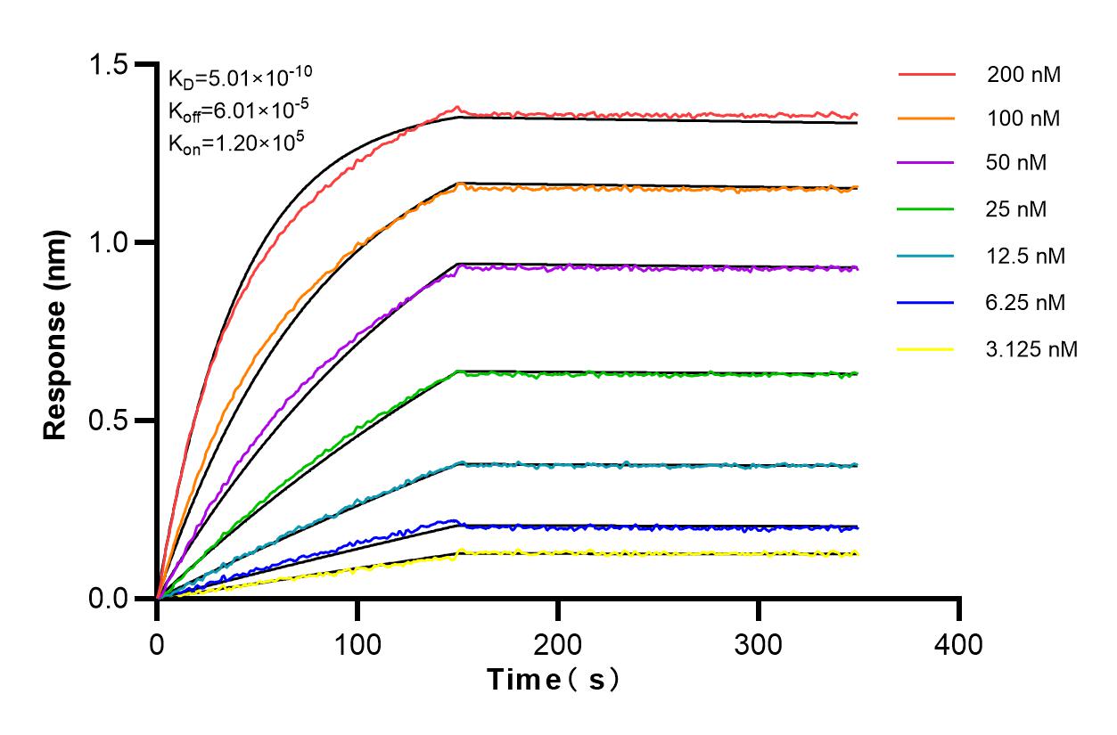
Affinity and Kinetic Characterization of 82016-1-RR
Biolayer interferometry (BLl) kinetic assays of 82016-1-RR against Human STAT1 were performed. The affinity constant is 0.501 nM.
The Proteintech guarantee covers Proteintech antibodies in any species and any application, including those not listed on the datasheet. If the antibody doesn’t perform, you can receive a hassle-free refund or credit note.
human ovary tumor tissue, mouse spleen tissue, mouse colon tissue, rat colon tissue, rat spleen tissue Note: suggested antigen retrieval with TE buffer pH 9.0; (*) Alternatively, antigen retrieval may be performed with citrate buffer pH 6.0
Positive IF/ICC detected in
IFN alpha treated HeLa cells
Positive ChIP-qPCR detected in
IFN-γ -HT-1080 (50 ng/ml, 30min) HT-1080 cells
Recommended dilution
Application
Dilution
Western Blot (WB)
WB : 1:5000-1:50000
Immunoprecipitation (IP)
IP : 0.5-4.0 ug for 1.0-3.0 mg of total protein lysate
Immunohistochemistry (IHC)
IHC : 1:200-1:800
Immunofluorescence (IF)/ICC
IF/ICC : 1:500-1:2000
CHIP-QPCR
CHIP-QPCR : 1:10-1:100
It is recommended that this reagent should be titrated in each testing system to obtain optimal results.
Sample-dependent, Check data in validation data gallery.
PBS with 0.02% sodium azide and 50% glycerol, pH 7.3.
Storage Conditions
Store at -20°C. Stable for one year after shipment. Aliquoting is unnecessary for -20oC storage. 20ul sizes contain 0.1% BSA.
Background Information
STAT1 (signal transducers and activators of transcription 1) is a member of the STAT protein family. In response to cytokines and growth factors, STAT family members are phosphorylated by the receptor associated kinases, and then form homo- or heterodimers that translocate to the cell nucleus where they act as transcription activators. Various ligands, including interferons- and - , EGF, PDGF and IL-6, can activate STAT1. STAT1 protein mediates the expression of a variety of genes, which is thought to be important for cell viability in response to different cell stimuli and pathogens.
Various lysates were subjected to SDS PAGE followed by western blot with 82016-1-RR (STAT1 antibody) at dilution of 1:20000 incubated at room temperature for 1.5 hours.
WB analysis of HEK-293 using 82016-1-RR
WB result of STAT1 antibody (82016-1-RR; 1:40000; incubated at room temperature for 1.5 hours) with sh-Control and sh-STAT1 transfected HEK-293 cells.
IHC Figures
IHC staining of human ovary tumor using 82016-1-RR
Immunohistochemical analysis of paraffin-embedded human ovary tumor tissue slide using 82016-1-RR (STAT1 antibody) at dilution of 1:400 (under 10x lens). Heat mediated antigen retrieval with Tris-EDTA buffer (pH 9.0).
IHC staining of human ovary tumor using 82016-1-RR
Immunohistochemical analysis of paraffin-embedded human ovary tumor tissue slide using 82016-1-RR (STAT1 antibody) at dilution of 1:400 (under 40x lens). Heat mediated antigen retrieval with Tris-EDTA buffer (pH 9.0).
IHC staining of mouse spleen using 82016-1-RR
Immunohistochemical analysis of paraffin-embedded mouse spleen tissue slide using 82016-1-RR (STAT1 antibody) at dilution of 1:1000 (under 10x lens). Heat mediated antigen retrieval with Tris-EDTA buffer (pH 9.0).
IHC staining of mouse colon using 82016-1-RR
Immunohistochemical analysis of paraffin-embedded mouse colon tissue slide using 82016-1-RR (STAT1 antibody) at dilution of 1:4000 (under 10x lens). Heat mediated antigen retrieval with Tris-EDTA buffer (pH 9.0).
IHC staining of rat colon using 82016-1-RR
Immunohistochemical analysis of paraffin-embedded rat colon tissue slide using 82016-1-RR (STAT1 antibody) at dilution of 1:4000 (under 10x lens). Heat mediated antigen retrieval with Tris-EDTA buffer (pH 9.0).
IHC staining of rat spleen using 82016-1-RR
Immunohistochemical analysis of paraffin-embedded rat spleen tissue slide using 82016-1-RR (STAT1 antibody) at dilution of 1:4000 (under 10x lens). Heat mediated antigen retrieval with Tris-EDTA buffer (pH 9.0).
IP Figures
IP experiment of PC-3 using 82016-1-RR
IP result of anti-STAT1 (IP:82016-1-RR, 4ug; Detection:82016-1-RR 1:10000) with PC-3 cells lysate 2000 ug.
IF/ICC Figures
IF Staining of HeLa using 82016-1-RR
Immunofluorescent analysis of (4% PFA) fixed IFN alpha treated HeLa cells using STAT1 antibody (82016-1-RR, Clone: 2F16 ) at dilution of 1:1000 and CoraLite®488-Conjugated Goat Anti-Rabbit IgG(H+L) (SA00013-2), CL594-phalloidin (red).
CHIP-QPCR Figures
ChIP experiment of HT-1080 using 82016-1-RR
Chromatin was prepared from HT-1080 cells either non-treated or treated with hIFN-γ (50 ng/ml, 30min). Cells were fixed with formaldehyde for 10 minutes. The ChIP was performed with 15 µg of cross-linked chromatin, 5 µg of STAT1 (82016-1-RR) or 5 ug of Normal Rabbit IgG (98136-1-RR), and 20 µl of Protein A Magarose Beads. The immunoprecipitated DNA was quantified by real-time PCR.
AFFINITY Figures
Affinity and Kinetic Characterization of 82016-1-RR
Biolayer interferometry (BLl) kinetic assays of 82016-1-RR against Human STAT1 were performed. The affinity constant is 0.501 nM.
The species listed in Tested Reactivity are in-house verified and applicable species. For unlisted species, please refer to the homology analysis of the immunogen sequence and related species. For rabbit polyclonal antibodies, homology >70% is recommended. For mouse monoclonal antibodies and rabbit recombinant antibodies, homology >90% is recommended. Generally, the higher the homology, the greater the applicability. However, there will be certain differences in protein expression in different species, tissues or cells. Therefore, the homology analysis results are for reference only and do not serve as a guarantee.
At Proteintech, we pride ourselves on our antibody quality, customer service and transparency. As such, we are comparing our antibodies with other vendors, enabling easy identification and comparisons of key data to help you choose the suitable antibody for your needs.
We have selected the top cited antibodies from these vendors for you to compare.
Proteintech
KD/KO VALIDATED
STAT1 Recombinant antibody
Catalog Number
82016-1-RR
Citations
1
Dilutions
WB : 1:5000-1:50000 IP : 0.5-4.0 ug for IP and 0.5-4.0 ug for 1.0-3.0 mg of total protein lysate for WB IHC : 1:200-1:800 IF/ICC : 1:500-1:2000 CHIP-QPCR : 1:10-1:100
Applications
WB, IHC, IF/ICC, IP, ELISA, ChIP-qPCR
Reactivity
human, mouse, rat
Product Guarantee
Covers any species including not listed on datasheet
Covers any applications including not listed on datasheet
 at dilution of 1:20000 incubated at room temperature for 1.5 hours.")
 with sh-Control and sh-STAT1 transfected HEK-293 cells.")
 with PC-3 cells lysate 2000 ug.")
 at dilution of 1:400 (under 10x lens). Heat mediated antigen retrieval with Tris-EDTA buffer (pH 9.0).")
 at dilution of 1:400 (under 40x lens). Heat mediated antigen retrieval with Tris-EDTA buffer (pH 9.0).")
 at dilution of 1:1000 (under 10x lens). Heat mediated antigen retrieval with Tris-EDTA buffer (pH 9.0).")
 at dilution of 1:4000 (under 10x lens). Heat mediated antigen retrieval with Tris-EDTA buffer (pH 9.0).")
 at dilution of 1:4000 (under 10x lens). Heat mediated antigen retrieval with Tris-EDTA buffer (pH 9.0).")
 at dilution of 1:4000 (under 10x lens). Heat mediated antigen retrieval with Tris-EDTA buffer (pH 9.0).")
 fixed IFN alpha treated HeLa cells using STAT1 antibody (82016-1-RR, Clone: 2F16 ) at dilution of 1:1000 and CoraLite®488-Conjugated Goat Anti-Rabbit IgG(H+L) (SA00013-2), CL594-phalloidin (red).")
. Cells were fixed with formaldehyde for 10 minutes. The ChIP was performed with 15 µg of cross-linked chromatin, 5 µg of STAT1 (82016-1-RR) or 5 ug of Normal Rabbit IgG (98136-1-RR), and 20 µl of Protein A Magarose Beads. The immunoprecipitated DNA was quantified by real-time PCR.")
 kinetic assays of 82016-1-RR against Human STAT1 were performed. The affinity constant is 0.501 nM.")