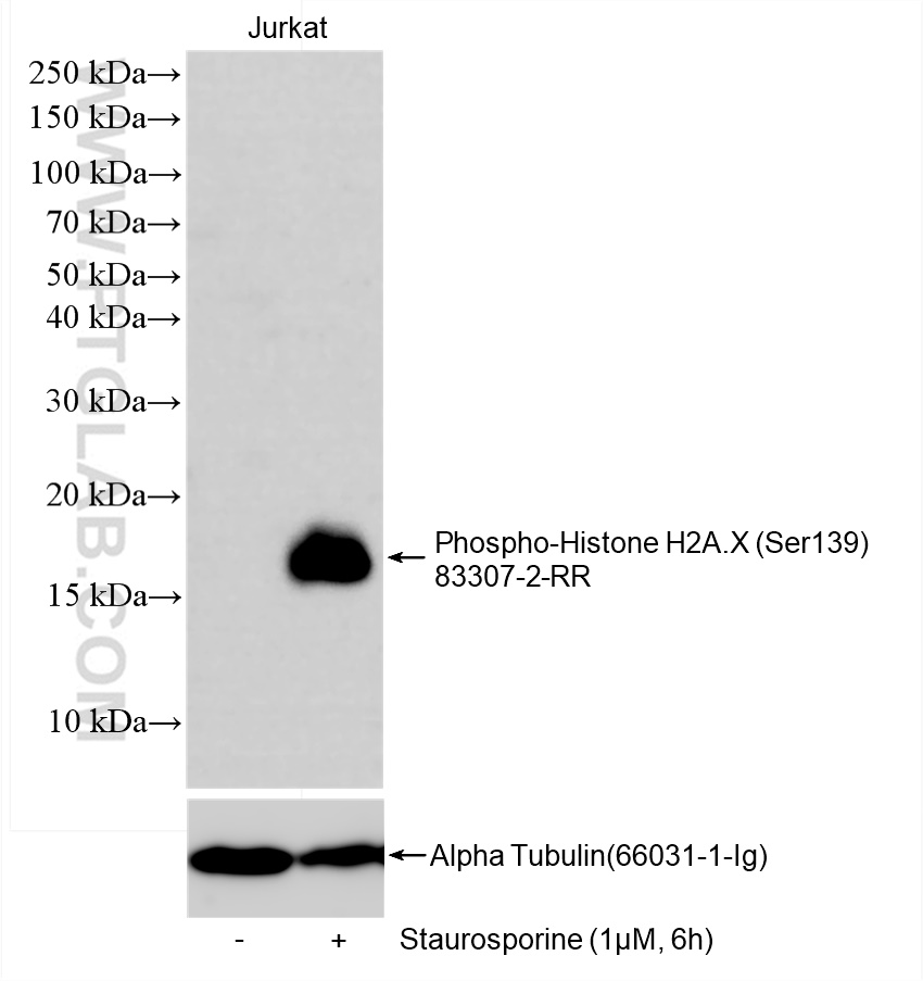
antibody) at dilution of 1:10000 incubated at room temperature for 1.5 hours. The membrane was stripped and re-blotted with Alpha Tubulin (66031-1-Ig) antibody as loading control.")
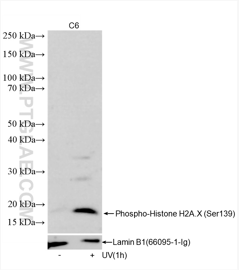
 antibody) at dilution of 1:2000 incubated at room temperature for 1.5 hours.")
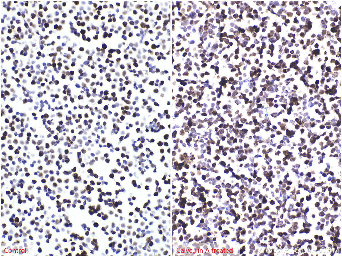
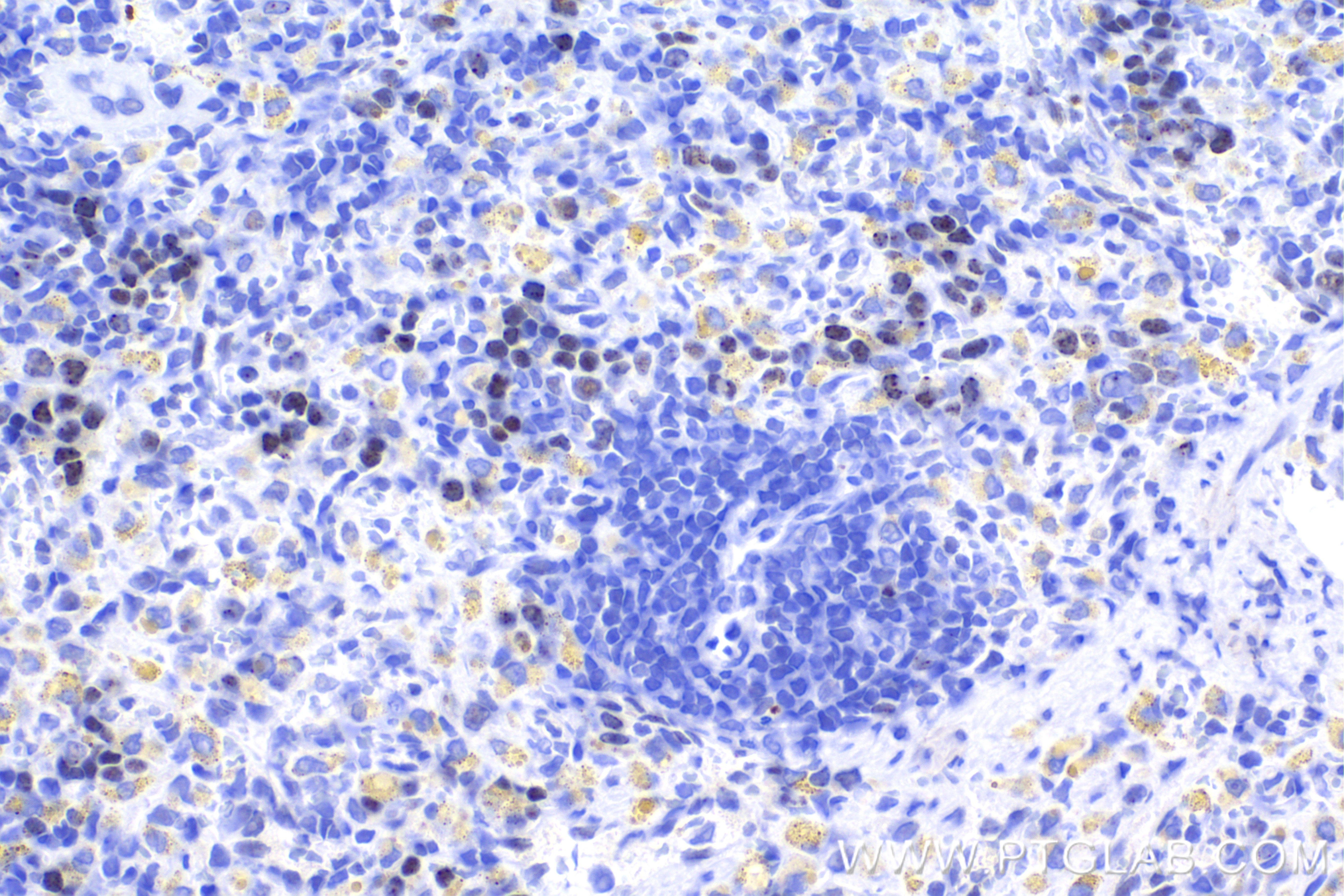
 antibody) at dilution of 1:4000 (under 40x lens). Heat mediated antigen retrieval with Tris-EDTA buffer (pH 9.0).")
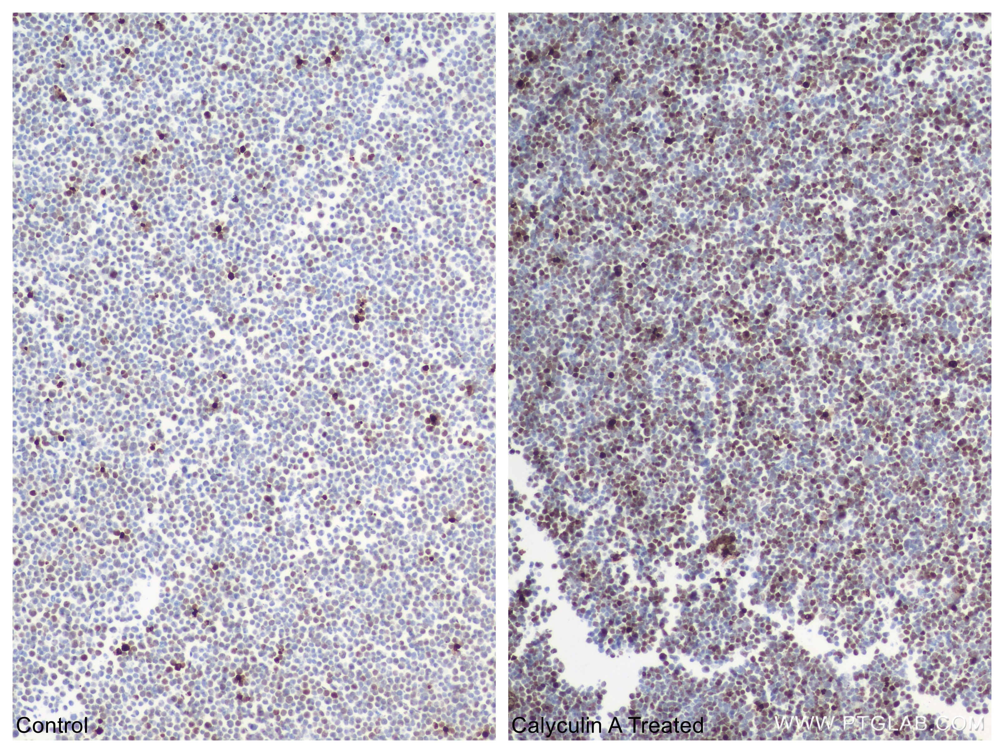
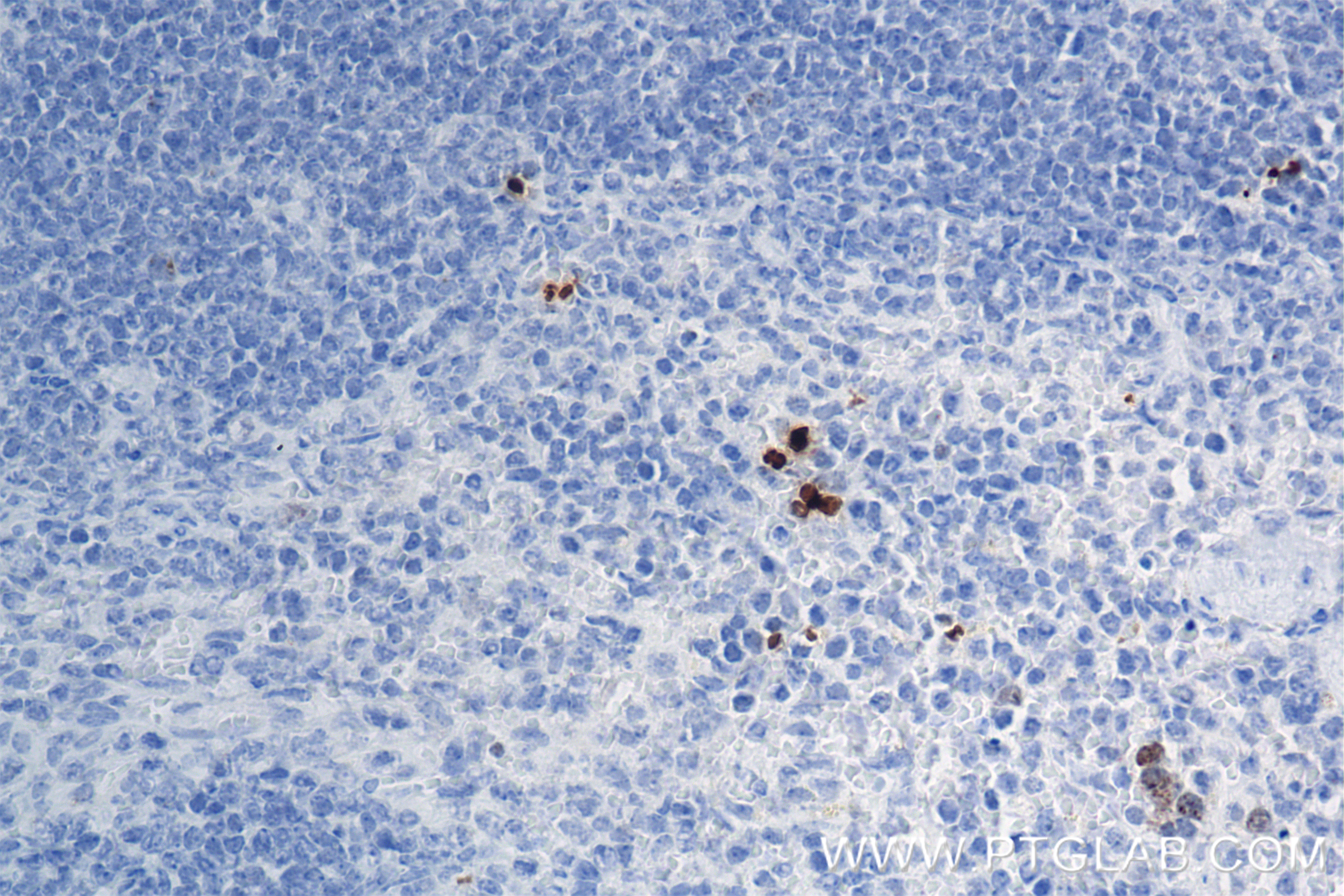
 antibody) at dilution of 1:4000 (under 10x lens). Heat mediated antigen retrieval with Tris-EDTA buffer (pH 9.0).")
 antibody) at dilution of 1:500 (under 40x lens). Heat mediated antigen retrieval with Tris-EDTA buffer (pH 9.0).")
 antibody) at dilution of 1:500 (under 40x lens). Heat mediated antigen retrieval with Tris-EDTA buffer (pH 9.0).")
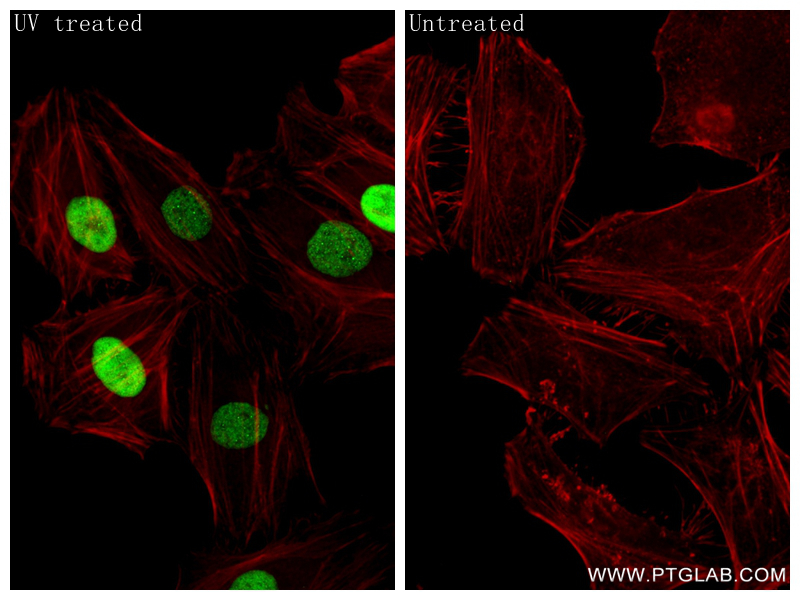
 fixed UV treated HeLa cells using Phospho-Histone H2A.X (Ser139) antibody (83307-2-RR, Clone: 5N19 ) at dilution of 1:400 and CoraLite®488-Conjugated Goat Anti-Rabbit IgG(H+L) (SA00013-2), CL594-Phalloidin (red).")
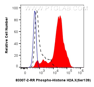
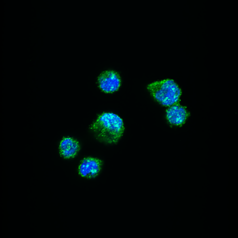
 or treated with Staurosporine which intracellularly stained with 0.06 ug Phospho-Histone H2A.X (Ser139) Recombinant antibody (83307-2-RR, Clone:5N19) and CoraLite®488-Conjugated Goat Anti-Rabbit IgG(H+L) (SA00013-2)(red), or 0.06 ug Rabbit IgG Isotype Control Recombinant Antibody (98136-1-RR, Clone: 240953C9) (blue). Cells were fixed with 4% PFA and permeabilized with 90% MeOH.")
Tested Applications
| Positive WB detected in | Staurosporine treated Jurkat cells, C6 cells |
| Positive IHC detected in | Jurkat cells, mouse spleen tissue, rat spleen tissue Note: suggested antigen retrieval with TE buffer pH 9.0; (*) Alternatively, antigen retrieval may be performed with citrate buffer pH 6.0 |
| Positive IF/ICC detected in | UV treated HeLa cells |
| Positive FC (Intra) detected in | Staurosporine treated Jurkat cells |
Recommended dilution
| Application | Dilution |
|---|---|
| Western Blot (WB) | WB : 1:5000-1:50000 |
| Immunohistochemistry (IHC) | IHC : 1:2000-1:8000 |
| Immunofluorescence (IF)/ICC | IF/ICC : 1:200-1:800 |
| Flow Cytometry (FC) (INTRA) | FC (INTRA) : 0.06 ug per 10^6 cells in a 100 µl suspension |
| It is recommended that this reagent should be titrated in each testing system to obtain optimal results. | |
| Sample-dependent, Check data in validation data gallery. | |
Published Applications
| WB | See 1 publications below |
| IF | See 1 publications below |
Product Information
83307-2-RR targets Phospho-Histone H2A.X (Ser139) in WB, IHC, IF/ICC, FC (Intra), ELISA applications and shows reactivity with human, mouse, rat samples.
| Tested Reactivity | human, mouse, rat |
| Cited Reactivity | human |
| Host / Isotype | Rabbit / IgG |
| Class | Recombinant |
| Type | Antibody |
| Immunogen |
Peptide Predict reactive species |
| Full Name | H2A histone family, member X |
| Calculated Molecular Weight | 15 kDa |
| Observed Molecular Weight | 15 kDa |
| GenBank Accession Number | BC013416 |
| Gene Symbol | Histone H2A.X |
| Gene ID (NCBI) | 3014 |
| RRID | AB_3670974 |
| Conjugate | Unconjugated |
| Form | Liquid |
| Purification Method | Protein A purfication |
| UNIPROT ID | P16104 |
| Storage Buffer | PBS with 0.02% sodium azide and 50% glycerol, pH 7.3. |
| Storage Conditions | Store at -20°C. Stable for one year after shipment. Aliquoting is unnecessary for -20oC storage. 20ul sizes contain 0.1% BSA. |
Background Information
The histone variant H2AX is a major component of the DNA damage response (DDR), especially functioning in amplifying DNA damage signals. In response to DNA double-strand breaks (DSBs), H2AX is instantaneously phosphorylated at Ser139 (a form called cH2AX) by the kinases ATM and ATR. The phosphorylation of H2AX at Ser139, resulting in the formation of gamma-H2AX puncta in the nuclei, is an early event in the cellular response to DNA damage. Therefore, phospho-Histone H2A. X (Ser139) is also known as γH2AX. The phosphorylation site of H2AX, Ser139, has also been described as Ser140 in other literature, and they recognize the same amino acid site. (PMID: 22908299, PMID: 30106130, PMID:22941631)
Protocols
| Product Specific Protocols | |
|---|---|
| IF protocol for Phospho-Histone H2A.X (Ser139) antibody 83307-2-RR | Download protocol |
| IHC protocol for Phospho-Histone H2A.X (Ser139) antibody 83307-2-RR | Download protocol |
| WB protocol for Phospho-Histone H2A.X (Ser139) antibody 83307-2-RR | Download protocol |
| Standard Protocols | |
|---|---|
| Click here to view our Standard Protocols |
Publications
| Species | Application | Title |
|---|---|---|
Transl Oncol PiR-hsa-23533 promotes malignancy in head and neck squamous cell carcinoma via USP7 | ||
Cancer Biol Ther SLX1 silencing overcomes Olaparib resistance in metastatic castration-resistant prostate cancer by disrupting SLX4-mediated DNA repair complexes | ||
J Microbiol Biotechnol BlingLife®-Marigold Extract Alleviates Blue Light-Induced Retinal Mitochondria Oxidative Stress and Senescence by Activating NRF2/HO-1 Signaling |
Reviews
The reviews below have been submitted by verified Proteintech customers who received an incentive for providing their feedback.
FH Eglantine (Verified Customer) (02-10-2026) | The H2AX antibody demonstrates low specificity with pronounced non-nuclear staining. There is also a lot of signal in control conditions (no irradiation, no IONPs) where there should not be any DNA damage.
 |