Various lysates were subjected to SDS PAGE followed by western blot with 68115-1-Ig (PACSIN1 antibody) at dilution of 1:2000 incubated at room temperature for 1.5 hours. The membrane was stripped and reblotted with HRP-conjugated GAPDH Monoclonal antibody (HRP-60004) as loading control.
Various lysates were subjected to SDS PAGE followed by western blot with 68115-1-Ig (PACSIN1 antibody) at dilution of 1:2000 incubated at room temperature for 1.5 hours. The membrane was stripped and reblotted with HRP-conjugated GAPDH Monoclonal antibody (HRP-60004) as loading control.
IHC staining of mouse cerebellum using 68115-1-Ig
Immunohistochemical analysis of paraffin-embedded mouse cerebellum tissue slide using 68115-1-Ig (PACSIN1 antibody) at dilution of 1:4000 (under 10x lens). Heat mediated antigen retrieval with Tris-EDTA buffer (pH 9.0).
Immunofluorescent analysis of (4% PFA) fixed paraffin-embedded mouse brain tissue using PACSIN1 antibody (68115-1-Ig, Clone: 1B7B4 ) at dilution of 1:400 and CoraLite®488-Conjugated Goat Anti-Mouse IgG(H+L). Heat mediated antigen retrieval with Tris-EDTA buffer (pH 9.0).
IF Staining of SH-SY5Y using 68115-1-Ig
Immunofluorescent analysis of (-20°C Ethanol) fixed SH-SY5Y cells using PACSIN1 antibody (68115-1-Ig, Clone: 1B7B4 ) at dilution of 1:800 and CoraLite®488-Conjugated Goat Anti-Mouse IgG(H+L).
Immunofluorescent analysis of (-20°C Ethanol) fixed SH-SY5Y cells using PACSIN1 antibody (68115-1-Ig, Clone: 1B7B4 ) at dilution of 1:800 and CoraLite®488-Conjugated Goat Anti-Mouse IgG(H+L).
FC experiment of SH-SY5Y using 68115-1-Ig
1X10^6 SH-SY5Y cells were intracellularly stained with 0.4 ug Anti-Human PACSIN1 (68115-1-Ig, Clone:1B7B4) and CoraLite®488-Conjugated Goat Anti-Mouse IgG(H+L) at dilution 1:1000 (red), or 0.4 ug Mouse IgG1 Isotype Control (MOPC-21) (65124-1-Ig, Clone: MOPC-21) (blue). Cells were fixed with 4% PFA and permeabilized with Flow Cytometry Perm Buffer (PF00011-C).
1X10^6 SH-SY5Y cells were intracellularly stained with 0.4 ug Anti-Human PACSIN1 (68115-1-Ig, Clone:1B7B4) and CoraLite®488-Conjugated Goat Anti-Mouse IgG(H+L) at dilution 1:1000 (red), or 0.4 ug Mouse IgG1 Isotype Control (MOPC-21) (65124-1-Ig, Clone: MOPC-21) (blue). Cells were fixed with 4% PFA and permeabilized with Flow Cytometry Perm Buffer (PF00011-C).
The Proteintech guarantee covers Proteintech antibodies in any species and any application, including those not listed on the datasheet. If the antibody doesn’t perform, you can receive a hassle-free refund or credit note.
mouse cerebellum tissue, mouse brain tissue Note: suggested antigen retrieval with TE buffer pH 9.0; (*) Alternatively, antigen retrieval may be performed with citrate buffer pH 6.0
Positive IF-P detected in
mouse brain tissue
Positive IF/ICC detected in
SH-SY5Y cells
Positive FC (Intra) detected in
SH-SY5Y cells
Recommended dilution
Application
Dilution
Western Blot (WB)
WB : 1:1000-1:4000
Immunohistochemistry (IHC)
IHC : 1:2000-1:8000
Immunofluorescence (IF)-P
IF-P : 1:200-1:800
Immunofluorescence (IF)/ICC
IF/ICC : 1:400-1:1600
Flow Cytometry (FC) (INTRA)
FC (INTRA) : 0.40 ug per 10^6 cells in a 100 µl suspension
It is recommended that this reagent should be titrated in each testing system to obtain optimal results.
Sample-dependent, Check data in validation data gallery.
Product Information
68115-1-Ig targets PACSIN1 in WB, IHC, IF/ICC, IF-P, FC (Intra), ELISA applications and shows reactivity with human, mouse, rat, pig, rabbit, chicken samples.
PBS with 0.02% sodium azide and 50% glycerol , pH 7.3
Storage Conditions
Store at -20°C. Stable for one year after shipment. Aliquoting is unnecessary for -20oC storage. 20ul sizes contain 0.1% BSA.
Background Information
PACSIN1 (also known as syndapin-1) is a member of the protein kinase C and casein kinase substrate in neurons (PACSIN) family. In mammals, the PACSIN family is comprised of three members, PACSIN1, PACSIN2, and PACSIN3 (PMID: 34990060). PACSIN1 is expressed mainly in neurons, whereas PACSIN2 is ubiquitously expressed in all tissues, and PACSIN3 is expressed mainly in skeletal muscle and the heart (PMID: 23668323). All of these three members contain an N-terminal F-BAR domain and a C-terminal SH3 domain. PACSIN1 plays a role in endocytosis and endosomal recycling. Meanwhile, it has a role in actin remodeling and microtubule nucleation and also plays a particular role in membrane shaping and reconstruction (PMID: 23035120; 34422904). PACSIN1 is involved in neuromorphogenesis and the regulation of the nervous system, and the inappropriate expression of PACSIN1 has been associated with some neurological diseases, including schizophrenia, Alzheimer's disease, and Huntington's disease (PMID: 34990060).
Various lysates were subjected to SDS PAGE followed by western blot with 68115-1-Ig (PACSIN1 antibody) at dilution of 1:2000 incubated at room temperature for 1.5 hours. The membrane was stripped and reblotted with HRP-conjugated GAPDH Monoclonal antibody (HRP-60004) as loading control.
IHC Figures
IHC staining of mouse cerebellum using 68115-1-Ig
Immunohistochemical analysis of paraffin-embedded mouse cerebellum tissue slide using 68115-1-Ig (PACSIN1 antibody) at dilution of 1:4000 (under 10x lens). Heat mediated antigen retrieval with Tris-EDTA buffer (pH 9.0).
IHC staining of mouse cerebellum using 68115-1-Ig
Immunohistochemical analysis of paraffin-embedded mouse cerebellum tissue slide using 68115-1-Ig (PACSIN1 antibody) at dilution of 1:4000 (under 40x lens). Heat mediated antigen retrieval with Tris-EDTA buffer (pH 9.0).
IHC staining of mouse brain using 68115-1-Ig
Immunohistochemical analysis of paraffin-embedded mouse brain tissue slide using 68115-1-Ig (PACSIN1 antibody) at dilution of 1:1000 (under 10x lens). Heat mediated antigen retrieval with Tris-EDTA buffer (pH 9.0).
IHC staining of mouse brain using 68115-1-Ig
Immunohistochemical analysis of paraffin-embedded mouse brain tissue slide using 68115-1-Ig (PACSIN1 antibody) at dilution of 1:1000 (under 40x lens). Heat mediated antigen retrieval with Tris-EDTA buffer (pH 9.0).
IF-P Figures
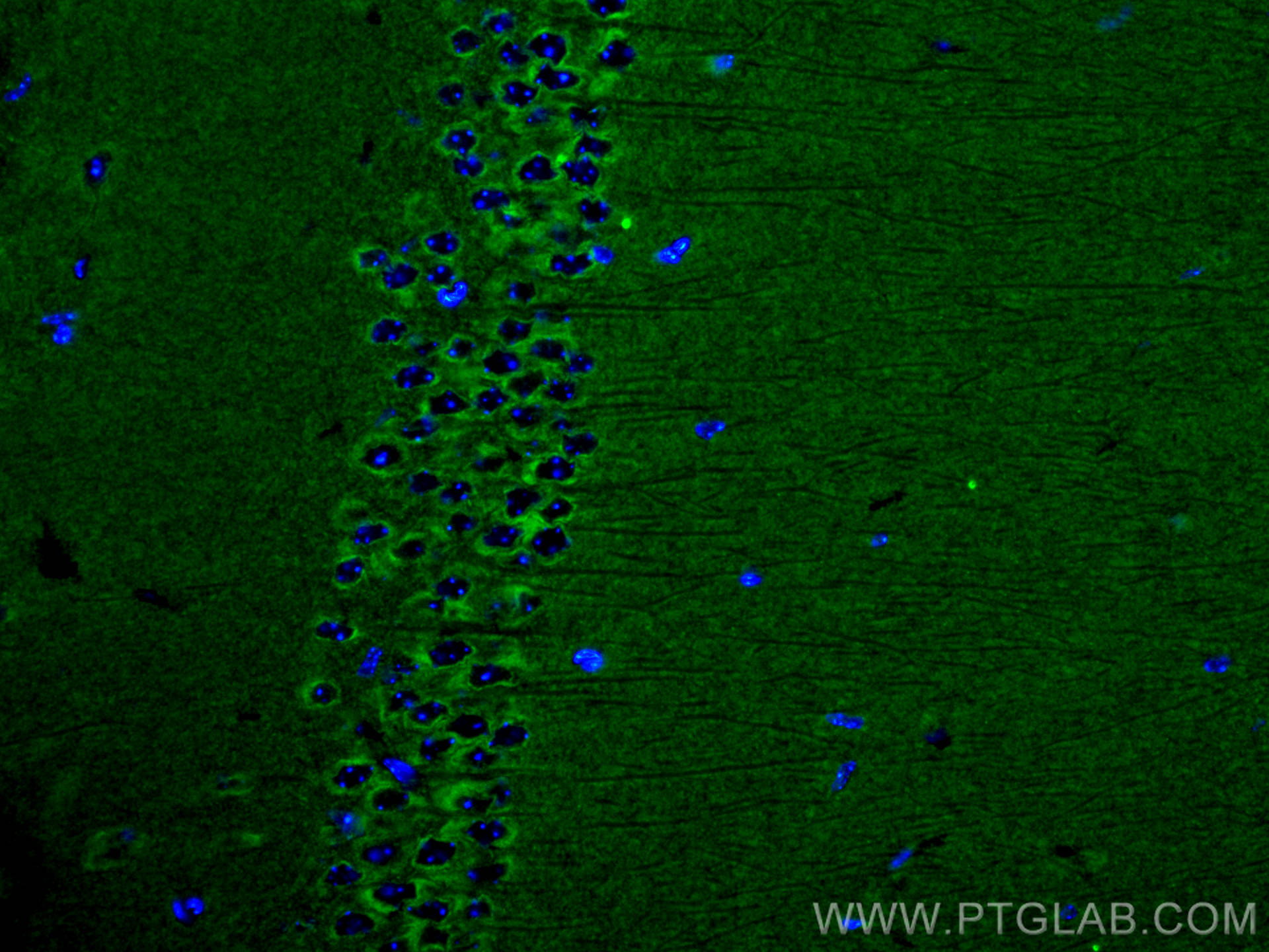
IF Staining of mouse brain using 68115-1-Ig
Immunofluorescent analysis of (4% PFA) fixed paraffin-embedded mouse brain tissue using PACSIN1 antibody (68115-1-Ig, Clone: 1B7B4 ) at dilution of 1:400 and CoraLite®488-Conjugated Goat Anti-Mouse IgG(H+L). Heat mediated antigen retrieval with Tris-EDTA buffer (pH 9.0).
IF/ICC Figures
IF Staining of SH-SY5Y using 68115-1-Ig
Immunofluorescent analysis of (-20°C Ethanol) fixed SH-SY5Y cells using PACSIN1 antibody (68115-1-Ig, Clone: 1B7B4 ) at dilution of 1:800 and CoraLite®488-Conjugated Goat Anti-Mouse IgG(H+L).
FC (INTRA) Figures
FC experiment of SH-SY5Y using 68115-1-Ig
1X10^6 SH-SY5Y cells were intracellularly stained with 0.4 ug Anti-Human PACSIN1 (68115-1-Ig, Clone:1B7B4) and CoraLite®488-Conjugated Goat Anti-Mouse IgG(H+L) at dilution 1:1000 (red), or 0.4 ug Mouse IgG1 Isotype Control (MOPC-21) (65124-1-Ig, Clone: MOPC-21) (blue). Cells were fixed with 4% PFA and permeabilized with Flow Cytometry Perm Buffer (PF00011-C).
The species listed in Tested Reactivity are in-house verified and applicable species. For unlisted species, please refer to the homology analysis of the immunogen sequence and related species. For rabbit polyclonal antibodies, homology >70% is recommended. For mouse monoclonal antibodies and rabbit recombinant antibodies, homology >90% is recommended. Generally, the higher the homology, the greater the applicability. However, there will be certain differences in protein expression in different species, tissues or cells. Therefore, the homology analysis results are for reference only and do not serve as a guarantee.
At Proteintech, we pride ourselves on our antibody quality, customer service and transparency. As such, we are comparing our antibodies with other vendors, enabling easy identification and comparisons of key data to help you choose the suitable antibody for your needs.
We have selected the top cited antibodies from these vendors for you to compare.
Proteintech
PACSIN1 Monoclonal antibody
Catalog Number
68115-1-Ig
Citations
-
Dilutions
WB : 1:1000-1:4000 IHC : 1:2000-1:8000 IF-P : 1:200-1:800 IF/ICC : 1:400-1:1600 FC (INTRA) : 0.40 ug per 10^6 cells in a 100 µl suspension
Applications
WB, IHC, IF/ICC, IF-P, FC (Intra), ELISA
Reactivity
human, mouse, rat, pig, rabbit, chicken
Product Guarantee
Covers any species including not listed on datasheet
Covers any applications including not listed on datasheet
 at dilution of 1:2000 incubated at room temperature for 1.5 hours. The membrane was stripped and reblotted with HRP-conjugated GAPDH Monoclonal antibody (HRP-60004) as loading control.")
 at dilution of 1:4000 (under 10x lens). Heat mediated antigen retrieval with Tris-EDTA buffer (pH 9.0).")
 at dilution of 1:4000 (under 40x lens). Heat mediated antigen retrieval with Tris-EDTA buffer (pH 9.0).")
 at dilution of 1:1000 (under 10x lens). Heat mediated antigen retrieval with Tris-EDTA buffer (pH 9.0).")
 at dilution of 1:1000 (under 40x lens). Heat mediated antigen retrieval with Tris-EDTA buffer (pH 9.0).")
 fixed paraffin-embedded mouse brain tissue using PACSIN1 antibody (68115-1-Ig, Clone: 1B7B4 ) at dilution of 1:400 and CoraLite®488-Conjugated Goat Anti-Mouse IgG(H+L). Heat mediated antigen retrieval with Tris-EDTA buffer (pH 9.0).")
 fixed SH-SY5Y cells using PACSIN1 antibody (68115-1-Ig, Clone: 1B7B4 ) at dilution of 1:800 and CoraLite®488-Conjugated Goat Anti-Mouse IgG(H+L).")
 and CoraLite®488-Conjugated Goat Anti-Mouse IgG(H+L) at dilution 1:1000 (red), or 0.4 ug Mouse IgG1 Isotype Control (MOPC-21) (65124-1-Ig, Clone: MOPC-21) (blue). Cells were fixed with 4% PFA and permeabilized with Flow Cytometry Perm Buffer (PF00011-C).")