Filter:
 at dilution of 1:10000 incubated at room temperature for 1.5 hours.")
 with sh-Control and sh-OXCT1 transfected HeLa cells.")
 at dilution of 1:10000 incubated at room temperature for 1.5 hours.")
 at dilution of 1:10000 incubated at room temperature for 1.5 hours.")
 at dilution of 1:10000 incubated at room temperature for 1.5 hours.")
 at dilution of 1:10000 incubated at room temperature for 1.5 hours.")
 at dilution of 1:10000 incubated at room temperature for 1.5 hours.")
 at dilution of 1:10000 incubated at room temperature for 1.5 hours.")
 at dilution of 1:1000 (under 40x lens). Heat mediated antigen retrieval with Tris-EDTA buffer (pH 9.0).")
 fixed MCF-7 cells using OXCT1 antibody (67836-1-Ig, Clone: 1G1B9 ) at dilution of 1:400 and CoraLite®488-Conjugated AffiniPure Goat Anti-Mouse IgG(H+L). CL594-Phalloidin (red).")
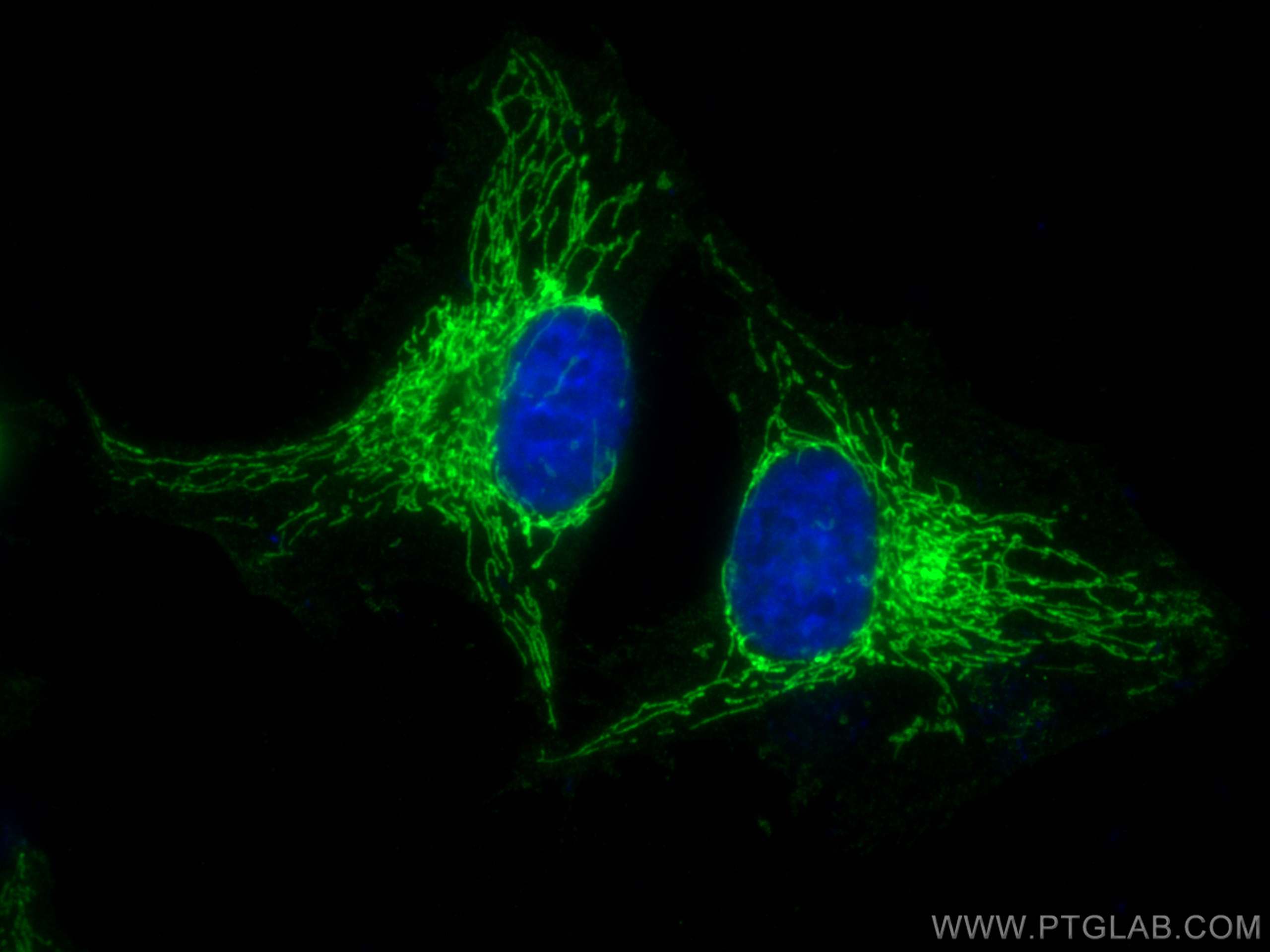
 fixed HeLa cells using OXCT1 antibody (67836-1-Ig, Clone: 1G1B9 ) at dilution of 1:500 and CoraLite®488-Conjugated AffiniPure Goat Anti-Mouse IgG(H+L), CL594-Phalloidin (red).")
 fixed HeLa cells using OXCT1 antibody (67836-1-Ig, Clone: 1G1B9 ) at dilution of 1:500 and CoraLite®488-Conjugated AffiniPure Goat Anti-Mouse IgG(H+L), CL594-Phalloidin (red).")
 fixed HeLa cells using OXCT1 antibody (67836-1-Ig, Clone: 1G1B9 ) at dilution of 1:800 and CoraLite®488-Conjugated AffiniPure Goat Anti-Mouse IgG(H+L).")
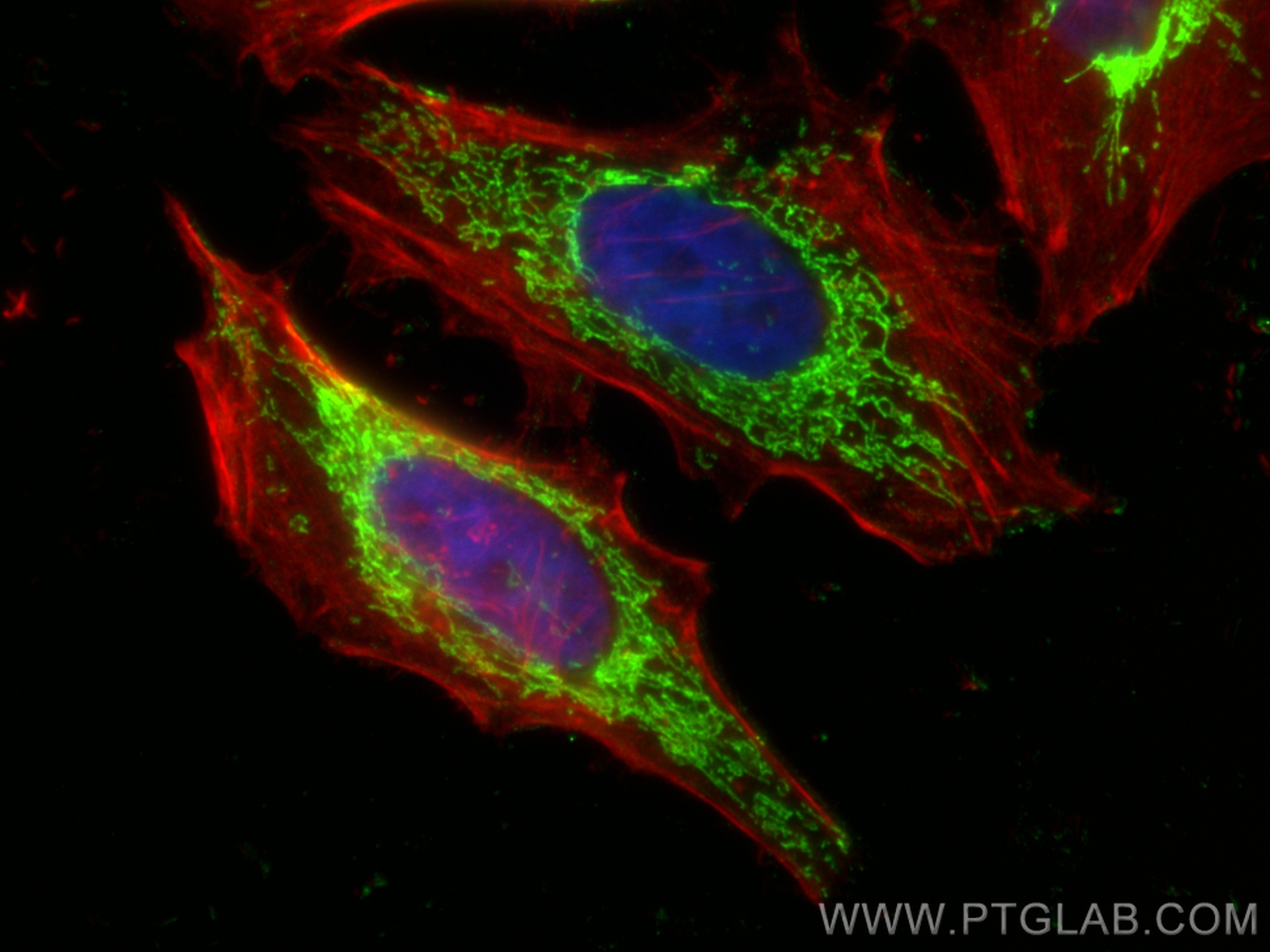
 fixed HeLa cells using OXCT1 antibody (67836-1-Ig, Clone: 1G1B9 ) at dilution of 1:800 and CoraLite®488-Conjugated Goat Anti-Mouse IgG(H+L) (SA00013-1), CL594-phalloidin (red).")
 and CoraLite®488-Conjugated AffiniPure Goat Anti-Mouse IgG(H+L) at dilution 1:1000 (red), or 0.4 ug Mouse IgG1 Isotype Control (MOPC-21) (65124-1-Ig, Clone: MOPC-21) (blue). Cells were fixed with 4% PFA and permeabilized with Flow Cytometry Perm Buffer (PF00011-C).")
Tested Applications
| Positive WB detected in | rat heart tissue, HeLa cells, Jurkat cells, pig brain tissue, rat brain tissue, mouse brain tissue, rabbit brain tissue |
| Positive IHC detected in | rat brain tissue Note: suggested antigen retrieval with TE buffer pH 9.0; (*) Alternatively, antigen retrieval may be performed with citrate buffer pH 6.0 |
| Positive IF/ICC detected in | MCF-7 cells, HeLa cells |
| Positive FC (Intra) detected in | HeLa cells |
Recommended dilution
| Application | Dilution |
|---|---|
| Western Blot (WB) | WB : 1:5000-1:50000 |
| Immunohistochemistry (IHC) | IHC : 1:500-1:2000 |
| Immunofluorescence (IF)/ICC | IF/ICC : 1:200-1:800 |
| Flow Cytometry (FC) (INTRA) | FC (INTRA) : 0.40 ug per 10^6 cells in a 100 µl suspension |
| It is recommended that this reagent should be titrated in each testing system to obtain optimal results. | |
| Sample-dependent, Check data in validation data gallery. | |
Published Applications
| WB | See 1 publications below |
Product Information
67836-1-Ig targets OXCT1 in WB, IHC, IF/ICC, FC (Intra), ELISA applications and shows reactivity with human, mouse, rat, pig, rabbit samples.
| Tested Reactivity | human, mouse, rat, pig, rabbit |
| Cited Reactivity | human |
| Host / Isotype | Mouse / IgG1 |
| Class | Monoclonal |
| Type | Antibody |
| Immunogen | OXCT1 fusion protein Ag24792 Predict reactive species |
| Full Name | 3-oxoacid CoA transferase 1 |
| Calculated Molecular Weight | 520 aa, 56 kDa |
| Observed Molecular Weight | 52-56 kDa |
| GenBank Accession Number | BC009001 |
| Gene Symbol | OXCT1 |
| Gene ID (NCBI) | 5019 |
| RRID | AB_2918598 |
| Conjugate | Unconjugated |
| Form | Liquid |
| Purification Method | Protein G purification |
| UNIPROT ID | P55809 |
| Storage Buffer | PBS with 0.02% sodium azide and 50% glycerol, pH 7.3. |
| Storage Conditions | Store at -20°C. Stable for one year after shipment. Aliquoting is unnecessary for -20oC storage. 20ul sizes contain 0.1% BSA. |
Background Information
OXCT1 (also know as SCOT), encoded by nuclear gene, is a mitochondrial CoA transferase required for ketone body degradation. It catalyzes the transfer of CoA from succinyl-CoA to acetoacetate, generating acetoacetyl-CoA. OXCT1 is expressed in brain, heart, and skeletal muscle, but not in liver. This antibody specifically recognizes endogenous OXCT1. (21209089)
Protocols
| Product Specific Protocols | |
|---|---|
| WB protocol for OXCT1 antibody 67836-1-Ig | Download protocol |
| IHC protocol for OXCT1 antibody 67836-1-Ig | Download protocol |
| IF protocol for OXCT1 antibody 67836-1-Ig | Download protocol |
| FC protocol for OXCT1 antibody 67836-1-Ig | Download protocol |
| Standard Protocols | |
|---|---|
| Click here to view our Standard Protocols |
Publications
| Species | Application | Title |
|---|---|---|
Mol Cell OXCT1 succinylation and activation by SUCLA2 promotes ketolysis and liver tumor growth | ||
Sci Rep Imaging phenotype reveals that disulfirams induce protein insolubility in the mitochondrial matrix | ||
Cancer Med Induction of resistance to neurotrophic tropomyosin-receptor kinase inhibitors by HMGCS2 via a mevalonate pathway |