at dilution of 1:8000 incubated at room temperature for 1.5 hours.")
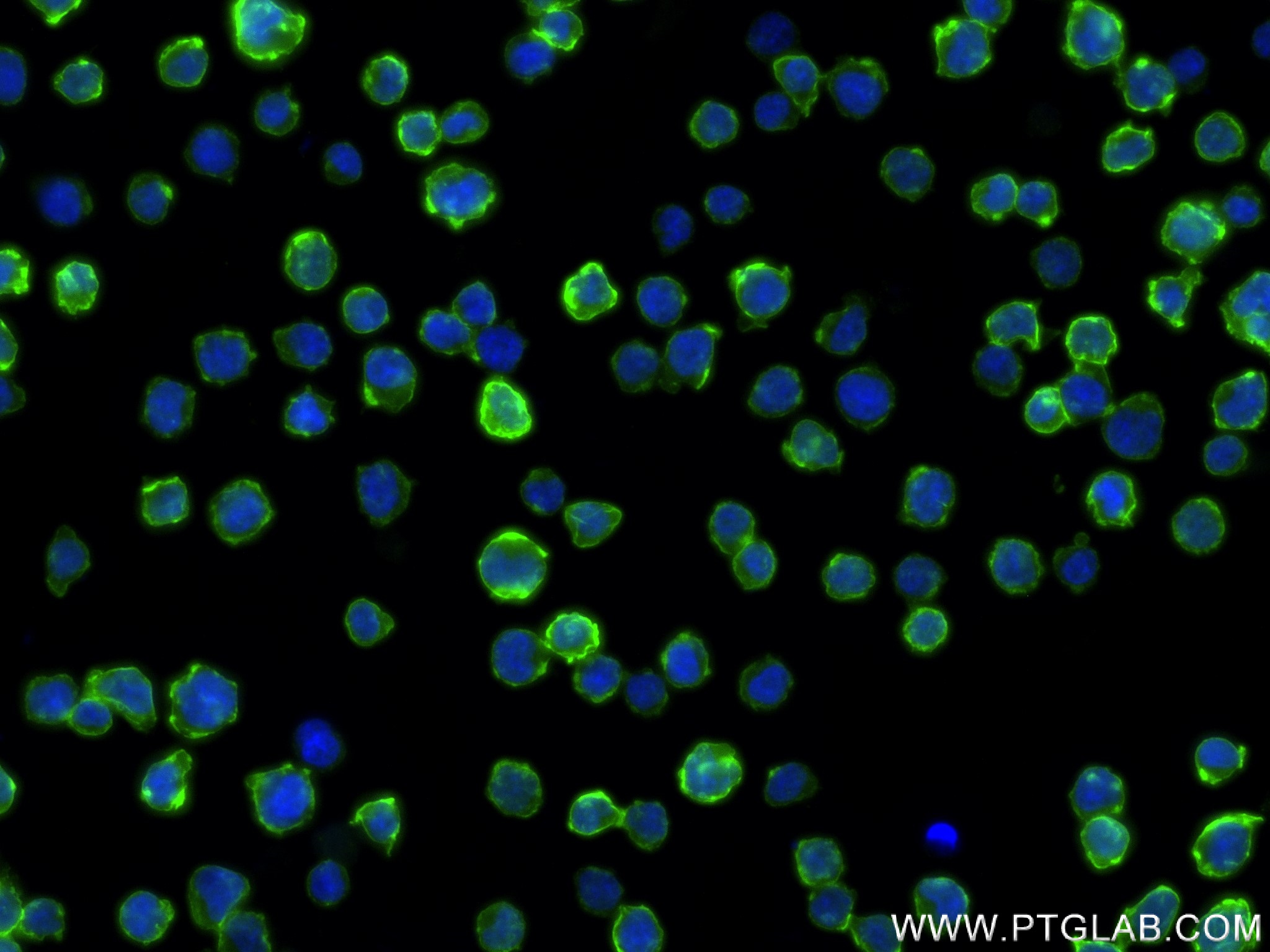
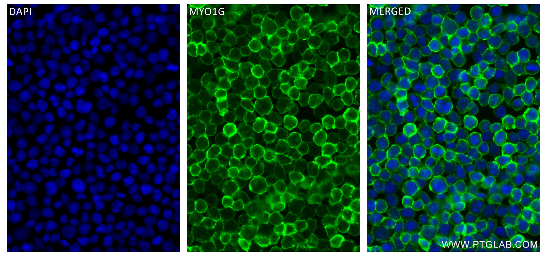
 fixed Jurkat cells using MYO1G antibody (83556-5-RR, Clone: 240503E1 ) at dilution of 1:200 and CoraLite®488-Conjugated AffiniPure Goat Anti-Rabbit IgG(H+L) (SA00013-2).")
 fixed Jurkat cells using MYO1G antibody (83556-5-RR, Clone: 240503E1 ) at dilution of 1:200 and CoraLite®488-Conjugated Goat Anti-Rabbit IgG(H+L) (SA00013-2).")
 and APC-Conjugated AffiniPure Goat Anti-Rabbit IgG(H+L)(red), or 0.25 ug Isotype Control (blue). Cells were fixed and permeabilized with Transcription Factor Staining Buffer Kit (PF00011).")
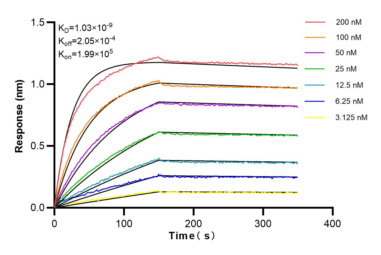
 kinetic assays of 83556-5-RR against Human MYO1G were performed. The affinity constant is 1.03 nM.")
Tested Applications
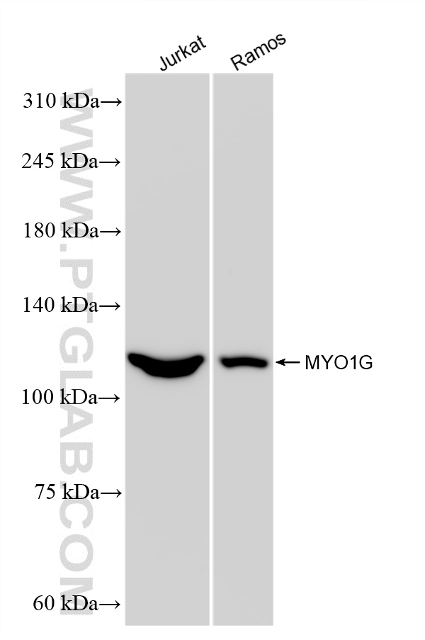
| Positive WB detected in | Jurkat cells, Ramos cells |
| Positive IF/ICC detected in | Jurkat cells |
| Positive FC (Intra) detected in | Jurkat cells |
Recommended dilution
| Application | Dilution |
|---|---|
| Western Blot (WB) | WB : 1:2000-1:16000 |
| Immunofluorescence (IF)/ICC | IF/ICC : 1:50-1:500 |
| Flow Cytometry (FC) (INTRA) | FC (INTRA) : 0.25 ug per 10^6 cells in a 100 µl suspension |
| It is recommended that this reagent should be titrated in each testing system to obtain optimal results. | |
| Sample-dependent, Check data in validation data gallery. | |
Product Information
83556-5-RR targets MYO1G in WB, IF/ICC, FC (Intra), ELISA applications and shows reactivity with human samples.
| Tested Reactivity | human |
| Host / Isotype | Rabbit / IgG |
| Class | Recombinant |
| Type | Antibody |
| Immunogen | MYO1G fusion protein Ag22698 Predict reactive species |
| Full Name | myosin IG |
| Calculated Molecular Weight | 1018 aa, 116 kDa |
| Observed Molecular Weight | 110-120 kDa |
| GenBank Accession Number | BC113544 |
| Gene Symbol | MYO1G |
| Gene ID (NCBI) | 64005 |
| RRID | AB_3671173 |
| Conjugate | Unconjugated |
| Form | Liquid |
| Purification Method | Protein A purfication |
| UNIPROT ID | B0I1T2 |
| Storage Buffer | PBS with 0.02% sodium azide and 50% glycerol , pH 7.3 |
| Storage Conditions | Store at -20°C. Stable for one year after shipment. Aliquoting is unnecessary for -20oC storage. 20ul sizes contain 0.1% BSA. |
Background Information
MYO1G is a plasma membrane-associated class I myosin that is abundant in T and B lymphocytes and mast cells and expressed specifically in the haematopoietic system. Class I myosins have been implicated in various cellular processes relying on actin-dependent membrane dynamics such as endocytosis, secretion, adhesion, motility and regulation of mechanosensitive channels (PMID: 19968988). MYO1G localizes to the plasma membrane linked to PIP2 and PIP3, is particularly enriched at cell-surface microvilli, and associates in an ATP-releasable manner to the actin cytoskeleton (PMID: 24310084). MYO1G can be detected as about 110-120 kDa.
Protocols
| Product Specific Protocols | |
|---|---|
| WB protocol for MYO1G antibody 83556-5-RR | Download protocol |
| IF protocol for MYO1G antibody 83556-5-RR | Download protocol |
| FC protocol for MYO1G antibody 83556-5-RR | Download protocol |
| Standard Protocols | |
|---|---|
| Click here to view our Standard Protocols |