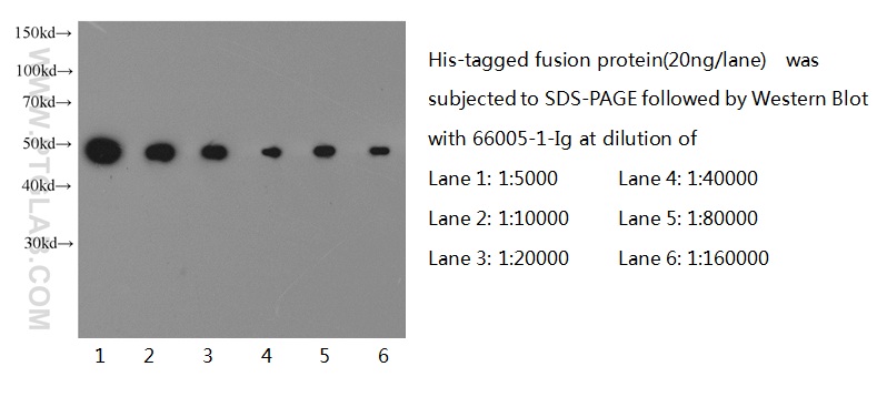
at various dilutions.")
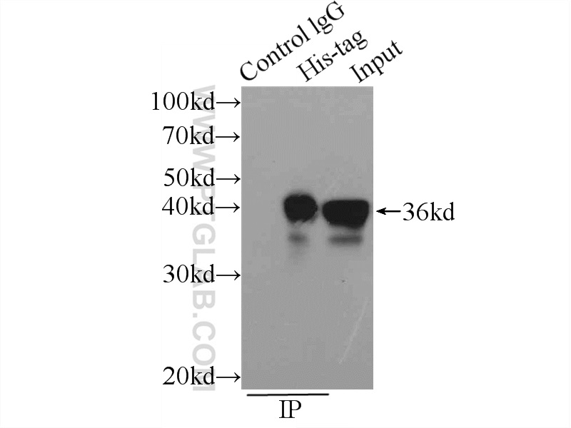
 with Transfected HEK-293 cells lysate 300ug.")
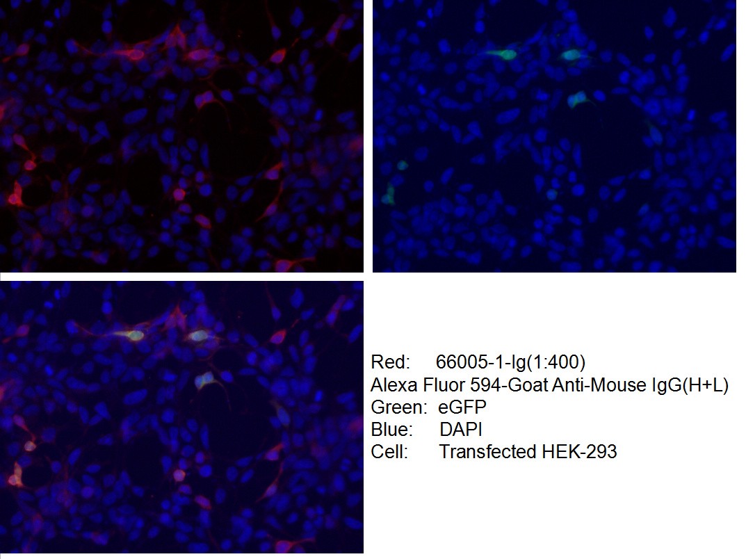
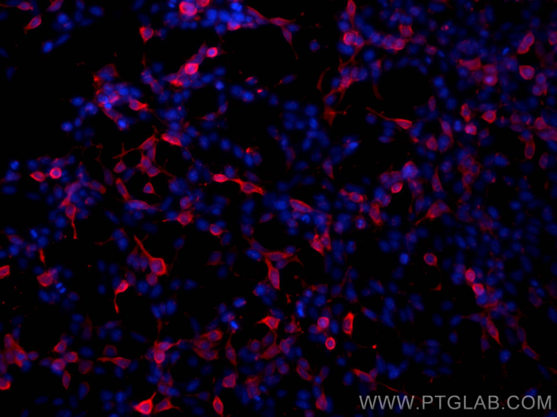
 fixed Transfected HEK-293 cells using 66005-1-Ig (6*His, His-Tag antibody) at dilution of 1:400 and Alexa Fluor 594-Conjugated AffiniPure Goat Anti-Mouse IgG(H+L).")
 fixed Transfected HEK-293 cells using 6*His, His-Tag antibody (66005-1-Ig, Clone: 1B7G5 ) at dilution of 1:800 and CoraLite®594-Conjugated AffiniPure Goat Anti-Mouse IgG(H+L) (SA00013-1).")
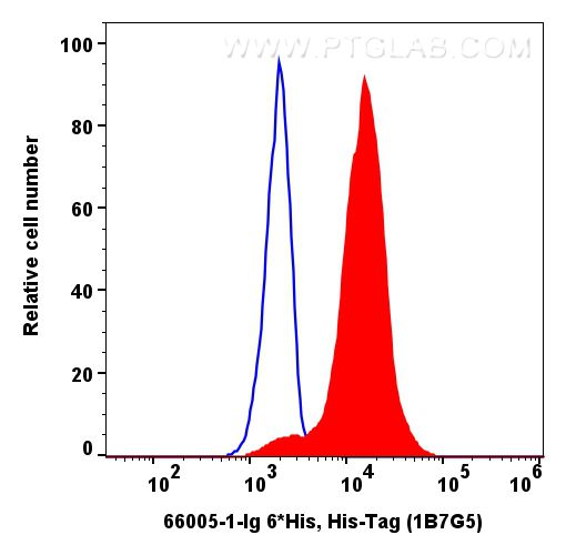
 and CoraLite488-conjugated Goat Anti-Mouse IgG(H+L) (Cat.NO. SA00013-1). Mouse IgG1 isotype control (66360-1-Ig, Clone: 1F8D3, blue) was parallel stained as control. Cells were not fixed.")
"6*His, His-Tag Antibodies" Comparison
View side-by-side comparison of 6*His, His-Tag antibodies from other vendors to find the one that best suits your research needs.
Tested Applications
| Positive WB detected in | recombinant protein |
| Positive IP detected in | Transfected HEK-293 cells |
| Positive IF/ICC detected in | Transfected HEK-293 cells |
| Positive FC detected in | Transfexted HEK-293 cells |
Recommended dilution
| Application | Dilution |
|---|---|
| Western Blot (WB) | WB : 1:5000-1:50000 |
| Immunoprecipitation (IP) | IP : 0.5-4.0 ug for 1.0-3.0 mg of total protein lysate |
| Immunofluorescence (IF)/ICC | IF/ICC : 1:200-1:800 |
| Flow Cytometry (FC) | FC : 0.20 ug per 10^6 cells in a 100 µl suspension |
| It is recommended that this reagent should be titrated in each testing system to obtain optimal results. | |
| Sample-dependent, Check data in validation data gallery. | |
Product Information
66005-1-Ig targets 6*His, His-Tag in WB, IHC, IF/ICC, FC, IP, CoIP, ChIP, ELISA applications and shows reactivity with recombinant protein samples.
| Tested Reactivity | recombinant protein |
| Cited Reactivity | human, mouse, rat, chicken, hamster, yeast |
| Host / Isotype | Mouse / IgG1 |
| Class | Monoclonal |
| Type | Antibody |
| Immunogen |
Peptide Predict reactive species |
| Full Name | 6*His, His-Tag |
| Calculated Molecular Weight | 0.84 kDa |
| Gene Symbol | |
| Gene ID (NCBI) | |
| RRID | AB_11232599 |
| Conjugate | Unconjugated |
| Form | Liquid |
| Purification Method | Protein G purification |
| UNIPROT ID | HISTAG |
| Storage Buffer | PBS with 0.02% sodium azide and 50% glycerol, pH 7.3. |
| Storage Conditions | Store at -20°C. Stable for one year after shipment. Aliquoting is unnecessary for -20oC storage. 20ul sizes contain 0.1% BSA. |
Background Information
Protein tags are a protein or peptide sequences located either on the C- or N- terminal of the target protein. His-tag is often used for affinity purification and binding assays. Expressed. The His-tag antibody is a useful tool for monitoring of the His-tagged proteins and recognizes His-tags placed at N-terminal, C-terminal, and internal regions of fusion proteins expressed in bacteria, insect, and mammalian cells. A His-tag (polyhistidine tag) consists of at least six histidine residues that are located at the N- or C-terminus of recombinant proteins. It is commonly used for affinity purification and protein binding experiments.
The recombinant protein has only 6 histidine residues as a tag. Is this sufficient to be detected by your antibody? Is the His-tag antibody able to detect N-terminal and C-terminal His tags?
The His-tag antibody was raised using a 6-His tag peptide as the immunogen, which is ~1 kDa. Some scientists use more than 6 histidine residues as a tag for recombinant proteins to increase affinity to metal ions. A 6-histidine tag is sufficient for specific recognition by the His-tag antibody. It is able to recognize both N-terminal and C-terminal His tags of recombinant proteins produced in various expression systems, including bacteria, yeasts, insect, and mammalian cells.
After purification, I can detect my protein by western blotting using the His-tag antibody but can also see additional bands running lower. What might they represent?
They may represent cleavage products of the recombinant protein. Unspecific protein cleavage can occur during protein production (e.g., in bacteria) or protein purification post the cell lysis steps. Changing expression conditions (temperature, time, induction methods, medium) and using protease inhibitors during purification helps to minimize protein degradation. Additional steps, such as ion exchange, size exclusion, or chromatography, allow isolation of the full-length product.
Can a His-tag antibody be used in applications other than western blotting?
Yes, this antibody has been successfully used for immunofluorescence (IF), immunoprecipitation (IP), and indirect ELISA analysis.
Protocols
| Product Specific Protocols | |
|---|---|
| FC protocol for 6*His, His-Tag antibody 66005-1-Ig | Download protocol |
| IF protocol for 6*His, His-Tag antibody 66005-1-Ig | Download protocol |
| IP protocol for 6*His, His-Tag antibody 66005-1-Ig | Download protocol |
| Standard Protocols | |
|---|---|
| Click here to view our Standard Protocols |
Publications
| Species | Application | Title |
|---|---|---|
Signal Transduct Target Ther Aberrant Notch-signaling promotes tumor angiogenesis in esophageal squamous-cell carcinoma | ||
Science Structural insight into the SAM-mediated assembly of the mitochondrial TOM core complex. |
Reviews
The reviews below have been submitted by verified Proteintech customers who received an incentive for providing their feedback.
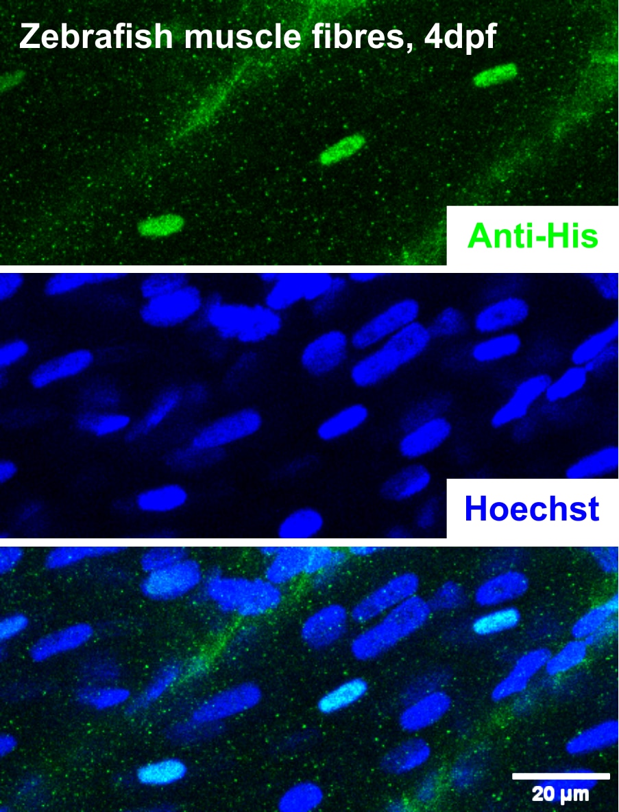
FH Jeffrey (Verified Customer) (01-30-2026) | Works very well in validating the mosaic expression of a nuclear-localised, His-tagged protein in zebrafish muscle fibres in vivo via immunostaining.
 |
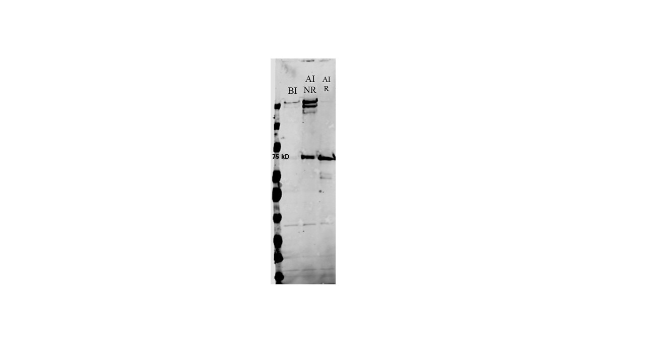
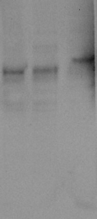
FH Vahid (Verified Customer) (07-10-2025) | Image caption: Western blot analysis of total lysate of E coli expressing recombinant His-tagged adenosine deaminase. The blot reveals a dimeric conformation of ADA2 under non-reducing conditions (After Induction NR) and a monomeric form under reducing conditions (After Induction R). BI:Befor indiction. The blot was probed with anti-His antibody (Proteintech)
 |
FH S (Verified Customer) (08-15-2024) | Excellent
 |
FH Xiaoyu (Verified Customer) (07-18-2024) | The antibody works well for Western Blot, and there are no unspecific bands.
 |
FH Raquel (Verified Customer) (04-13-2023) | Nice selective staining. No background detected when looking for our 6xHis tagged recombinant protein within yeast supernatant. Good quality western blot image. Great value for money and would recommend.
|
FH Ken (Verified Customer) (01-30-2023) | This is a specific antibody. It detects His-tagged proteins expressed in 293T cells with low background.
|
FH Samruddhi (Verified Customer) (02-22-2022) | The antibody works well for Western Blot, and there are no unspecific bands.
|
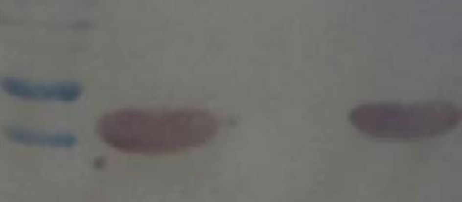
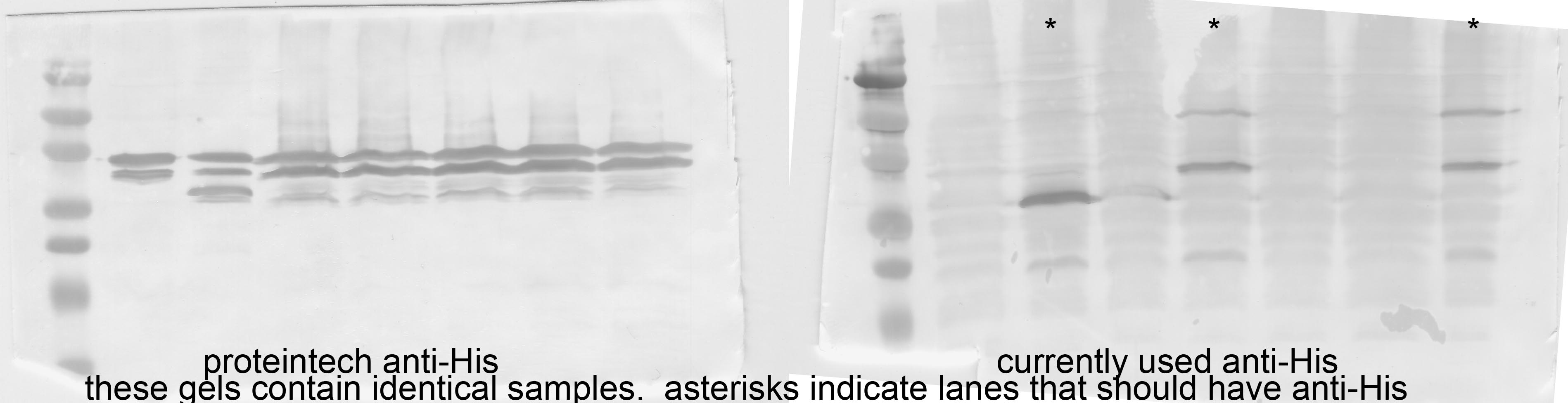
FH Amie (Verified Customer) (07-27-2021) | I ran two identical gels, with two different sized His-tagged proteins on them, and some lanes that should contain no His-tagged protein. As you can see in the image, the Proteintech antibody had much more background and less specificity than the anti-His antibody I currently use (I cannot recall what vendor it is from). It is not useful for my purposes given the cross-reactivity/lack of specificity.
 |
FH Barry (Verified Customer) (01-07-2021) | Suitable
|
FH Adrian (Verified Customer) (04-22-2020) | We tested anti-His tag Ab's in our immunometric analyses using recombinant his-tagged nickel binding protein NikR as a primary Ab. We had no issue obtaining a signal, but the paper eventually was left out of this part because our primary Ab had stability issues. It was a few years ago, though.
|
FH Paul (Verified Customer) (01-15-2020) | Works well for Western Blot.
|
FH Laura (Verified Customer) (01-14-2020) | Good sensitivity for Western blot.
|
FH Aamir (Verified Customer) (01-08-2020) | Works well for WB
|
FH Benjamin (Verified Customer) (01-07-2020) | Works very well. Detects a his-tagged fusion protein with high sensitivity and very little background in western blot.
|
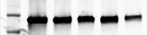
FH Nikhil (Verified Customer) (10-16-2019) | Recombinant His tagged p53 expressed in Bacteria. Lane 1 marker, Lane2-lane 6: Elutions 1-5
 |
FH Shashirekha (Verified Customer) (07-09-2019) | The antibody is good but it stains ladder as well.
 |
FH Chun (Verified Customer) (07-03-2019) | This antibody is excellent.
|
FH John (Verified Customer) (02-27-2019) | We unfortunately saw unexpected fluorescence in negative controls when using this antibody, so were unable to draw conclusions from our initial experiments. On a more positive note, after running through attempts to resolve the issue Proteintech did offer us an alternative antibody to try.
|
FH Marisa (Verified Customer) (02-05-2019) | Highly dependent on sample type. Works very nice and shurly up to 1:50,000 detecting recombinant His-proteins of E. coli. Difficult to analyze proteins produced in insect cells. Please use 1:1000 as a dilution.
|
FH Youjun (Verified Customer) (11-27-2018) |
|