Various lysates were subjected to SDS PAGE followed by western blot with 67813-1-Ig (FXR1 antibody) at dilution of 1:10000 incubated at room temperature for 1.5 hours. The membrane was stripped and reblotted with HRP-conjugated Beta Actin Monoclonal antibody (HRP-66009) as loading control.
Various lysates were subjected to SDS PAGE followed by western blot with 67813-1-Ig (FXR1 antibody) at dilution of 1:10000 incubated at room temperature for 1.5 hours. The membrane was stripped and reblotted with HRP-conjugated Beta Actin Monoclonal antibody (HRP-66009) as loading control.
IP experiment of K-562 using 67813-1-Ig
IP result of anti-FXR1 (IP:67813-1-Ig, 5ug; Detection:67813-1-Ig 1:1000) with K-562 cells lysate 3680 ug.
Immunohistochemical analysis of paraffin-embedded mouse brain tissue slide using 67813-1-Ig (FXR1 antibody) at dilution of 1:1000 (under 10x lens). Heat mediated antigen retrieval with Tris-EDTA buffer (pH 9.0).
IHC staining of human gliomas using 67813-1-Ig
Immunohistochemical analysis of paraffin-embedded human gliomas tissue slide using 67813-1-Ig (FXR1 antibody) at dilution of 1:1000 (under 10x lens). Heat mediated antigen retrieval with Tris-EDTA buffer (pH 9.0).
Immunohistochemical analysis of paraffin-embedded human gliomas tissue slide using 67813-1-Ig (FXR1 antibody) at dilution of 1:1000 (under 10x lens). Heat mediated antigen retrieval with Tris-EDTA buffer (pH 9.0).
IHC staining of human gliomas using 67813-1-Ig
Immunohistochemical analysis of paraffin-embedded human gliomas tissue slide using 67813-1-Ig (FXR1 antibody) at dilution of 1:1000 (under 40x lens). Heat mediated antigen retrieval with Tris-EDTA buffer (pH 9.0).
Immunohistochemical analysis of paraffin-embedded human gliomas tissue slide using 67813-1-Ig (FXR1 antibody) at dilution of 1:1000 (under 40x lens). Heat mediated antigen retrieval with Tris-EDTA buffer (pH 9.0).
IF Staining of mouse brain using 67813-1-Ig
Immunofluorescent analysis of (4% PFA) fixed mouse brain tissue using FXR1 antibody (67813-1-Ig, Clone: 1E12E8 ) at dilution of 1:400 and CoraLite®488-Conjugated Goat Anti-Mouse IgG(H+L).
Immunofluorescent analysis of (4% PFA) fixed mouse brain tissue using FXR1 antibody (67813-1-Ig, Clone: 1E12E8 ) at dilution of 1:400 and CoraLite®488-Conjugated Goat Anti-Mouse IgG(H+L).
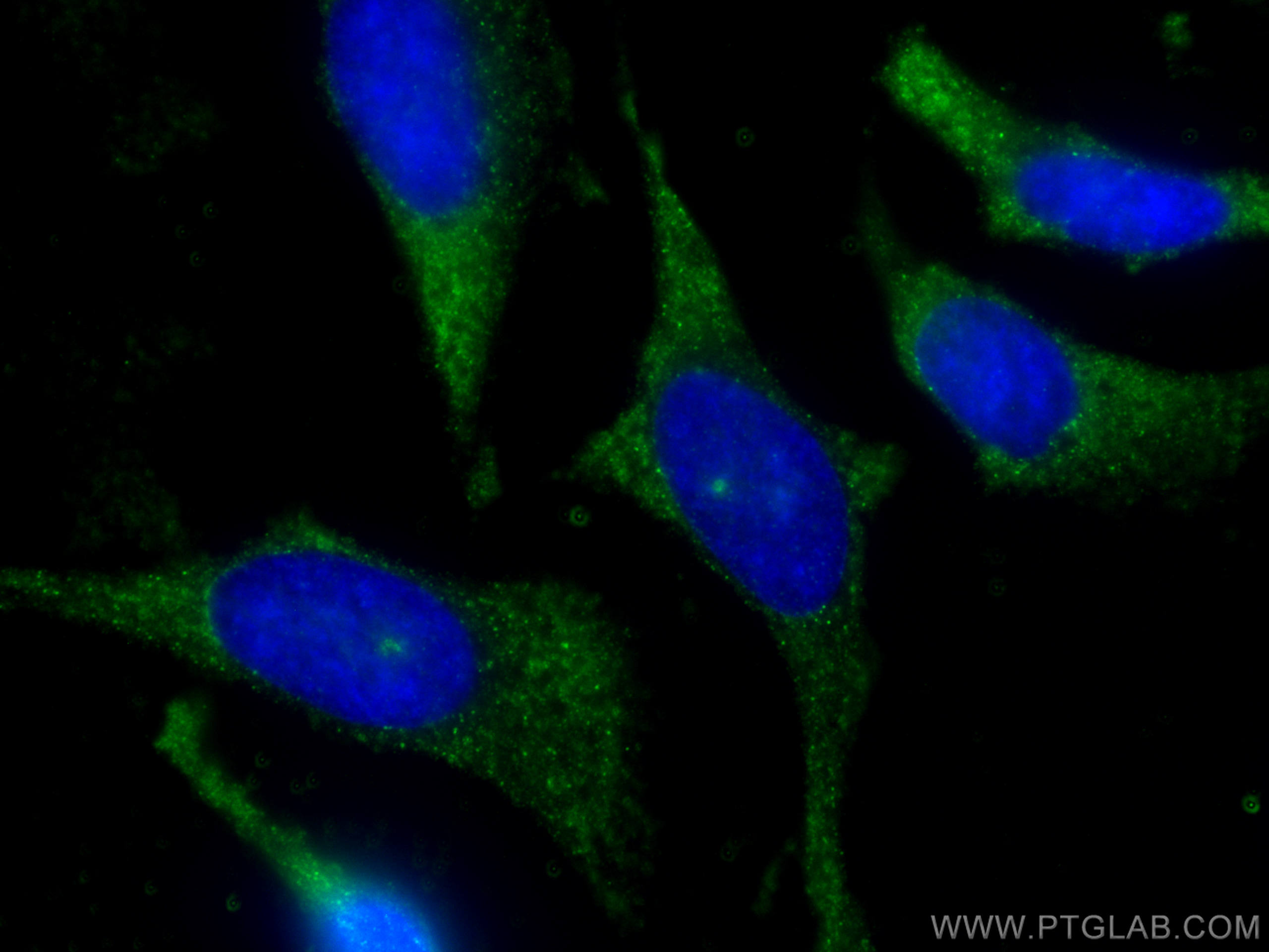
IF Staining of HeLa using 67813-1-Ig
Immunofluorescent analysis of (-20°C Methanol) fixed HeLa cells using FXR1 antibody (67813-1-Ig, Clone: 1E12E8 ) at dilution of 1:400 and CoraLite®488-Conjugated Goat Anti-Mouse IgG(H+L).
Immunofluorescent analysis of (-20°C Methanol) fixed HeLa cells using FXR1 antibody (67813-1-Ig, Clone: 1E12E8 ) at dilution of 1:400 and CoraLite®488-Conjugated Goat Anti-Mouse IgG(H+L).
The Proteintech guarantee covers Proteintech antibodies in any species and any application, including those not listed on the datasheet. If the antibody doesn’t perform, you can receive a hassle-free refund or credit note.
mouse brain tissue, human gliomas tissue Note: suggested antigen retrieval with TE buffer pH 9.0; (*) Alternatively, antigen retrieval may be performed with citrate buffer pH 6.0
Positive IF-P detected in
mouse brain tissue
Positive IF/ICC detected in
HeLa cells
Recommended dilution
Application
Dilution
Western Blot (WB)
WB : 1:5000-1:50000
Immunoprecipitation (IP)
IP : 0.5-4.0 ug for 1.0-3.0 mg of total protein lysate
Immunohistochemistry (IHC)
IHC : 1:500-1:2000
Immunofluorescence (IF)-P
IF-P : 1:200-1:800
Immunofluorescence (IF)/ICC
IF/ICC : 1:200-1:800
It is recommended that this reagent should be titrated in each testing system to obtain optimal results.
Sample-dependent, Check data in validation data gallery.
PBS with 0.02% sodium azide and 50% glycerol, pH 7.3.
Storage Conditions
Store at -20°C. Stable for one year after shipment. Aliquoting is unnecessary for -20oC storage. 20ul sizes contain 0.1% BSA.
Background Information
Tumour necrosis factor-alpha (TNF-alpha) is a key mediator of inflammation in host defence against infection and in autoimmune disease. Its production is controlled post-transcriptionally by multiple RNA-binding proteins that interact with the TNF-alpha AU-rich element and regulate its expression; Fragile X mental retardation-related protein 1 (FXR1) is one of these. FXR1 (fragile-X-mental retardation-related protein 1) are RNA-binding proteins that have been demonstrated to impact miRNA-mediated, post-transcriptional gene regulation, and required for embryonic and postnatal development of muscle tissue. It can regulate intracellular transport and local translation of certain mRNAs. The FXR1 exists some isoforms with the MW 69 kDa and 60 kDa.
Silibinin, a commonly used therapeutic agent for non-alcohol fatty liver disease, functions through upregulating intestinal expression of fibroblast growth factor 15/19
Various lysates were subjected to SDS PAGE followed by western blot with 67813-1-Ig (FXR1 antibody) at dilution of 1:10000 incubated at room temperature for 1.5 hours. The membrane was stripped and reblotted with HRP-conjugated Beta Actin Monoclonal antibody (HRP-66009) as loading control.
IHC Figures
IHC staining of mouse brain using 67813-1-Ig
Immunohistochemical analysis of paraffin-embedded mouse brain tissue slide using 67813-1-Ig (FXR1 antibody) at dilution of 1:1000 (under 40x lens). Heat mediated antigen retrieval with Tris-EDTA buffer (pH 9.0).
IHC staining of mouse brain using 67813-1-Ig
Immunohistochemical analysis of paraffin-embedded mouse brain tissue slide using 67813-1-Ig (FXR1 antibody) at dilution of 1:1000 (under 10x lens). Heat mediated antigen retrieval with Tris-EDTA buffer (pH 9.0).
IHC staining of human gliomas using 67813-1-Ig
Immunohistochemical analysis of paraffin-embedded human gliomas tissue slide using 67813-1-Ig (FXR1 antibody) at dilution of 1:1000 (under 10x lens). Heat mediated antigen retrieval with Tris-EDTA buffer (pH 9.0).
IHC staining of human gliomas using 67813-1-Ig
Immunohistochemical analysis of paraffin-embedded human gliomas tissue slide using 67813-1-Ig (FXR1 antibody) at dilution of 1:1000 (under 40x lens). Heat mediated antigen retrieval with Tris-EDTA buffer (pH 9.0).
IP Figures
IP experiment of K-562 using 67813-1-Ig
IP result of anti-FXR1 (IP:67813-1-Ig, 5ug; Detection:67813-1-Ig 1:1000) with K-562 cells lysate 3680 ug.
IF-P Figures
IF Staining of mouse brain using 67813-1-Ig
Immunofluorescent analysis of (4% PFA) fixed mouse brain tissue using FXR1 antibody (67813-1-Ig, Clone: 1E12E8 ) at dilution of 1:400 and CoraLite®488-Conjugated Goat Anti-Mouse IgG(H+L).
IF/ICC Figures
IF Staining of HeLa using 67813-1-Ig
Immunofluorescent analysis of (-20°C Methanol) fixed HeLa cells using FXR1 antibody (67813-1-Ig, Clone: 1E12E8 ) at dilution of 1:400 and CoraLite®488-Conjugated Goat Anti-Mouse IgG(H+L).
IF Staining of HeLa using 67813-1-Ig
Immunofluorescent analysis of (-20°C Methanol) fixed HeLa cells using FXR1 antibody (67813-1-Ig, Clone: 1E12E8 ) at dilution of 1:400 and CoraLite®488-Conjugated Goat Anti-Mouse IgG(H+L).
The species listed in Tested Reactivity are in-house verified and applicable species. For unlisted species, please refer to the homology analysis of the immunogen sequence and related species. For rabbit polyclonal antibodies, homology >70% is recommended. For mouse monoclonal antibodies and rabbit recombinant antibodies, homology >90% is recommended. Generally, the higher the homology, the greater the applicability. However, there will be certain differences in protein expression in different species, tissues or cells. Therefore, the homology analysis results are for reference only and do not serve as a guarantee.
At Proteintech, we pride ourselves on our antibody quality, customer service and transparency. As such, we are comparing our antibodies with other vendors, enabling easy identification and comparisons of key data to help you choose the suitable antibody for your needs.
We have selected the top cited antibodies from these vendors for you to compare.
Proteintech
KD/KO VALIDATED
FXR1 Monoclonal antibody
Catalog Number
67813-1-Ig
Citations
2
Dilutions
WB : 1:5000-1:50000 IP : 0.5-4.0 ug for IP and 0.5-4.0 ug for 1.0-3.0 mg of total protein lysate for WB IHC : 1:500-1:2000 IF-P : 1:200-1:800 IF/ICC : 1:200-1:800
Applications
WB, IHC, IF/ICC, IF-P, IP, ELISA
Reactivity
human, mouse, rat
Product Guarantee
Covers any species including not listed on datasheet
Covers any applications including not listed on datasheet
 at dilution of 1:10000 incubated at room temperature for 1.5 hours. The membrane was stripped and reblotted with HRP-conjugated Beta Actin Monoclonal antibody (HRP-66009) as loading control.")
 with K-562 cells lysate 3680 ug.")
 at dilution of 1:1000 (under 40x lens). Heat mediated antigen retrieval with Tris-EDTA buffer (pH 9.0).")
 at dilution of 1:1000 (under 10x lens). Heat mediated antigen retrieval with Tris-EDTA buffer (pH 9.0).")
 at dilution of 1:1000 (under 10x lens). Heat mediated antigen retrieval with Tris-EDTA buffer (pH 9.0).")
 at dilution of 1:1000 (under 40x lens). Heat mediated antigen retrieval with Tris-EDTA buffer (pH 9.0).")
 fixed mouse brain tissue using FXR1 antibody (67813-1-Ig, Clone: 1E12E8 ) at dilution of 1:400 and CoraLite®488-Conjugated Goat Anti-Mouse IgG(H+L).")
 fixed HeLa cells using FXR1 antibody (67813-1-Ig, Clone: 1E12E8 ) at dilution of 1:400 and CoraLite®488-Conjugated Goat Anti-Mouse IgG(H+L).")
 fixed HeLa cells using FXR1 antibody (67813-1-Ig, Clone: 1E12E8 ) at dilution of 1:400 and CoraLite®488-Conjugated Goat Anti-Mouse IgG(H+L).")