Filter:
 at dilution of 1:10000 incubated at room temperature for 1.5 hours.")
 at dilution of 1:3000 incubated at room temperature for 1.5 hours.")
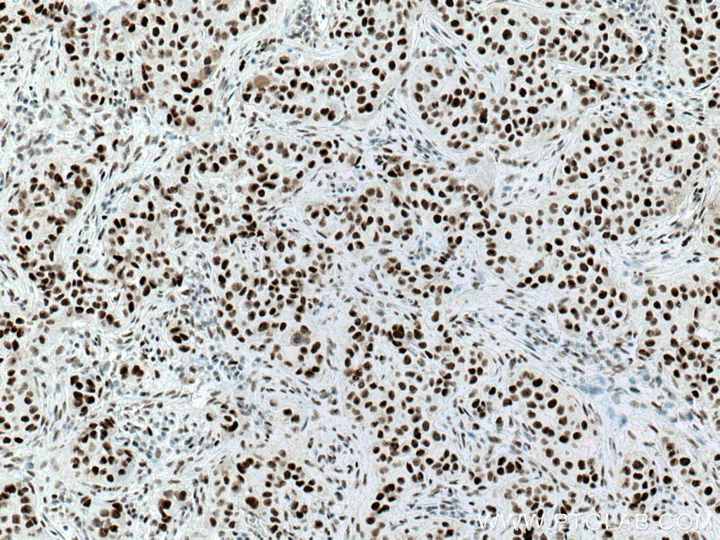
 at dilution of 1:800 (under 10x lens. Heat mediated antigen retrieval with Tris-EDTA buffer (pH 9.0).")
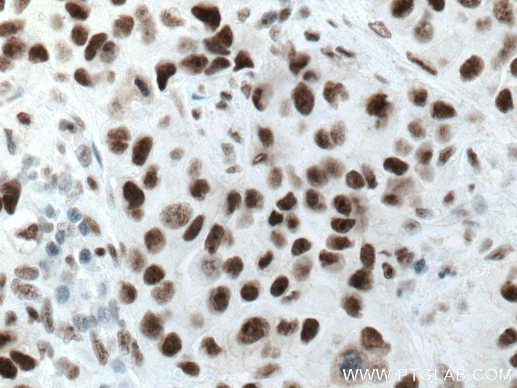
 at dilution of 1:800 (under 40x lens. Heat mediated antigen retrieval with Tris-EDTA buffer (pH 9.0).")
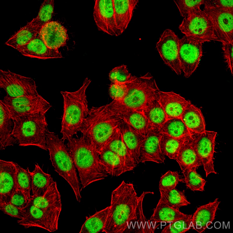
 fixed PC-3 cells using Cyclin H antibody (67065-1-Ig, Clone: 2C5G11 ) at dilution of 1:400 and CoraLite®488-Conjugated Goat Anti-Mouse IgG(H+L), CL594-Phalloidin (red).")
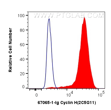
 and CoraLite®488-Conjugated Goat Anti-Mouse IgG(H+L) at dilution 1:1000 (red), or 0.4 ug Mouse IgG2a Isotype Control (C1.18.4) (65208-1-Ig, Clone: C1.18.4) (blue). Cells were fixed and permeabilized with Transcription Factor Staining Buffer Kit (PF00011).")
Tested Applications
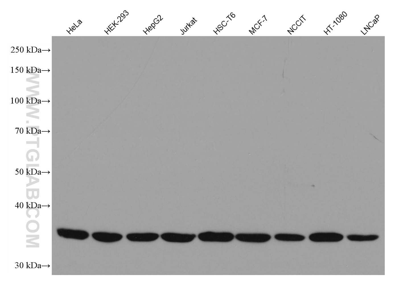
| Positive WB detected in | HeLa cells, HEK-293 cells, HepG2 cells, Jurkat cells, HSC-T6 cells, MCF-7 cells, NCCIT cells, HT-1080 cells, LNCaP cells |
| Positive IHC detected in | human breast cancer tissue Note: suggested antigen retrieval with TE buffer pH 9.0; (*) Alternatively, antigen retrieval may be performed with citrate buffer pH 6.0 |
| Positive IF/ICC detected in | PC-3 cells |
| Positive FC (Intra) detected in | PC-3 cells |
Recommended dilution
| Application | Dilution |
|---|---|
| Western Blot (WB) | WB : 1:2000-1:10000 |
| Immunohistochemistry (IHC) | IHC : 1:500-1:1000 |
| Immunofluorescence (IF)/ICC | IF/ICC : 1:200-1:800 |
| Flow Cytometry (FC) (INTRA) | FC (INTRA) : 0.40 ug per 10^6 cells in a 100 µl suspension |
| It is recommended that this reagent should be titrated in each testing system to obtain optimal results. | |
| Sample-dependent, Check data in validation data gallery. | |
Published Applications
| WB | See 2 publications below |
Product Information
67065-1-Ig targets Cyclin H in WB, IHC, IF/ICC, FC (Intra), ELISA applications and shows reactivity with human, mouse, rat samples.
| Tested Reactivity | human, mouse, rat |
| Cited Reactivity | mouse |
| Host / Isotype | Mouse / IgG2a |
| Class | Monoclonal |
| Type | Antibody |
| Immunogen |
CatNo: Ag28556 Product name: Recombinant human CCNH protein Source: e coli.-derived, PET28a Tag: 6*His Domain: 1-323 aa of BC005280 Sequence: MYHNSSQKRHWTFSSEEQLARLRADANRKFRCKAVANGKVLPNDPVFLEPHEEMTLCKYYEKRLLEFCSVFKPAMPRSVVGTACMYFKRFYLNNSVMEYHPRIIMLTCAFLACKVDEFNVSSPQFVGNLRESPLGQEKALEQILEYELLLIQQLNFHLIVHNPYRPFEGFLIDLKTRYPILENPEILRKTADDFLNRIALTDAYLLYTPSQIALTAILSSASRAGITMESYLSESLMLKENRTCLSQLLDIMKSMRNLVKKYEPPRSEEVAVLKQKLERCHSAELALNVITKKRKGYEDDDYVSKKSKHEEEEWTDDDLVESL Predict reactive species |
| Full Name | cyclin H |
| Calculated Molecular Weight | 38 kDa |
| Observed Molecular Weight | 38 kDa |
| GenBank Accession Number | BC005280 |
| Gene Symbol | Cyclin H |
| Gene ID (NCBI) | 902 |
| RRID | AB_2882375 |
| Conjugate | Unconjugated |
| Form | Liquid |
| Purification Method | Protein A purification |
| UNIPROT ID | P51946 |
| Storage Buffer | PBS with 0.02% sodium azide and 50% glycerol, pH 7.3. |
| Storage Conditions | Store at -20°C. Stable for one year after shipment. Aliquoting is unnecessary for -20oC storage. 20ul sizes contain 0.1% BSA. |
Protocols
| Product Specific Protocols | |
|---|---|
| IF protocol for Cyclin H antibody 67065-1-Ig | Download protocol |
| IHC protocol for Cyclin H antibody 67065-1-Ig | Download protocol |
| WB protocol for Cyclin H antibody 67065-1-Ig | Download protocol |
| Standard Protocols | |
|---|---|
| Click here to view our Standard Protocols |
Publications
| Species | Application | Title |
|---|---|---|
Adv Sci (Weinh) m6A Reader hnRNPA2B1 Modulates Late Pachytene Progression in Male Meiosis Through Post-Transcriptional Control |