
 fixed human tonsillitis tissue using CoraLite®594-conjugated CD63 antibody (CL594-67605, Clone: 3D4D1 ) at dilution of 1:200.")
 fixed human tonsillitis tissue using CoraLite®594-conjugated CD63 antibody (CL594-67605, Clone: 3D4D1 ) at dilution of 1:200.")
 fixed human tonsillitis tissue using CoraLite®594-conjugated CD63 antibody (CL594-67605, Clone: 3D4D1 ) at dilution of 1:200.")
Tested Applications
| Positive IF-P detected in | human tonsillitis tissue |
Recommended dilution
| Application | Dilution |
|---|---|
| Immunofluorescence (IF)-P | IF-P : 1:50-1:500 |
| It is recommended that this reagent should be titrated in each testing system to obtain optimal results. | |
| Sample-dependent, Check data in validation data gallery. | |
Product Information
CL594-67605 targets CD63 in IF-P applications and shows reactivity with Human samples.
| Tested Reactivity | Human |
| Host / Isotype | Mouse / IgG1 |
| Class | Monoclonal |
| Type | Antibody |
| Immunogen |
CatNo: Ag19690 Product name: Recombinant human CD63 protein Source: e coli.-derived, PET28a Tag: 6*His Domain: 104-209 aa of BC002349 Sequence: GYVFRDKVMSEFNNNFRQQMENYPKNNHTASILDRMQADFKCCGAANYTDWEKIPSMSKNRVPDSCCINVTVGCGINFNEKAIHKEGCVEKIGGWLRKNVLVVAAA Predict reactive species |
| Full Name | CD63 molecule |
| Calculated Molecular Weight | 26 kDa |
| Observed Molecular Weight | 35 kDa |
| GenBank Accession Number | BC002349 |
| Gene Symbol | CD63 |
| Gene ID (NCBI) | 967 |
| RRID | AB_2920164 |
| Conjugate | CoraLite®594 Fluorescent Dye |
| Excitation/Emission Maxima Wavelengths | 588 nm / 604 nm |
| Form | Liquid |
| Purification Method | Protein G purification |
| UNIPROT ID | P08962 |
| Storage Buffer | PBS with 50% glycerol, 0.05% Proclin300, 0.5% BSA, pH 7.3. |
| Storage Conditions | Store at -20°C. Avoid exposure to light. Stable for one year after shipment. Aliquoting is unnecessary for -20oC storage. |
Background Information
CD63 is a 30-60 kDa lysosomal membrane protein that belongs to the tetraspanin family. This protein plays many important roles in immuno-physiological functions. It mediate signal transduction events that play a role in the regulation of cell development, activation and motility. CD63 is expressed on activated platelets, thus it may function as a blood platelet activation marker. CD63 is a lysosomal membrane glycoprotein that is translocated to plasma membrane after platelet activation. The CD63 tetraspanin is highly expressed in the early stages of melanoma and decreases in advanced lesions, suggesting it as a possible suppressor of tumor progression. Deficiency of this protein is associated with Hermansky-Pudlak syndrome.
Protocols
| Product Specific Protocols | |
|---|---|
| IF protocol for CL594 CD63 antibody CL594-67605 | Download protocol |
| Standard Protocols | |
|---|---|
| Click here to view our Standard Protocols |
Reviews
The reviews below have been submitted by verified Proteintech customers who received an incentive for providing their feedback.
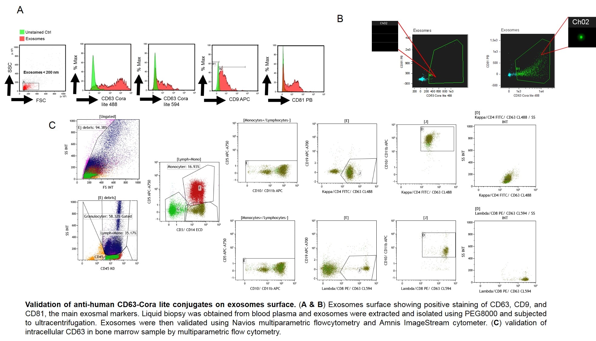
FH Amr (Verified Customer) (10-07-2021) | Anti CD63 CL488 and CL594 were validated in Blood plasma exosomes and in bone marrow. The antibodies seem to recogize their specific epitopes very well both extra and intracellularly. Strongly recommended.!
 |


