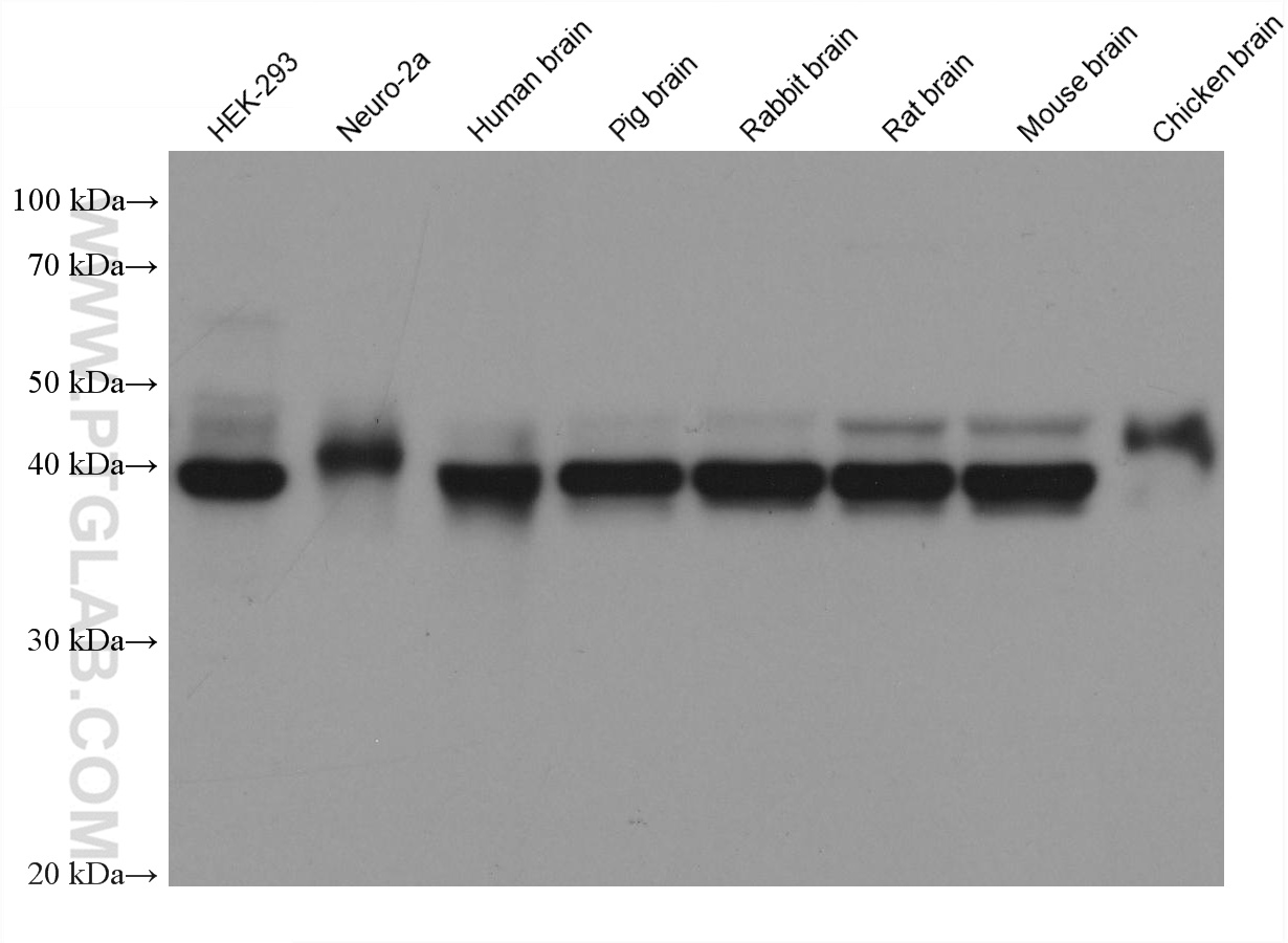
Various lysates were subjected to SDS PAGE followed by western blot with 66346-1-Ig (ASNA1 antibody) at dilution of 1:10000 incubated at room temperature for 1.5 hours.
Various lysates were subjected to SDS PAGE followed by western blot with 66346-1-Ig (ASNA1 antibody) at dilution of 1:10000 incubated at room temperature for 1.5 hours.
WB analysis using 66346-1-Ig
Various lysates were subjected to SDS PAGE followed by western blot with 66346-1-Ig (ASNA1 antibody) at dilution of 1:10000 incubated at room temperature for 1.5 hours.
Various lysates were subjected to SDS PAGE followed by western blot with 66346-1-Ig (ASNA1 antibody) at dilution of 1:10000 incubated at room temperature for 1.5 hours.
WB analysis of Neuro-2a using 66346-1-Ig
Neuro-2a cells were subjected to SDS PAGE followed by western blot with 66346-1-Ig (ASNA1 Antibody) at dilution of 1:1000 incubated at room temperature for 1.5 hours.
Neuro-2a cells were subjected to SDS PAGE followed by western blot with 66346-1-Ig (ASNA1 Antibody) at dilution of 1:1000 incubated at room temperature for 1.5 hours.
WB analysis of fetal human brain using 66346-1-Ig
fetal human brain tissue were subjected to SDS PAGE followed by western blot with 66346-1-Ig (ASNA1 Antibody) at dilution of 1:1000 incubated at room temperature for 1.5 hours.
fetal human brain tissue were subjected to SDS PAGE followed by western blot with 66346-1-Ig (ASNA1 Antibody) at dilution of 1:1000 incubated at room temperature for 1.5 hours.
WB analysis of pig brain using 66346-1-Ig
pig brain tissue were subjected to SDS PAGE followed by western blot with 66346-1-Ig (ASNA1 Antibody) at dilution of 1:1000 incubated at room temperature for 1.5 hours.
pig brain tissue were subjected to SDS PAGE followed by western blot with 66346-1-Ig (ASNA1 Antibody) at dilution of 1:1000 incubated at room temperature for 1.5 hours.
WB analysis of HEK-293 using 66346-1-Ig
HEK-293 cells were subjected to SDS PAGE followed by western blot with 66346-1-Ig (ASNA1 Antibody) at dilution of 1:1000 incubated at room temperature for 1.5 hours.
HEK-293 cells were subjected to SDS PAGE followed by western blot with 66346-1-Ig (ASNA1 Antibody) at dilution of 1:1000 incubated at room temperature for 1.5 hours.
IHC staining of human lung cancer using 66346-1-Ig
Immunohistochemical analysis of paraffin-embedded human lung cancer tissue slide using 66346-1-Ig (ASNA1 Antibody) at dilution of 1:200 (under 10x lens). Heat mediated antigen retrieval with Tris-EDTA buffer (pH 9.0).
Immunohistochemical analysis of paraffin-embedded human lung cancer tissue slide using 66346-1-Ig (ASNA1 Antibody) at dilution of 1:200 (under 10x lens). Heat mediated antigen retrieval with Tris-EDTA buffer (pH 9.0).
IHC staining of human lung cancer using 66346-1-Ig
Immunohistochemical analysis of paraffin-embedded human lung cancer tissue slide using 66346-1-Ig (ASNA1 Antibody) at dilution of 1:200 (under 40x lens). Heat mediated antigen retrieval with Tris-EDTA buffer (pH 9.0).
Immunohistochemical analysis of paraffin-embedded human lung cancer tissue slide using 66346-1-Ig (ASNA1 Antibody) at dilution of 1:200 (under 40x lens). Heat mediated antigen retrieval with Tris-EDTA buffer (pH 9.0).
IF Staining of HEK-293 using 66346-1-Ig
Immunofluorescent analysis of (-20°C Ethanol) fixed HEK-293 cells using ASNA1 antibody (66346-1-Ig, Clone: 1B7C11 ) at dilution of 1:400 and CoraLite®488-Conjugated Goat Anti-Mouse IgG(H+L).
Immunofluorescent analysis of (-20°C Ethanol) fixed HEK-293 cells using ASNA1 antibody (66346-1-Ig, Clone: 1B7C11 ) at dilution of 1:400 and CoraLite®488-Conjugated Goat Anti-Mouse IgG(H+L).
IF Staining of HEK-293 using 66346-1-Ig
Immunofluorescent analysis of (4% PFA) fixed HEK-293 cells using ASNA1 antibody (66346-1-Ig, Clone: 1B7C11 ) at dilution of 1:400 and CoraLite®488-Conjugated Goat Anti-Mouse IgG(H+L), CL594-Phalloidin (red).
The Proteintech guarantee covers Proteintech antibodies in any species and any application, including those not listed on the datasheet. If the antibody doesn’t perform, you can receive a hassle-free refund or credit note.
Neuro-2a cells, fetal human brain tissue, pig brain tissue, HEK-293 cells, HeLa cells, rat brain tissue, rabbit brain tissue, mouse brain tissue, chicken brain tissue
Positive IHC detected in
human lung cancer tissue Note: suggested antigen retrieval with TE buffer pH 9.0; (*) Alternatively, antigen retrieval may be performed with citrate buffer pH 6.0
Positive IF/ICC detected in
HEK-293 cells
Recommended dilution
Application
Dilution
Western Blot (WB)
WB : 1:5000-1:50000
Immunohistochemistry (IHC)
IHC : 1:50-1:500
Immunofluorescence (IF)/ICC
IF/ICC : 1:200-1:800
It is recommended that this reagent should be titrated in each testing system to obtain optimal results.
Sample-dependent, Check data in validation data gallery.
Product Information
66346-1-Ig targets ASNA1 in WB, IHC, IF/ICC, ELISA applications and shows reactivity with human, mouse, rat, pig, chicken samples.
PBS with 0.02% sodium azide and 50% glycerol , pH 7.3
Storage Conditions
Store at -20°C. Stable for one year after shipment. Aliquoting is unnecessary for -20oC storage. 20ul sizes contain 0.1% BSA.
Background Information
ASNA1 (also known as TRC40) is a highly conserved ATPase involved in efflux of arsenite and antimonite. Reduced ASNA1 expression is associated with significant inhibition of cell growth, increased apoptosis and increased sensitivity to arsenite.Thus ASNA1 is proposed to be a target to overcome resistance to cancer chemotherapy. In addition, ASNA1 has been identified as an ER targeting factor for tail-anchored proteins in the posttranslational membrane insertion pathway.
Various lysates were subjected to SDS PAGE followed by western blot with 66346-1-Ig (ASNA1 antibody) at dilution of 1:10000 incubated at room temperature for 1.5 hours.
WB analysis using 66346-1-Ig
Various lysates were subjected to SDS PAGE followed by western blot with 66346-1-Ig (ASNA1 antibody) at dilution of 1:10000 incubated at room temperature for 1.5 hours.
WB analysis using 66346-1-Ig
Various lysates were subjected to SDS PAGE followed by western blot with 66346-1-Ig (ASNA1 antibody) at dilution of 1:10000 incubated at room temperature for 1.5 hours.
WB analysis using 66346-1-Ig
Various lysates were subjected to SDS PAGE followed by western blot with 66346-1-Ig (ASNA1 antibody) at dilution of 1:10000 incubated at room temperature for 1.5 hours.
WB analysis of Neuro-2a using 66346-1-Ig
Neuro-2a cells were subjected to SDS PAGE followed by western blot with 66346-1-Ig (ASNA1 Antibody) at dilution of 1:1000 incubated at room temperature for 1.5 hours.
WB analysis of fetal human brain using 66346-1-Ig
fetal human brain tissue were subjected to SDS PAGE followed by western blot with 66346-1-Ig (ASNA1 Antibody) at dilution of 1:1000 incubated at room temperature for 1.5 hours.
WB analysis of pig brain using 66346-1-Ig
pig brain tissue were subjected to SDS PAGE followed by western blot with 66346-1-Ig (ASNA1 Antibody) at dilution of 1:1000 incubated at room temperature for 1.5 hours.
WB analysis of HEK-293 using 66346-1-Ig
HEK-293 cells were subjected to SDS PAGE followed by western blot with 66346-1-Ig (ASNA1 Antibody) at dilution of 1:1000 incubated at room temperature for 1.5 hours.
IHC Figures
IHC staining of human lung cancer using 66346-1-Ig
Immunohistochemical analysis of paraffin-embedded human lung cancer tissue slide using 66346-1-Ig (ASNA1 Antibody) at dilution of 1:200 (under 10x lens). Heat mediated antigen retrieval with Tris-EDTA buffer (pH 9.0).
IHC staining of human lung cancer using 66346-1-Ig
Immunohistochemical analysis of paraffin-embedded human lung cancer tissue slide using 66346-1-Ig (ASNA1 Antibody) at dilution of 1:200 (under 40x lens). Heat mediated antigen retrieval with Tris-EDTA buffer (pH 9.0).
IF/ICC Figures
IF Staining of HEK-293 using 66346-1-Ig
Immunofluorescent analysis of (-20°C Ethanol) fixed HEK-293 cells using ASNA1 antibody (66346-1-Ig, Clone: 1B7C11 ) at dilution of 1:400 and CoraLite®488-Conjugated Goat Anti-Mouse IgG(H+L).
IF Staining of HEK-293 using 66346-1-Ig
Immunofluorescent analysis of (4% PFA) fixed HEK-293 cells using ASNA1 antibody (66346-1-Ig, Clone: 1B7C11 ) at dilution of 1:400 and CoraLite®488-Conjugated Goat Anti-Mouse IgG(H+L), CL594-Phalloidin (red).
The species listed in Tested Reactivity are in-house verified and applicable species. For unlisted species, please refer to the homology analysis of the immunogen sequence and related species. For rabbit polyclonal antibodies, homology >70% is recommended. For mouse monoclonal antibodies and rabbit recombinant antibodies, homology >90% is recommended. Generally, the higher the homology, the greater the applicability. However, there will be certain differences in protein expression in different species, tissues or cells. Therefore, the homology analysis results are for reference only and do not serve as a guarantee.
At Proteintech, we pride ourselves on our antibody quality, customer service and transparency. As such, we are comparing our antibodies with other vendors, enabling easy identification and comparisons of key data to help you choose the suitable antibody for your needs.
We have selected the top cited antibodies from these vendors for you to compare.
 at dilution of 1:10000 incubated at room temperature for 1.5 hours.")
 at dilution of 1:10000 incubated at room temperature for 1.5 hours.")
 at dilution of 1:10000 incubated at room temperature for 1.5 hours.")
 at dilution of 1:10000 incubated at room temperature for 1.5 hours.")
 at dilution of 1:1000 incubated at room temperature for 1.5 hours.")
 at dilution of 1:1000 incubated at room temperature for 1.5 hours.")
 at dilution of 1:1000 incubated at room temperature for 1.5 hours.")
 at dilution of 1:1000 incubated at room temperature for 1.5 hours.")
 at dilution of 1:200 (under 10x lens). Heat mediated antigen retrieval with Tris-EDTA buffer (pH 9.0).")
 at dilution of 1:200 (under 40x lens). Heat mediated antigen retrieval with Tris-EDTA buffer (pH 9.0).")
 fixed HEK-293 cells using ASNA1 antibody (66346-1-Ig, Clone: 1B7C11 ) at dilution of 1:400 and CoraLite®488-Conjugated Goat Anti-Mouse IgG(H+L).")
 fixed HEK-293 cells using ASNA1 antibody (66346-1-Ig, Clone: 1B7C11 ) at dilution of 1:400 and CoraLite®488-Conjugated Goat Anti-Mouse IgG(H+L), CL594-Phalloidin (red).")