human milk were subjected to SDS PAGE followed by western blot with 68018-1-Ig (ALDH1L1 antibody) at dilution of 1:20000 incubated at room temperature for 1.5 hours.
human milk were subjected to SDS PAGE followed by western blot with 68018-1-Ig (ALDH1L1 antibody) at dilution of 1:20000 incubated at room temperature for 1.5 hours.
WB analysis of pig liver using 68018-1-Ig
pig liver tissue were subjected to SDS PAGE followed by western blot with 68018-1-Ig (ALDH1L1 antibody) at dilution of 1:20000 incubated at room temperature for 1.5 hours.
pig liver tissue were subjected to SDS PAGE followed by western blot with 68018-1-Ig (ALDH1L1 antibody) at dilution of 1:20000 incubated at room temperature for 1.5 hours.
WB analysis of rat liver using 68018-1-Ig
rat liver tissue were subjected to SDS PAGE followed by western blot with 68018-1-Ig (ALDH1L1 antibody) at dilution of 1:20000 incubated at room temperature for 1.5 hours.
rat liver tissue were subjected to SDS PAGE followed by western blot with 68018-1-Ig (ALDH1L1 antibody) at dilution of 1:20000 incubated at room temperature for 1.5 hours.
WB analysis of mouse liver using 68018-1-Ig
mouse liver tissue were subjected to SDS PAGE followed by western blot with 68018-1-Ig (ALDH1L1 antibody) at dilution of 1:20000 incubated at room temperature for 1.5 hours.
mouse liver tissue were subjected to SDS PAGE followed by western blot with 68018-1-Ig (ALDH1L1 antibody) at dilution of 1:20000 incubated at room temperature for 1.5 hours.
IHC staining of mouse brain using 68018-1-Ig
Immunohistochemical analysis of paraffin-embedded mouse brain tissue slide using 68018-1-Ig (ALDH1L1 antibody) at dilution of 1:32000 (under 40x lens). Heat mediated antigen retrieval with Tris-EDTA buffer (pH 9.0).
The Proteintech guarantee covers Proteintech antibodies in any species and any application, including those not listed on the datasheet. If the antibody doesn’t perform, you can receive a hassle-free refund or credit note.
human colostrum, pig liver tissue, rat liver tissue, mouse liver tissue
Positive IHC detected in
mouse brain tissue Note: suggested antigen retrieval with TE buffer pH 9.0; (*) Alternatively, antigen retrieval may be performed with citrate buffer pH 6.0
Positive IF-Fro detected in
mouse brain tissue
Recommended dilution
Application
Dilution
Western Blot (WB)
WB : 1:5000-1:50000
Immunohistochemistry (IHC)
IHC : 1:16000-1:64000
Immunofluorescence (IF)-FRO
IF-FRO : 1:200-1:800
It is recommended that this reagent should be titrated in each testing system to obtain optimal results.
Sample-dependent, Check data in validation data gallery.
Product Information
68018-1-Ig targets ALDH1L1 in WB, IHC, IF-Fro, ELISA applications and shows reactivity with human, mouse, rat, pig samples.
PBS with 0.02% sodium azide and 50% glycerol , pH 7.3
Storage Conditions
Store at -20°C. Stable for one year after shipment. Aliquoting is unnecessary for -20oC storage. 20ul sizes contain 0.1% BSA.
Background Information
Aldehyde dehydrogenase 1 family member L1 (ALDH1L1), belongs to the aldehyde dehydrogenase family of proteins and is responsible for formate oxidation in vivo. Loss of ALDH1L1 is associated with decreased apoptosis, increased cell motility, and cancer progression. ALDH1L1 catalyzes the conversion of 10-formyltetrahydrofolate, nicotinamide adenine dinucleotide phosphate (NADP+), and water to tetrahydrofolate, NADPH, and carbon dioxide. ALDH1L1 has some isoforms with MW of 99, 88 and 55 kDa.
human milk were subjected to SDS PAGE followed by western blot with 68018-1-Ig (ALDH1L1 antibody) at dilution of 1:20000 incubated at room temperature for 1.5 hours.
WB analysis of pig liver using 68018-1-Ig
pig liver tissue were subjected to SDS PAGE followed by western blot with 68018-1-Ig (ALDH1L1 antibody) at dilution of 1:20000 incubated at room temperature for 1.5 hours.
WB analysis of rat liver using 68018-1-Ig
rat liver tissue were subjected to SDS PAGE followed by western blot with 68018-1-Ig (ALDH1L1 antibody) at dilution of 1:20000 incubated at room temperature for 1.5 hours.
WB analysis of mouse liver using 68018-1-Ig
mouse liver tissue were subjected to SDS PAGE followed by western blot with 68018-1-Ig (ALDH1L1 antibody) at dilution of 1:20000 incubated at room temperature for 1.5 hours.
IHC Figures
IHC staining of mouse brain using 68018-1-Ig
Immunohistochemical analysis of paraffin-embedded mouse brain tissue slide using 68018-1-Ig (ALDH1L1 antibody) at dilution of 1:32000 (under 40x lens). Heat mediated antigen retrieval with Tris-EDTA buffer (pH 9.0).
IHC staining of mouse brain using 68018-1-Ig
Immunohistochemical analysis of paraffin-embedded mouse brain tissue slide using 68018-1-Ig (ALDH1L1 antibody) at dilution of 1:32000 (under 4x lens). Heat mediated antigen retrieval with Tris-EDTA buffer (pH 9.0).
IF-FRO Figures
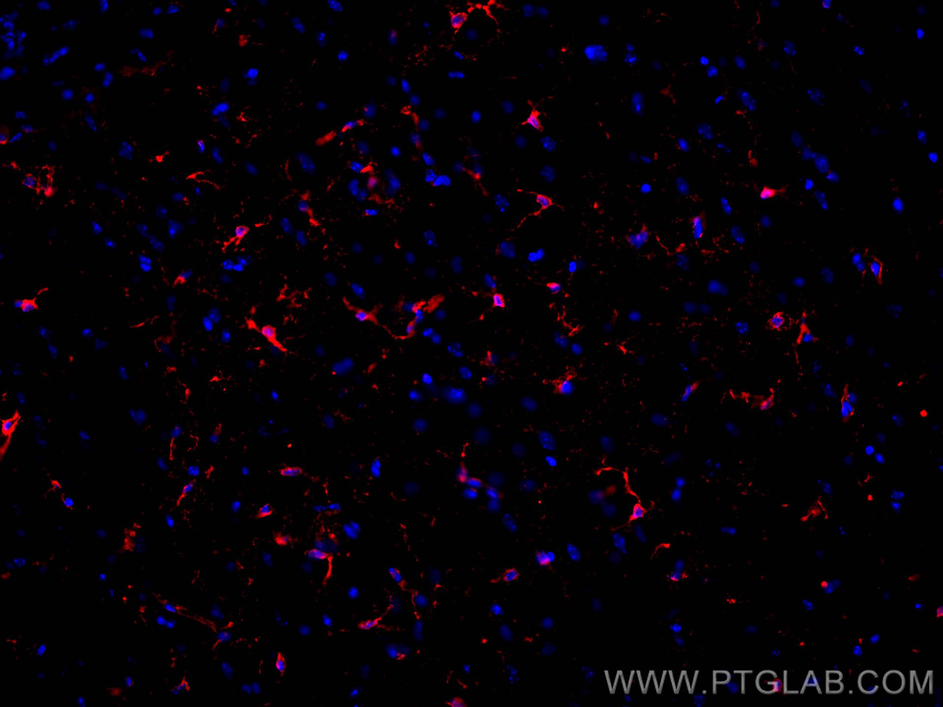
IF Staining of mouse brain using 68018-1-Ig
Immunofluorescent analysis of (4% PFA) fixed frozen OCT-embedded mouse brain tissue using ALDH1L1 antibody (68018-1-Ig, Clone: 1C1A3 ) at dilution of 1:400 and CoraLite®594-Conjugated AffiniPure Goat Anti-Mouse IgG(H+L) (SA00013-3).
The species listed in Tested Reactivity are in-house verified and applicable species. For unlisted species, please refer to the homology analysis of the immunogen sequence and related species. For rabbit polyclonal antibodies, homology >70% is recommended. For mouse monoclonal antibodies and rabbit recombinant antibodies, homology >90% is recommended. Generally, the higher the homology, the greater the applicability. However, there will be certain differences in protein expression in different species, tissues or cells. Therefore, the homology analysis results are for reference only and do not serve as a guarantee.
At Proteintech, we pride ourselves on our antibody quality, customer service and transparency. As such, we are comparing our antibodies with other vendors, enabling easy identification and comparisons of key data to help you choose the suitable antibody for your needs.
We have selected the top cited antibodies from these vendors for you to compare.
 at dilution of 1:20000 incubated at room temperature for 1.5 hours.")
 at dilution of 1:20000 incubated at room temperature for 1.5 hours.")
 at dilution of 1:20000 incubated at room temperature for 1.5 hours.")
 at dilution of 1:20000 incubated at room temperature for 1.5 hours.")
 at dilution of 1:32000 (under 40x lens). Heat mediated antigen retrieval with Tris-EDTA buffer (pH 9.0).")
 at dilution of 1:32000 (under 4x lens). Heat mediated antigen retrieval with Tris-EDTA buffer (pH 9.0).")
 fixed frozen OCT-embedded mouse brain tissue using ALDH1L1 antibody (68018-1-Ig, Clone: 1C1A3 ) at dilution of 1:400 and CoraLite®594-Conjugated AffiniPure Goat Anti-Mouse IgG(H+L) (SA00013-3).")