pig stomach tissue were subjected to SDS PAGE followed by western blot with 66768-1-Ig (AGR2 antibody) at dilution of 1:3000 incubated at room temperature for 1.5 hours.
pig stomach tissue were subjected to SDS PAGE followed by western blot with 66768-1-Ig (AGR2 antibody) at dilution of 1:3000 incubated at room temperature for 1.5 hours.
WB analysis of T-47D using 66768-1-Ig
T-47D cells were subjected to SDS PAGE followed by western blot with 66768-1-Ig (AGR2 antibody) at dilution of 1:3000 incubated at room temperature for 1.5 hours.
T-47D cells were subjected to SDS PAGE followed by western blot with 66768-1-Ig (AGR2 antibody) at dilution of 1:3000 incubated at room temperature for 1.5 hours.
WB analysis of HT-29 using 66768-1-Ig
HT-29 cells were subjected to SDS PAGE followed by western blot with 66768-1-Ig (AGR2 antibody) at dilution of 1:3000 incubated at room temperature for 1.5 hours.
HT-29 cells were subjected to SDS PAGE followed by western blot with 66768-1-Ig (AGR2 antibody) at dilution of 1:3000 incubated at room temperature for 1.5 hours.
IHC staining of human breast cancer using 66768-1-Ig
Immunohistochemical analysis of paraffin-embedded human breast cancer tissue slide using 66768-1-Ig (AGR2 antibody) at dilution of 1:300 (under 10x lens. Heat mediated antigen retrieval with Tris-EDTA buffer (pH 9.0).
Immunohistochemical analysis of paraffin-embedded human breast cancer tissue slide using 66768-1-Ig (AGR2 antibody) at dilution of 1:300 (under 10x lens. Heat mediated antigen retrieval with Tris-EDTA buffer (pH 9.0).
IHC staining of human breast cancer using 66768-1-Ig
Immunohistochemical analysis of paraffin-embedded human breast cancer tissue slide using 66768-1-Ig (AGR2 antibody) at dilution of 1:300 (under 40x lens. Heat mediated antigen retrieval with Tris-EDTA buffer (pH 9.0).
Immunohistochemical analysis of paraffin-embedded human breast cancer tissue slide using 66768-1-Ig (AGR2 antibody) at dilution of 1:300 (under 40x lens. Heat mediated antigen retrieval with Tris-EDTA buffer (pH 9.0).
IF Staining of human colon cancer using 66768-1-Ig
Immunofluorescent analysis of (4% PFA) fixed human colon cancer tissue using AGR2 antibody (66768-1-Ig, Clone: 1A8A8 ) at dilution of 1:400 and CoraLite®488-Conjugated AffiniPure Goat Anti-Mouse IgG(H+L).
Immunofluorescent analysis of (4% PFA) fixed human colon cancer tissue using AGR2 antibody (66768-1-Ig, Clone: 1A8A8 ) at dilution of 1:400 and CoraLite®488-Conjugated AffiniPure Goat Anti-Mouse IgG(H+L).
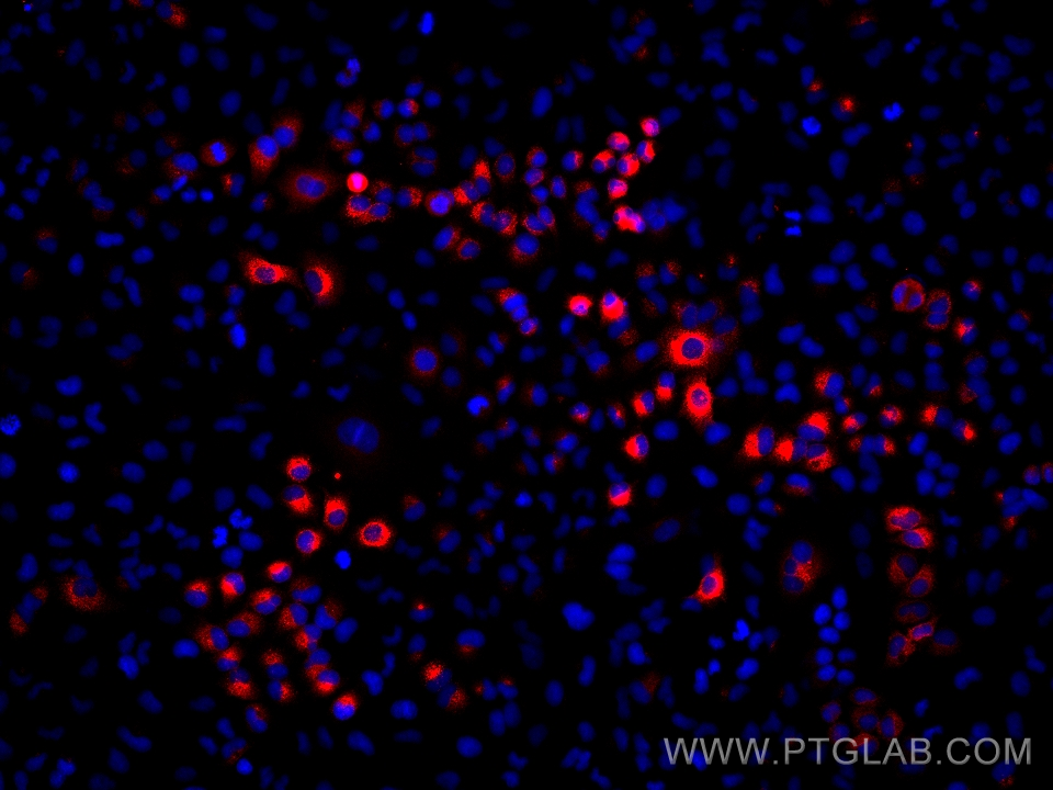
IF Staining of A549 using 66768-1-Ig
Immunofluorescent analysis of (4% PFA) fixed A549 cells using AGR2 antibody (66768-1-Ig, Clone: 1A8A8 ) at dilution of 1:1500 and Multi-rAb CoraLite® Plus 594-Goat Anti-Mouse Recombinant Secondary Antibody (H+L) (Cat.NO. RGAM004).
Immunofluorescent analysis of (4% PFA) fixed A549 cells using AGR2 antibody (66768-1-Ig, Clone: 1A8A8 ) at dilution of 1:1500 and Multi-rAb CoraLite® Plus 594-Goat Anti-Mouse Recombinant Secondary Antibody (H+L) (Cat.NO. RGAM004).
IF Staining of HT-29 using 66768-1-Ig
Immunofluorescent analysis of (-20°C Ethanol) fixed HT-29 cells using AGR2 antibody (66768-1-Ig, Clone: 1A8A8 ) at dilution of 1:800 and CoraLite®488-Conjugated AffiniPure Goat Anti-Mouse IgG(H+L).
Immunofluorescent analysis of (-20°C Ethanol) fixed HT-29 cells using AGR2 antibody (66768-1-Ig, Clone: 1A8A8 ) at dilution of 1:800 and CoraLite®488-Conjugated AffiniPure Goat Anti-Mouse IgG(H+L).
FC experiment of HT-29 using 66768-1-Ig
1X10^6 HT-29 cells were intracellularly stained with 0.2 ug Anti-Human AGR2 (66768-1-Ig, Clone:1A8A8) and CoraLite®488-Conjugated AffiniPure Goat Anti-Mouse IgG(H+L) at dilution 1:1000 (red), and 0.2 ug Mouse IgG2b Isotype Control (66360-3-Ig, Clone: K11B8C4B5) (blue). Cells were fixed with 4% PFA and permeabilized with 0.1% TritonX-100.
1X10^6 HT-29 cells were intracellularly stained with 0.2 ug Anti-Human AGR2 (66768-1-Ig, Clone:1A8A8) and CoraLite®488-Conjugated AffiniPure Goat Anti-Mouse IgG(H+L) at dilution 1:1000 (red), and 0.2 ug Mouse IgG2b Isotype Control (66360-3-Ig, Clone: K11B8C4B5) (blue). Cells were fixed with 4% PFA and permeabilized with 0.1% TritonX-100.
The Proteintech guarantee covers Proteintech antibodies in any species and any application, including those not listed on the datasheet. If the antibody doesn’t perform, you can receive a hassle-free refund or credit note.
human breast cancer tissue Note: suggested antigen retrieval with TE buffer pH 9.0; (*) Alternatively, antigen retrieval may be performed with citrate buffer pH 6.0
Positive IF-P detected in
human colon cancer tissue
Positive IF/ICC detected in
HT-29 cells, A549 cells
Positive FC (Intra) detected in
HT-29 cells
Recommended dilution
Application
Dilution
Western Blot (WB)
WB : 1:1000-1:6000
Immunohistochemistry (IHC)
IHC : 1:150-1:600
Immunofluorescence (IF)-P
IF-P : 1:200-1:800
Immunofluorescence (IF)/ICC
IF/ICC : 1:400-1:1600
Flow Cytometry (FC) (INTRA)
FC (INTRA) : 0.20 ug per 10^6 cells in a 100 µl suspension
It is recommended that this reagent should be titrated in each testing system to obtain optimal results.
Sample-dependent, Check data in validation data gallery.
PBS with 0.02% sodium azide and 50% glycerol, pH 7.3.
Storage Conditions
Store at -20°C. Stable for one year after shipment. Aliquoting is unnecessary for -20oC storage. 20ul sizes contain 0.1% BSA.
Background Information
AGR2, also named AG2 or HPC8, encodes anterior gradient protein 2 homolog which belongs to the AGR family. It is a secreted protein localized in endoplasmic reticulum. AGR2 plays roles in MUC2 post-transcriptional synthesis,secretion and production of mucus by intestinal cells. AGR2 was significantly elevated in the pancreatic juice from patients with pre-malignant conditions as well as pancreatic cancer compared to control pancreatic juice samples. AGR2 levels in pancreatic juice could potentially be used to aide in assessment of high-risk patients undergoing endoscopic procedures.
pig stomach tissue were subjected to SDS PAGE followed by western blot with 66768-1-Ig (AGR2 antibody) at dilution of 1:3000 incubated at room temperature for 1.5 hours.
WB analysis of T-47D using 66768-1-Ig
T-47D cells were subjected to SDS PAGE followed by western blot with 66768-1-Ig (AGR2 antibody) at dilution of 1:3000 incubated at room temperature for 1.5 hours.
WB analysis of HT-29 using 66768-1-Ig
HT-29 cells were subjected to SDS PAGE followed by western blot with 66768-1-Ig (AGR2 antibody) at dilution of 1:3000 incubated at room temperature for 1.5 hours.
IHC Figures
IHC staining of human breast cancer using 66768-1-Ig
Immunohistochemical analysis of paraffin-embedded human breast cancer tissue slide using 66768-1-Ig (AGR2 antibody) at dilution of 1:300 (under 10x lens. Heat mediated antigen retrieval with Tris-EDTA buffer (pH 9.0).
IHC staining of human breast cancer using 66768-1-Ig
Immunohistochemical analysis of paraffin-embedded human breast cancer tissue slide using 66768-1-Ig (AGR2 antibody) at dilution of 1:300 (under 40x lens. Heat mediated antigen retrieval with Tris-EDTA buffer (pH 9.0).
IF-P Figures
IF Staining of human colon cancer using 66768-1-Ig
Immunofluorescent analysis of (4% PFA) fixed human colon cancer tissue using AGR2 antibody (66768-1-Ig, Clone: 1A8A8 ) at dilution of 1:400 and CoraLite®488-Conjugated AffiniPure Goat Anti-Mouse IgG(H+L).
IF Staining of human colon cancer using 66768-1-Ig
Immunofluorescent analysis of (4% PFA) fixed human colon cancer tissue using AGR2 antibody (66768-1-Ig, Clone: 1A8A8 ) at dilution of 1:400 and CoraLite®488-Conjugated AffiniPure Goat Anti-Mouse IgG(H+L).
IF/ICC Figures
IF Staining of A549 using 66768-1-Ig
Immunofluorescent analysis of (4% PFA) fixed A549 cells using AGR2 antibody (66768-1-Ig, Clone: 1A8A8 ) at dilution of 1:1500 and Multi-rAb CoraLite® Plus 594-Goat Anti-Mouse Recombinant Secondary Antibody (H+L) (Cat.NO. RGAM004).
IF Staining of HT-29 using 66768-1-Ig
Immunofluorescent analysis of (-20°C Ethanol) fixed HT-29 cells using AGR2 antibody (66768-1-Ig, Clone: 1A8A8 ) at dilution of 1:800 and CoraLite®488-Conjugated AffiniPure Goat Anti-Mouse IgG(H+L).
FC (INTRA) Figures
FC experiment of HT-29 using 66768-1-Ig
1X10^6 HT-29 cells were intracellularly stained with 0.2 ug Anti-Human AGR2 (66768-1-Ig, Clone:1A8A8) and CoraLite®488-Conjugated AffiniPure Goat Anti-Mouse IgG(H+L) at dilution 1:1000 (red), and 0.2 ug Mouse IgG2b Isotype Control (66360-3-Ig, Clone: K11B8C4B5) (blue). Cells were fixed with 4% PFA and permeabilized with 0.1% TritonX-100.
The species listed in Tested Reactivity are in-house verified and applicable species. For unlisted species, please refer to the homology analysis of the immunogen sequence and related species. For rabbit polyclonal antibodies, homology >70% is recommended. For mouse monoclonal antibodies and rabbit recombinant antibodies, homology >90% is recommended. Generally, the higher the homology, the greater the applicability. However, there will be certain differences in protein expression in different species, tissues or cells. Therefore, the homology analysis results are for reference only and do not serve as a guarantee.
At Proteintech, we pride ourselves on our antibody quality, customer service and transparency. As such, we are comparing our antibodies with other vendors, enabling easy identification and comparisons of key data to help you choose the suitable antibody for your needs.
We have selected the top cited antibodies from these vendors for you to compare.
Proteintech
AGR2 Monoclonal antibody
$149 (20ul) $449 (150ul)
Catalog Number
66768-1-Ig
Citations
2
Dilutions
WB : 1:1000-1:6000 IHC : 1:150-1:600 IF-P : 1:200-1:800 IF/ICC : 1:400-1:1600 FC (INTRA) : 0.20 ug per 10^6 cells in a 100 µl suspension
Applications
WB, IHC, IF/ICC, IF-P, FC (Intra), ELISA
Reactivity
human, pig
Product Guarantee
Covers any species including not listed on datasheet
Covers any applications including not listed on datasheet
 at dilution of 1:3000 incubated at room temperature for 1.5 hours.")
 at dilution of 1:3000 incubated at room temperature for 1.5 hours.")
 at dilution of 1:3000 incubated at room temperature for 1.5 hours.")
 at dilution of 1:300 (under 10x lens. Heat mediated antigen retrieval with Tris-EDTA buffer (pH 9.0).")
 at dilution of 1:300 (under 40x lens. Heat mediated antigen retrieval with Tris-EDTA buffer (pH 9.0).")
 fixed human colon cancer tissue using AGR2 antibody (66768-1-Ig, Clone: 1A8A8 ) at dilution of 1:400 and CoraLite®488-Conjugated AffiniPure Goat Anti-Mouse IgG(H+L).")
 fixed human colon cancer tissue using AGR2 antibody (66768-1-Ig, Clone: 1A8A8 ) at dilution of 1:400 and CoraLite®488-Conjugated AffiniPure Goat Anti-Mouse IgG(H+L).")
 fixed A549 cells using AGR2 antibody (66768-1-Ig, Clone: 1A8A8 ) at dilution of 1:1500 and Multi-rAb CoraLite® Plus 594-Goat Anti-Mouse Recombinant Secondary Antibody (H+L) (Cat.NO. RGAM004).")
 fixed HT-29 cells using AGR2 antibody (66768-1-Ig, Clone: 1A8A8 ) at dilution of 1:800 and CoraLite®488-Conjugated AffiniPure Goat Anti-Mouse IgG(H+L).")
 and CoraLite®488-Conjugated AffiniPure Goat Anti-Mouse IgG(H+L) at dilution 1:1000 (red), and 0.2 ug Mouse IgG2b Isotype Control (66360-3-Ig, Clone: K11B8C4B5) (blue). Cells were fixed with 4% PFA and permeabilized with 0.1% TritonX-100.")