Various lysates were subjected to SDS PAGE followed by western blot with 67338-1-Ig (PSMD9 antibody) at dilution of 1:5000 incubated at room temperature for 1.5 hours.
Various lysates were subjected to SDS PAGE followed by western blot with 67338-1-Ig (PSMD9 antibody) at dilution of 1:5000 incubated at room temperature for 1.5 hours.
WB analysis of A549 using 67338-1-Ig
WB result of PSMD9 antibody (67338-1-Ig; 1:1000; incubated at room temperature for 1.5 hours) with sh-Control and sh-PSMD9 transfected A549 cells.
WB result of PSMD9 antibody (67338-1-Ig; 1:1000; incubated at room temperature for 1.5 hours) with sh-Control and sh-PSMD9 transfected A549 cells.
WB analysis using 67338-1-Ig
Various lysates were subjected to SDS PAGE followed by western blot with 67338-1-Ig (PSMD9 antibody) at dilution of 1:2800 incubated at room temperature for 1.5 hours.
Various lysates were subjected to SDS PAGE followed by western blot with 67338-1-Ig (PSMD9 antibody) at dilution of 1:2800 incubated at room temperature for 1.5 hours.
IHC staining of human cervical cancer using 67338-1-Ig
Immunohistochemical analysis of paraffin-embedded human cervical cancer tissue slide using 67338-1-Ig (PSMD9 antibody) at dilution of 1:800 (under 40x lens). Heat mediated antigen retrieval with Tris-EDTA buffer (pH 9.0).
Immunohistochemical analysis of paraffin-embedded human cervical cancer tissue slide using 67338-1-Ig (PSMD9 antibody) at dilution of 1:800 (under 40x lens). Heat mediated antigen retrieval with Tris-EDTA buffer (pH 9.0).
IHC staining of human cervical cancer using 67338-1-Ig
Immunohistochemical analysis of paraffin-embedded human cervical cancer tissue slide using 67338-1-Ig (PSMD9 antibody) at dilution of 1:800 (under 10x lens). Heat mediated antigen retrieval with Tris-EDTA buffer (pH 9.0).
Immunohistochemical analysis of paraffin-embedded human cervical cancer tissue slide using 67338-1-Ig (PSMD9 antibody) at dilution of 1:800 (under 10x lens). Heat mediated antigen retrieval with Tris-EDTA buffer (pH 9.0).
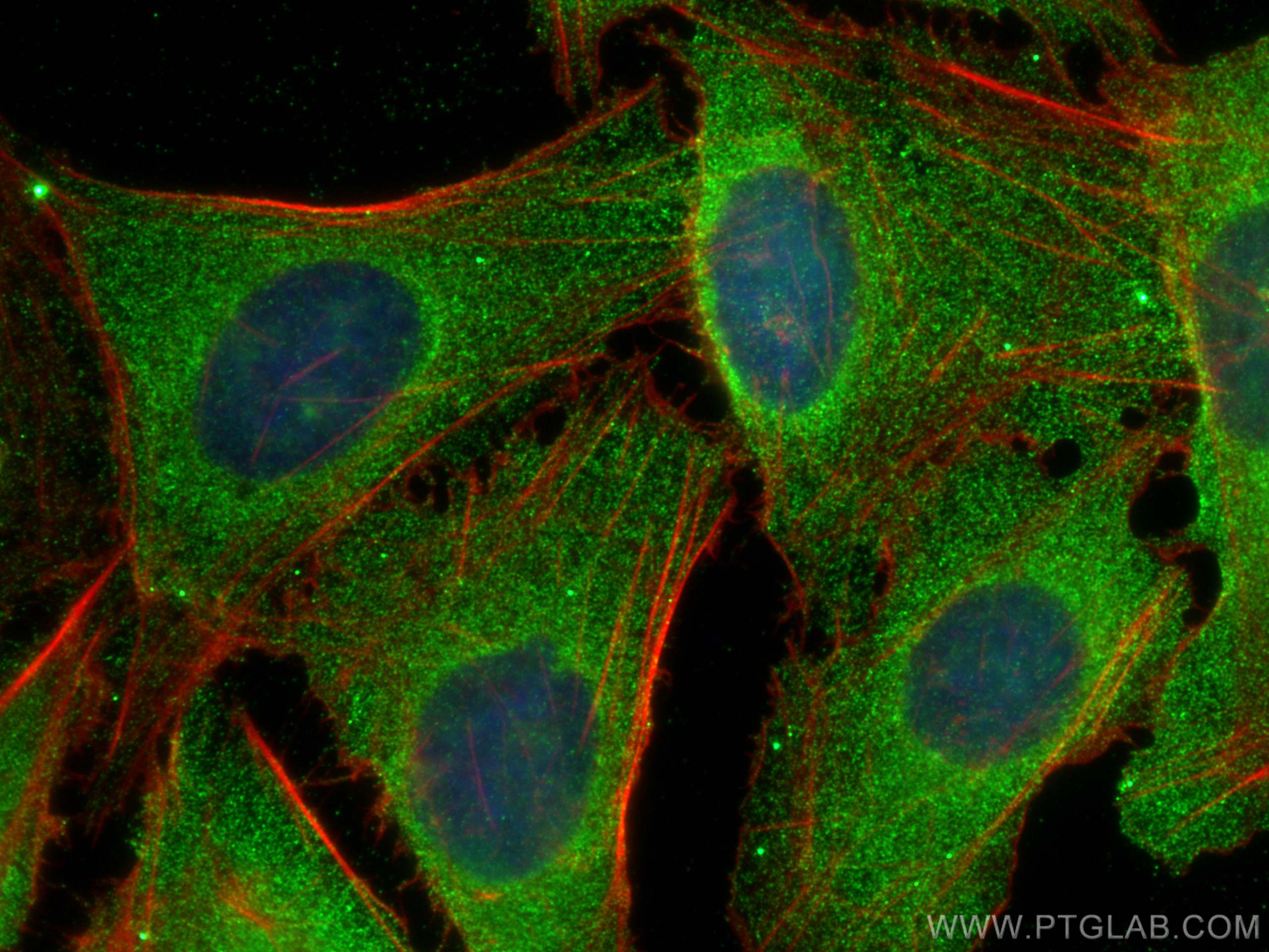
IF Staining of U2OS using 67338-1-Ig
Immunofluorescent analysis of (4% PFA) fixed U2OS cells using PSMD9 antibody (67338-1-Ig, Clone: 1H2G1 ) at dilution of 1:800 and CoraLite®488-Conjugated AffiniPure Goat Anti-Mouse IgG(H+L), CL594-Phalloidin (red).
Immunofluorescent analysis of (4% PFA) fixed U2OS cells using PSMD9 antibody (67338-1-Ig, Clone: 1H2G1 ) at dilution of 1:800 and CoraLite®488-Conjugated AffiniPure Goat Anti-Mouse IgG(H+L), CL594-Phalloidin (red).
IF Staining of HeLa using 67338-1-Ig
Immunofluorescent analysis of (-20°C Ethanol) fixed HeLa cells using PSMD9 antibody (67338-1-Ig, Clone: 1H2G1 ) at dilution of 1:400 and CoraLite®488-Conjugated AffiniPure Goat Anti-Mouse IgG(H+L).
Immunofluorescent analysis of (-20°C Ethanol) fixed HeLa cells using PSMD9 antibody (67338-1-Ig, Clone: 1H2G1 ) at dilution of 1:400 and CoraLite®488-Conjugated AffiniPure Goat Anti-Mouse IgG(H+L).
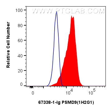
FC experiment of HeLa using 67338-1-Ig
1x10^6 HeLa cells were intracellularly stained with 0.25 ug PSMD9 Monoclonal antibody (67338-1-Ig, Clone:1H2G1) and CoraLite®488-Conjugated Goat Anti-Mouse IgG(H+L) (SA00013-1)(red), or 0.25 ug Mouse IgG1 isotype control Mouse McAb (66360-1-Ig, Clone: 1F8D3) (blue). Cells were fixed with 4% PFA and permeabilized with Flow Cytometry Perm Buffer (PF00011-C).
1x10^6 HeLa cells were intracellularly stained with 0.25 ug PSMD9 Monoclonal antibody (67338-1-Ig, Clone:1H2G1) and CoraLite®488-Conjugated Goat Anti-Mouse IgG(H+L) (SA00013-1)(red), or 0.25 ug Mouse IgG1 isotype control Mouse McAb (66360-1-Ig, Clone: 1F8D3) (blue). Cells were fixed with 4% PFA and permeabilized with Flow Cytometry Perm Buffer (PF00011-C).
The Proteintech guarantee covers Proteintech antibodies in any species and any application, including those not listed on the datasheet. If the antibody doesn’t perform, you can receive a hassle-free refund or credit note.
human cervical cancer tissue Note: suggested antigen retrieval with TE buffer pH 9.0; (*) Alternatively, antigen retrieval may be performed with citrate buffer pH 6.0
Positive IF/ICC detected in
U2OS cells, HeLa cells
Positive FC (Intra) detected in
HeLa cells
Recommended dilution
Application
Dilution
Western Blot (WB)
WB : 1:2000-1:10000
Immunohistochemistry (IHC)
IHC : 1:400-1:1600
Immunofluorescence (IF)/ICC
IF/ICC : 1:400-1:1600
Flow Cytometry (FC) (INTRA)
FC (INTRA) : 0.25 ug per 10^6 cells in a 100 µl suspension
It is recommended that this reagent should be titrated in each testing system to obtain optimal results.
Sample-dependent, Check data in validation data gallery.
PBS with 0.02% sodium azide and 50% glycerol , pH 7.3
Storage Conditions
Store at -20°C. Stable for one year after shipment. Aliquoting is unnecessary for -20oC storage. 20ul sizes contain 0.1% BSA.
Background Information
PSMD9 is a ubiquitous protein of eukaryotic cells and is a chaperon of the 26S proteasome complex, which degrades ubiquitinated proteins in eukaryotic cells and contributes to the degradation of intracellular proteins into antigenic peptides for antigen presentation by MHC class I cells. The 26S mammalian base sub-complex involves three distinct modules which have ATPase subunits distinctly associated to three chaperones, one of which is PSMD9 regulating the modules assembly. The PSMD9 ubiquitous regulatory role within the proteasome implies its potential pleiotropic effects within different physio-pathological systems. PSMD9 is known to form a stable subcomplex with PSMC3 and PSMC6, two of the AAA-ATPases, assisting in the assembly of the 20S and 19S particles to form the holo complex.
Various lysates were subjected to SDS PAGE followed by western blot with 67338-1-Ig (PSMD9 antibody) at dilution of 1:5000 incubated at room temperature for 1.5 hours.
WB analysis of A549 using 67338-1-Ig
WB result of PSMD9 antibody (67338-1-Ig; 1:1000; incubated at room temperature for 1.5 hours) with sh-Control and sh-PSMD9 transfected A549 cells.
WB analysis using 67338-1-Ig
Various lysates were subjected to SDS PAGE followed by western blot with 67338-1-Ig (PSMD9 antibody) at dilution of 1:2800 incubated at room temperature for 1.5 hours.
IHC Figures
IHC staining of human cervical cancer using 67338-1-Ig
Immunohistochemical analysis of paraffin-embedded human cervical cancer tissue slide using 67338-1-Ig (PSMD9 antibody) at dilution of 1:800 (under 40x lens). Heat mediated antigen retrieval with Tris-EDTA buffer (pH 9.0).
IHC staining of human cervical cancer using 67338-1-Ig
Immunohistochemical analysis of paraffin-embedded human cervical cancer tissue slide using 67338-1-Ig (PSMD9 antibody) at dilution of 1:800 (under 10x lens). Heat mediated antigen retrieval with Tris-EDTA buffer (pH 9.0).
IF/ICC Figures
IF Staining of U2OS using 67338-1-Ig
Immunofluorescent analysis of (4% PFA) fixed U2OS cells using PSMD9 antibody (67338-1-Ig, Clone: 1H2G1 ) at dilution of 1:800 and CoraLite®488-Conjugated AffiniPure Goat Anti-Mouse IgG(H+L), CL594-Phalloidin (red).
IF Staining of HeLa using 67338-1-Ig
Immunofluorescent analysis of (-20°C Ethanol) fixed HeLa cells using PSMD9 antibody (67338-1-Ig, Clone: 1H2G1 ) at dilution of 1:400 and CoraLite®488-Conjugated AffiniPure Goat Anti-Mouse IgG(H+L).
FC (INTRA) Figures
FC experiment of HeLa using 67338-1-Ig
1x10^6 HeLa cells were intracellularly stained with 0.25 ug PSMD9 Monoclonal antibody (67338-1-Ig, Clone:1H2G1) and CoraLite®488-Conjugated Goat Anti-Mouse IgG(H+L) (SA00013-1)(red), or 0.25 ug Mouse IgG1 isotype control Mouse McAb (66360-1-Ig, Clone: 1F8D3) (blue). Cells were fixed with 4% PFA and permeabilized with Flow Cytometry Perm Buffer (PF00011-C).
The species listed in Tested Reactivity are in-house verified and applicable species. For unlisted species, please refer to the homology analysis of the immunogen sequence and related species. For rabbit polyclonal antibodies, homology >70% is recommended. For mouse monoclonal antibodies and rabbit recombinant antibodies, homology >90% is recommended. Generally, the higher the homology, the greater the applicability. However, there will be certain differences in protein expression in different species, tissues or cells. Therefore, the homology analysis results are for reference only and do not serve as a guarantee.
At Proteintech, we pride ourselves on our antibody quality, customer service and transparency. As such, we are comparing our antibodies with other vendors, enabling easy identification and comparisons of key data to help you choose the suitable antibody for your needs.
We have selected the top cited antibodies from these vendors for you to compare.
Proteintech
KD/KO VALIDATED
PSMD9 Monoclonal antibody
Catalog Number
67338-1-Ig
Citations
2
Dilutions
WB : 1:2000-1:10000 IHC : 1:400-1:1600 IF/ICC : 1:400-1:1600 FC (INTRA) : 0.25 ug per 10^6 cells in a 100 µl suspension
Applications
WB, IHC, IF/ICC, FC (Intra), ELISA
Reactivity
human, mouse, rat, pig
Product Guarantee
Covers any species including not listed on datasheet
Covers any applications including not listed on datasheet
 at dilution of 1:5000 incubated at room temperature for 1.5 hours.")
 with sh-Control and sh-PSMD9 transfected A549 cells.")
 at dilution of 1:2800 incubated at room temperature for 1.5 hours.")
 at dilution of 1:800 (under 40x lens). Heat mediated antigen retrieval with Tris-EDTA buffer (pH 9.0).")
 at dilution of 1:800 (under 10x lens). Heat mediated antigen retrieval with Tris-EDTA buffer (pH 9.0).")
 fixed U2OS cells using PSMD9 antibody (67338-1-Ig, Clone: 1H2G1 ) at dilution of 1:800 and CoraLite®488-Conjugated AffiniPure Goat Anti-Mouse IgG(H+L), CL594-Phalloidin (red).")
 fixed HeLa cells using PSMD9 antibody (67338-1-Ig, Clone: 1H2G1 ) at dilution of 1:400 and CoraLite®488-Conjugated AffiniPure Goat Anti-Mouse IgG(H+L).")
 and CoraLite®488-Conjugated Goat Anti-Mouse IgG(H+L) (SA00013-1)(red), or 0.25 ug Mouse IgG1 isotype control Mouse McAb (66360-1-Ig, Clone: 1F8D3) (blue). Cells were fixed with 4% PFA and permeabilized with Flow Cytometry Perm Buffer (PF00011-C).")