- Phare
- Validé par KD/KO
Anticorps Polyclonal de lapin anti-Rubicon
Rubicon Polyclonal Antibody for WB, IF, IHC, ELISA
Hôte / Isotype
Lapin / IgG
Réactivité testée
Humain, rat, souris
Applications
WB, IHC, IF/ICC, IP, ELISA
Conjugaison
Non conjugué
N° de cat : 21444-1-AP
Synonymes
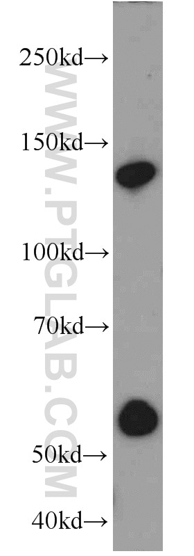
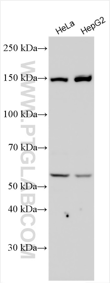
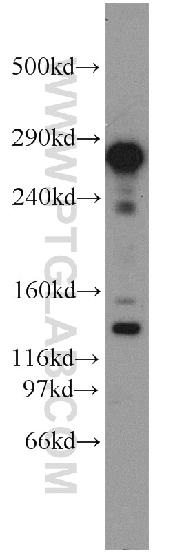
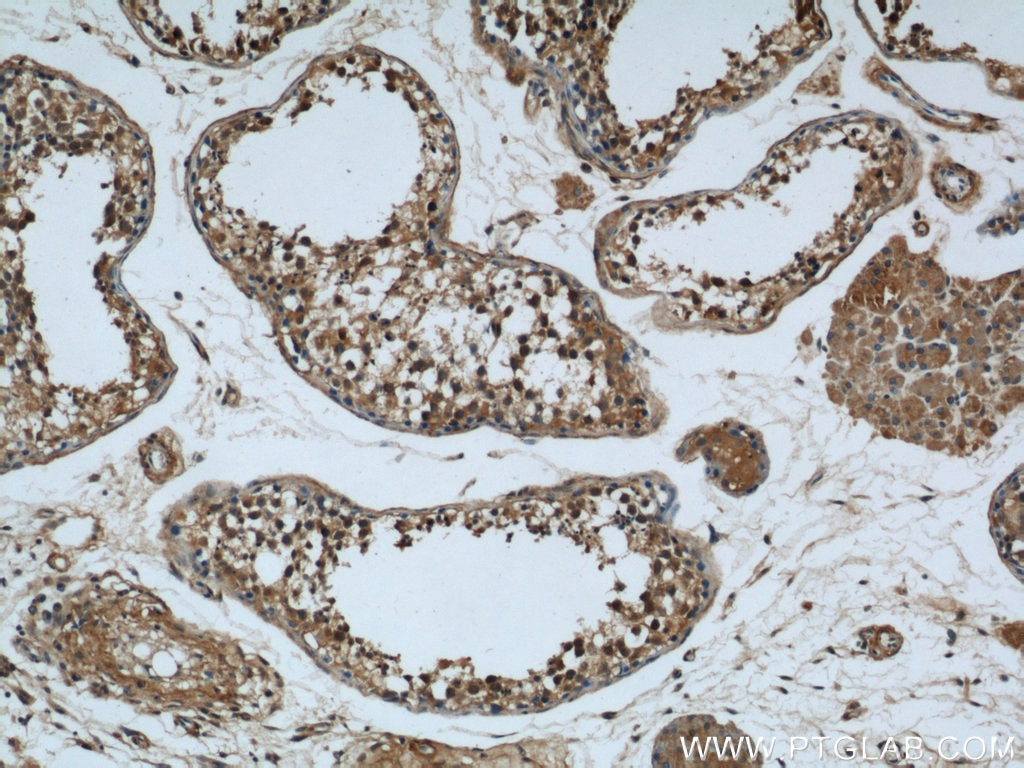
Galerie de données de validation
Applications testées
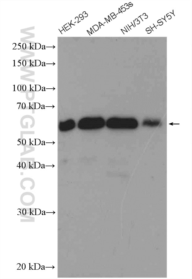
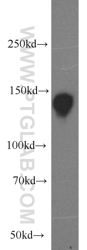
| Résultats positifs en WB | tissu hépatique de souris, cellules HeLa, cellules HepG2 |
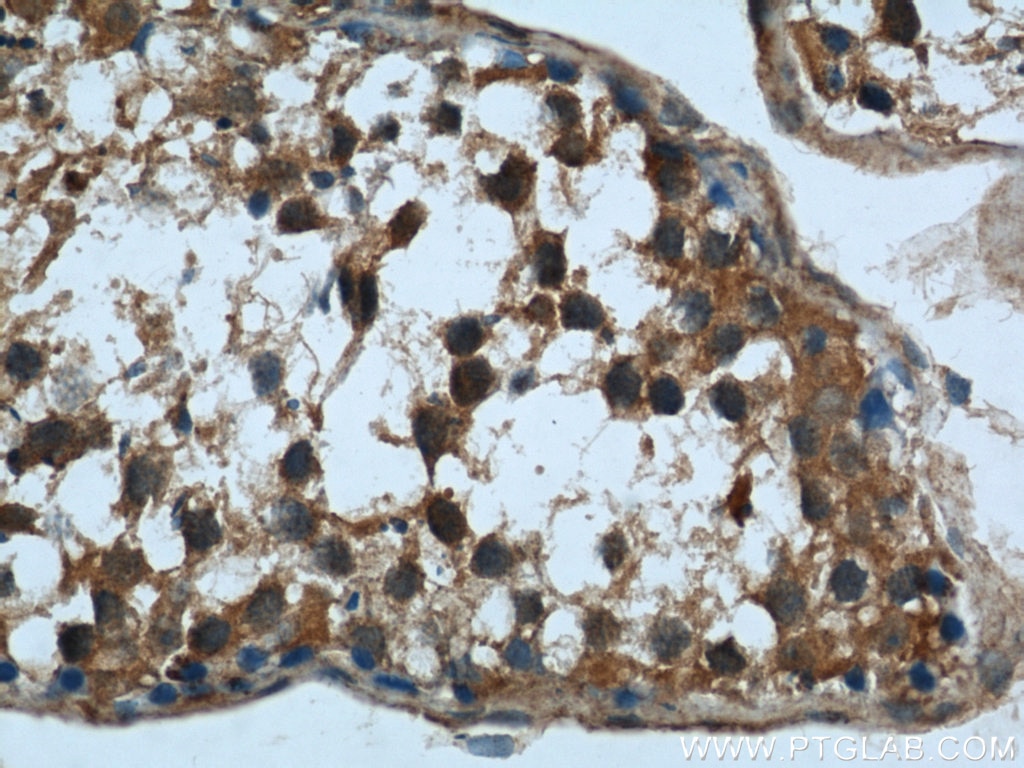
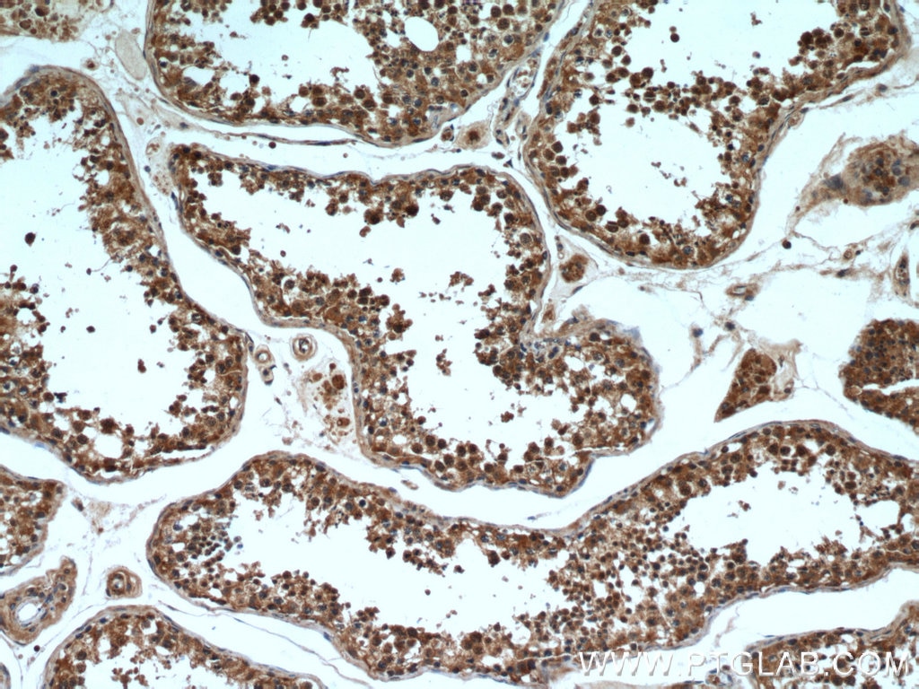
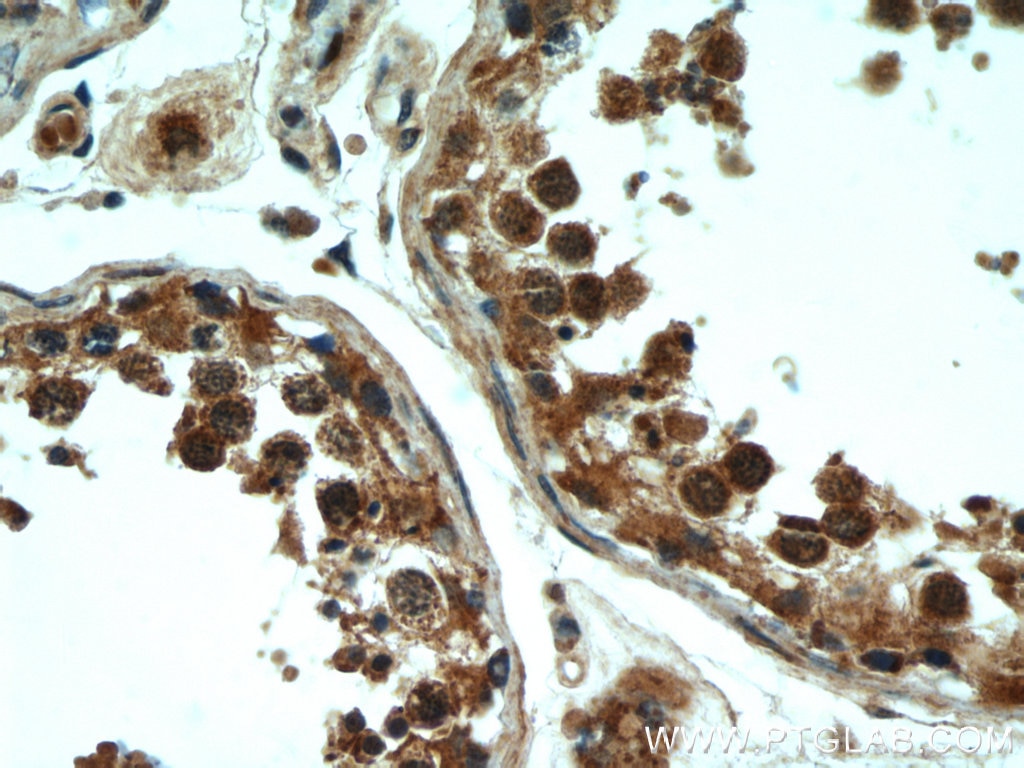
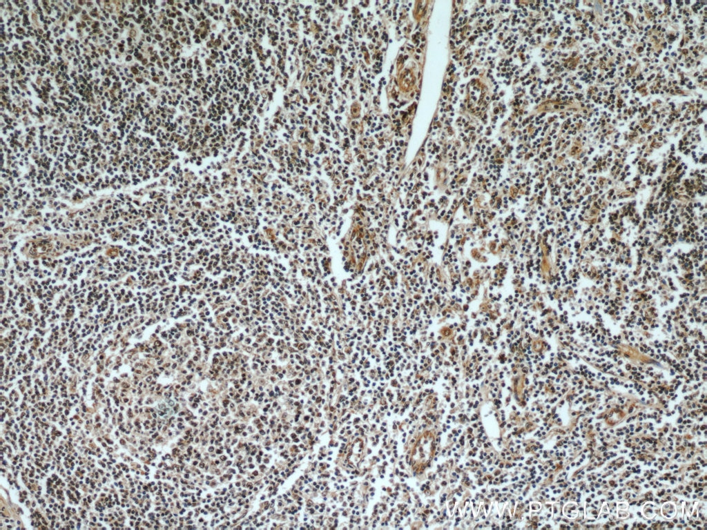
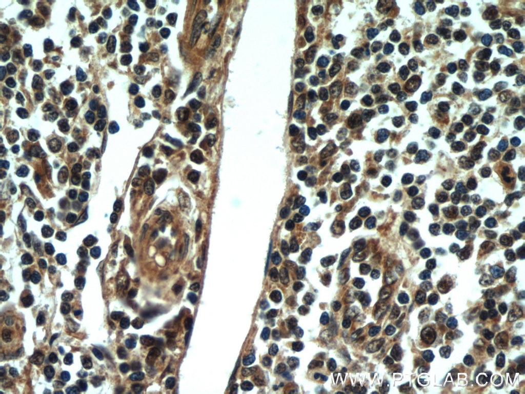
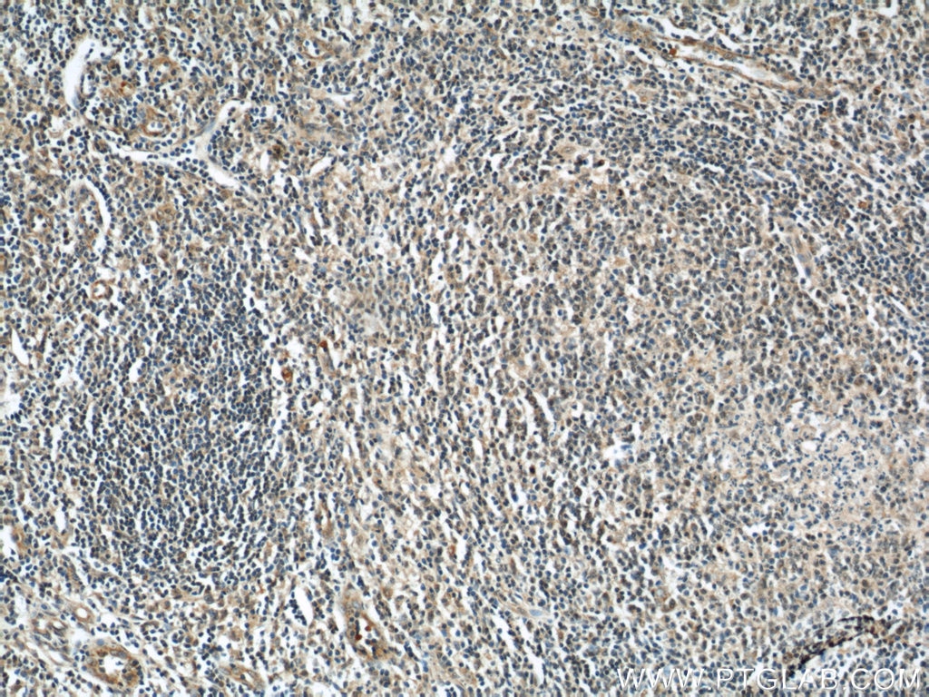
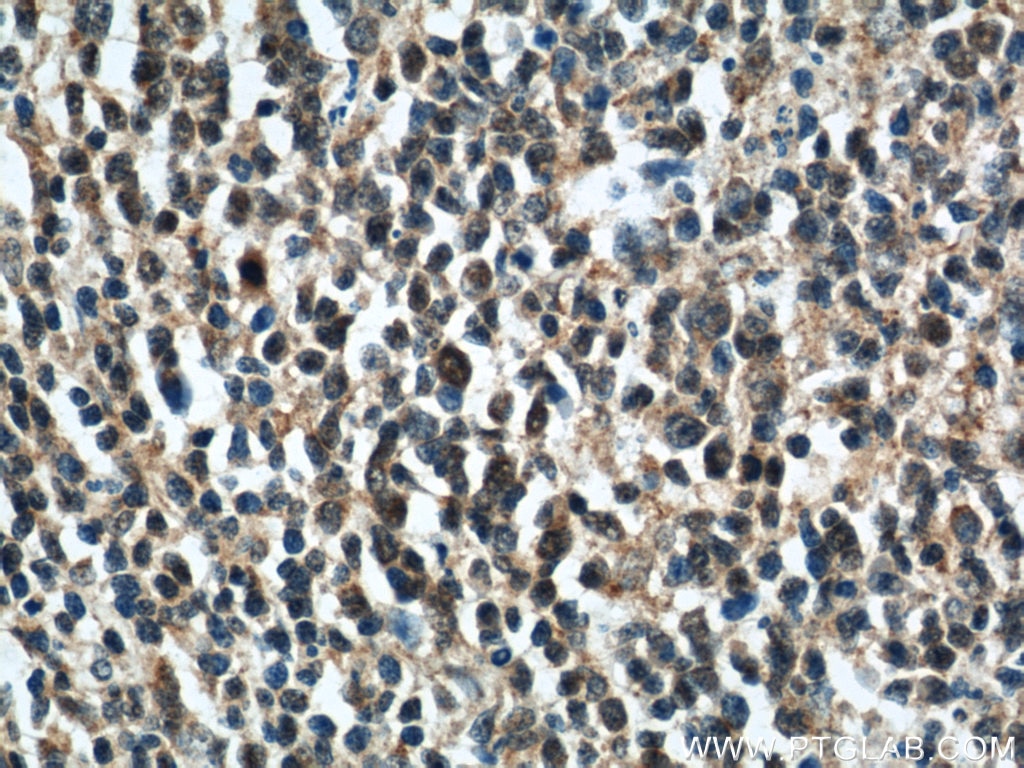
| Résultats positifs en IHC | tissu testiculaire humain, tissu d'amygdalite humain il est suggéré de démasquer l'antigène avec un tampon de TE buffer pH 9.0; (*) À défaut, 'le démasquage de l'antigène peut être 'effectué avec un tampon citrate pH 6,0. |
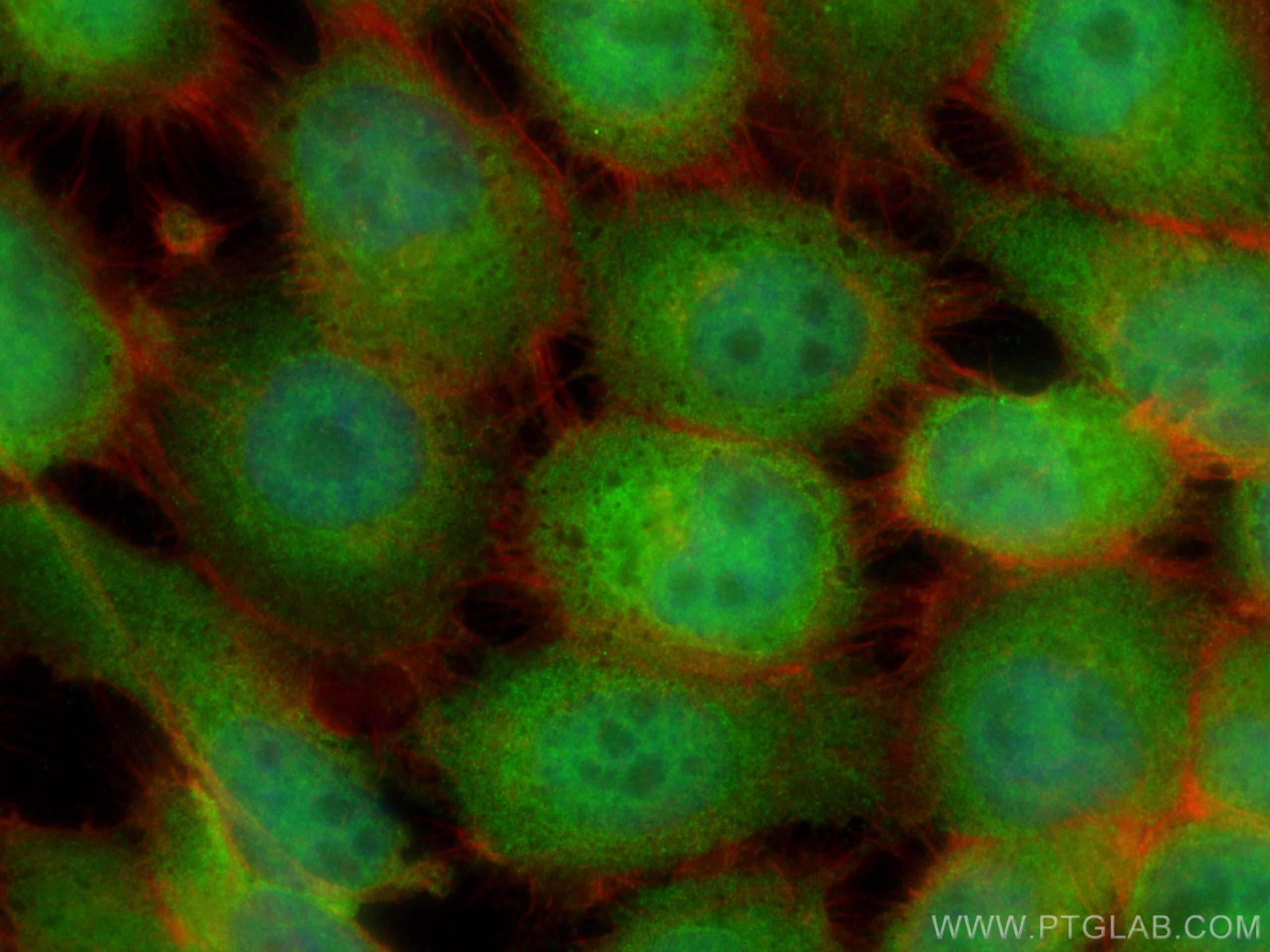
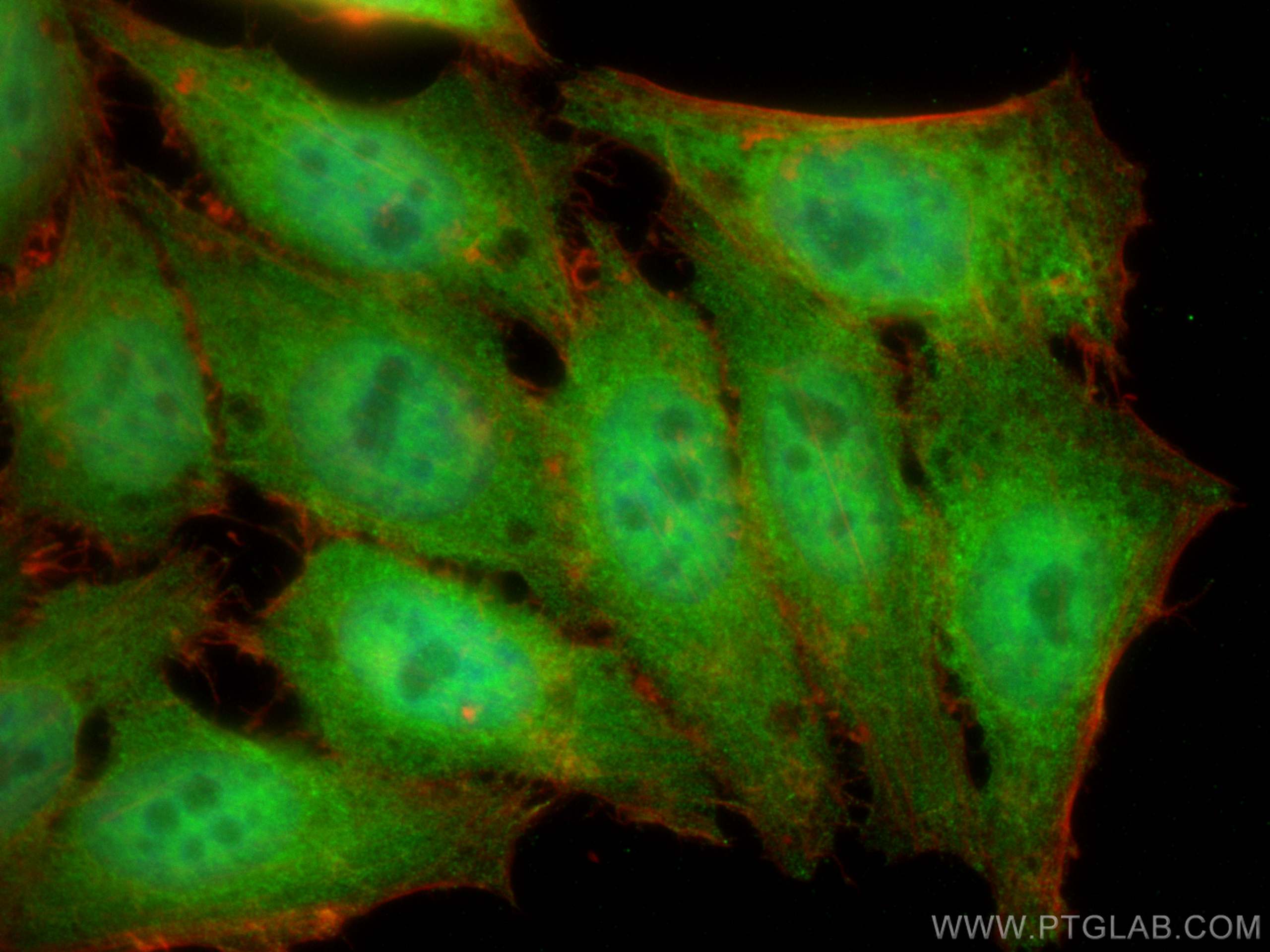
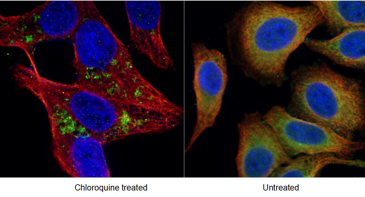
| Résultats positifs en IF/ICC | cellules HepG2, cellules A431 |
Dilution recommandée
| Application | Dilution |
|---|---|
| Western Blot (WB) | WB : 1:500-1:2000 |
| Immunohistochimie (IHC) | IHC : 1:20-1:200 |
| Immunofluorescence (IF)/ICC | IF/ICC : 1:50-1:500 |
| It is recommended that this reagent should be titrated in each testing system to obtain optimal results. | |
| Sample-dependent, check data in validation data gallery | |
Applications publiées
| KD/KO | See 4 publications below |
| WB | See 15 publications below |
| IHC | See 3 publications below |
| IF | See 8 publications below |
| IP | See 2 publications below |
Informations sur le produit
21444-1-AP cible Rubicon dans les applications de WB, IHC, IF/ICC, IP, ELISA et montre une réactivité avec des échantillons Humain, rat, souris
| Réactivité | Humain, rat, souris |
| Réactivité citée | rat, Humain, souris |
| Hôte / Isotype | Lapin / IgG |
| Clonalité | Polyclonal |
| Type | Anticorps |
| Immunogène | Rubicon Protéine recombinante Ag13715 |
| Nom complet | KIAA0226 |
| Masse moléculaire calculée | 972 aa, 109 kDa |
| Poids moléculaire observé | 110-130 kDa |
| Numéro d’acquisition GenBank | BC033615 |
| Symbole du gène | Rubicon |
| Identification du gène (NCBI) | 9711 |
| Conjugaison | Non conjugué |
| Forme | Liquide |
| Méthode de purification | Purification par affinité contre l'antigène |
| Tampon de stockage | PBS avec azoture de sodium à 0,02 % et glycérol à 50 % pH 7,3 |
| Conditions de stockage | Stocker à -20°C. Stable pendant un an après l'expédition. L'aliquotage n'est pas nécessaire pour le stockage à -20oC Les 20ul contiennent 0,1% de BSA. |
Informations générales
Rubicon (Run domain Beclin-1 interacting and cysteine-rich containing protein) consists of an N-terminal RUN domain, a middle region that includes its PI3K-binding domain (PIKBD), a coiled-coil domain (CCD), a serine-rich region (S-R), a helix-coil-rich domain (H-C) and a C-terminal Rubicon homology domain (RH) (PMID: 36288306). Rubicon is ubiquitously expressed in most tissue and organs.
Protocole
| Product Specific Protocols | |
|---|---|
| WB protocol for Rubicon antibody 21444-1-AP | Download protocol |
| IHC protocol for Rubicon antibody 21444-1-AP | Download protocol |
| IF protocol for Rubicon antibody 21444-1-AP | Download protocol |
| Standard Protocols | |
|---|---|
| Click here to view our Standard Protocols |
Publications
| Species | Application | Title |
|---|---|---|
ACS Nano Neutrophil Nanovesicle Protects against Experimental Autoimmune Encephalomyelitis through Enhancing Myelin Clearance by Microglia | ||
Adv Sci (Weinh) Bone Marrow Mesenchymal Stem Cell-Derived Dermcidin-Containing Migrasomes enhance LC3-Associated Phagocytosis of Pulmonary Macrophages and Protect against Post-Stroke Pneumonia | ||
Autophagy STYK1 promotes autophagy through enhancing the assembly of autophagy-specific class III phosphatidylinositol 3-kinase complex I. | ||
Autophagy RUBCN/rubicon and EGFR regulate lysosomal degradative processes in the retinal pigment epithelium (RPE) of the eye.
| ||
Avis
The reviews below have been submitted by verified Proteintech customers who received an incentive forproviding their feedback.
FH Daniel (Verified Customer) (07-21-2021) | The antibody seemed to work okay, although I had issues differentiating between autofluorescence and a positive signal. However, that is not the fault of the antibody, but rather my cells.
|
