Histone-H3 Polyklonaler Antikörper
Histone-H3 Polyklonal Antikörper für FC, IF, IHC, IP, WB, ELISA
Wirt / Isotyp
Kaninchen / IgG
Getestete Reaktivität
human, Maus, Ratte und mehr (6)
Anwendung
WB, IP, IHC, IF, FC, CoIP, ChIP, ELISA
Konjugation
Unkonjugiert
Kat-Nr. : 17168-1-AP
Synonyme
 at dilution of 1:8000 incubated at room temperature for 1.5 hours.")
 at dilution of 1:1000 incubated at room temperature for 1.5 hours.")
 at dilution of 1:300 incubated at room temperature for 1.5 hours.")
 at dilution of 1:300 incubated at room temperature for 1.5 hours.")
 at dilution of 1:300 incubated at room temperature for 1.5 hours.")
 with MCF-7 cells lysate 2120 ug.")
 at dilution of 1:200 (under 40x lens). Heat mediated antigen retrieval with Tris-EDTA buffer (pH 9.0).")
 at dilution of 1:200 (under 10x lens). Heat mediated antigen retrieval with Tris-EDTA buffer (pH 9.0).")
 at dilution of 1:200 (under 40x lens).")
 at dilution of 1:200 (under 10x lens).")
 at dilution of 1:2000 (under 10x lens). Heat mediated antigen retrieval with Tris-EDTA buffer (pH 9.0).")
 at dilution of 1:2000 (under 40x lens). Heat mediated antigen retrieval with Tris-EDTA buffer (pH 9.0).")
 fixed HeLa cells using Histone-H3 antibody (17168-1-AP) at dilution of 1:200 and CoraLite®488-Conjugated AffiniPure Goat Anti-Rabbit IgG(H+L).")
 at dilution of 1:50 and Rhodamine-Goat anti-Rabbit IgG.")
 fixed HeLa cells using Histone-H3 antibody (17168-1-AP) at dilution of 1:200 and CoraLite®488-Conjugated AffiniPure Goat Anti-Rabbit IgG(H+L), CL594-Phalloidin (red).")
 at dilution of 1:50 and Rhodamine-Goat anti-Rabbit IgG.")
 and control antibody (blue). Fixed with 90% MeOH.")
"Histone-H3 Antibodies" Comparison
View side-by-side comparison of Histone-H3 antibodies from other vendors to find the one that best suits your research needs.
Geprüfte Anwendungen
| Erfolgreiche Detektion in WB | HEK-293-Zellen, A549-Zellen, HeLa-Zellen, HepG-Zellen, Maushirngewebe, Mauslebergewebe, Mausnierengewebe, Maus-Skelettmuskelgewebe, MCF-7-Zellen, NIH/3T3-Zellen, Rattennierengewebe |
| Erfolgreiche IP | MCF-7-Zellen |
| Erfolgreiche Detektion in IHC | humanes Ösophaguskarzinomgewebe, humanes Hautkrebsgewebe, humanes Mammakarzinomgewebe Hinweis: Antigendemaskierung mit TE-Puffer pH 9,0 empfohlen. (*) Wahlweise kann die Antigendemaskierung auch mit Citratpuffer pH 6,0 erfolgen. |
| Erfolgreiche Detektion in IF | HEK-293-Zellen, HeLa-Zellen |
| Erfolgreiche Detektion in FC | HeLa-Zellen |
Empfohlene Verdünnung
| Anwendung | Verdünnung |
|---|---|
| Western Blot (WB) | WB : 1:2000-1:16000 |
| Immunpräzipitation (IP) | IP : 0.5-4.0 ug for 1.0-3.0 mg of total protein lysate |
| Immunhistochemie (IHC) | IHC : 1:50-1:500 |
| Immunfluoreszenz (IF) | IF : 1:20-1:200 |
| Durchflusszytometrie (FC) | FC : 1:10-1:100 |
| It is recommended that this reagent should be titrated in each testing system to obtain optimal results. | |
| Sample-dependent, check data in validation data gallery | |
Veröffentlichte Anwendungen
| KD/KO | See 2 publications below |
| WB | See 720 publications below |
| IHC | See 1 publications below |
| IF | See 8 publications below |
| IP | See 1 publications below |
| CoIP | See 1 publications below |
| ChIP | See 7 publications below |
Produktinformation
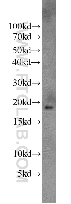
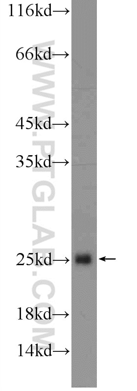
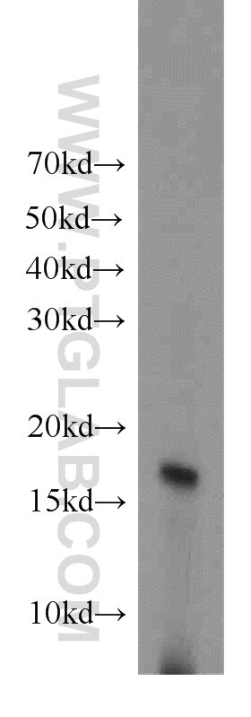
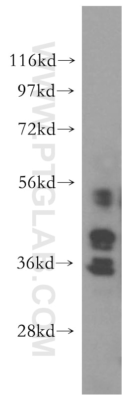
17168-1-AP bindet in WB, IP, IHC, IF, FC, CoIP, ChIP, ELISA Histone-H3 und zeigt Reaktivität mit human, Maus, Ratten
| Getestete Reaktivität | human, Maus, Ratte |
| In Publikationen genannte Reaktivität | human, Affe, arabidopsis, Fisch, Hausschwein, Huhn, Maus, Ratte, Ziege |
| Wirt / Isotyp | Kaninchen / IgG |
| Klonalität | Polyklonal |
| Typ | Antikörper |
| Immunogen | Histone-H3 fusion protein Ag10644 |
| Vollständiger Name | histone cluster 2, H3a |
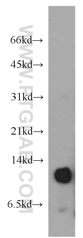
| Berechnetes Molekulargewicht | 136 aa, 15 kDa |
| Beobachtetes Molekulargewicht | 15-17 kDa |
| GenBank-Zugangsnummer | BC015544 |
| Gene symbol | HIST2H3A |
| Gene ID (NCBI) | 333932 |
| Konjugation | Unkonjugiert |
| Form | Liquid |
| Reinigungsmethode | Antigen-Affinitätsreinigung |
| Lagerungspuffer | PBS mit 0.02% Natriumazid und 50% Glycerin pH 7.3. |
| Lagerungsbedingungen | Bei -20°C lagern. Nach dem Versand ein Jahr lang stabil Aliquotieren ist bei -20oC Lagerung nicht notwendig. 20ul Größen enthalten 0,1% BSA. |
Hintergrundinformationen
Histone-H3, histone cluster 2, H3a is the core component of nucleosome. Nucleosomes wrap and compact DNA into chromatin, limiting DNA accessibility to the cellular machinery which requires DNA as a template. Histones thereby play a central role in transcription regulation, DNA repair, DNA replication and chromosomal stability. DNA accessibility is regulated via a complex set of post-translational modifications of histones, also called histone code, and nucleosome remodeling. Histone-H3 is expressed during S phase; then expression strongly decreases as cell division slows down during the process of differentiation.
Protokolle
| Produktspezifische Protokolle | |
|---|---|
| WB protocol for Histone-H3 antibody 17168-1-AP | Protokoll herunterladen |
| IHC protocol for Histone-H3 antibody 17168-1-AP | Protokoll herunterladen |
| IF protocol for Histone-H3 antibody 17168-1-AP | Protokoll herunterladen |
| IP protocol for Histone-H3 antibody 17168-1-AP | Protokoll herunterladen |
| Standard-Protokolle | |
|---|---|
| Klicken Sie hier, um unsere Standardprotokolle anzuzeigen |
Publikationen
| Species | Application | Title |
|---|---|---|
Signal Transduct Target Ther TRAF3 activates STING-mediated suppression of EV-A71 and target of viral evasion | ||
Cell Targeting Epigenetic Crosstalk as a Therapeutic Strategy for EZH2-Aberrant Solid Tumors. | ||
Adv Mater A Physiologically Responsive Nanocomposite Hydrogel for Treatment of Head and Neck Squamous Cell Carcinoma via Proteolysis-targeting Chimeras Enhanced Immunotherapy | ||
Nat Genet N6-Methyladenosine co-transcriptionally directs the demethylation of histone H3K9me2. |
Rezensionen
The reviews below have been submitted by verified Proteintech customers who received an incentive forproviding their feedback.
FH Mi (Verified Customer) (06-25-2023) | It works very well in nuclear extracts from human brown adipocyte cells.
|
FH Veda (Verified Customer) (04-29-2022) | IF staining test in cryo sections of skin. It does not seem to work. Maybe it only stains the nuclear membrane.
|
FH Pradeep (Verified Customer) (09-19-2020) | Good antibody for Western blot. Selvaraju, V., Thirunavukkarasu, M., Joshi, M. et al. Deletion of newly described pro-survival molecule Pellino-1 increases oxidative stress, downregulates cIAP2/NF-κB cell survival pathway, reduces angiogenic response, and thereby aggravates tissue function in mouse ischemic models. Basic Res Cardiol 115, 45 (2020). https://doi.org/10.1007/s00395-020-0804-4
|
FH Tanusree (Verified Customer) (12-03-2019) | The antibody works great in Western blotting analysis.
|
FH Ruiting (Verified Customer) (11-11-2019) | I use the Proteintech antibodies almost every week, the antibodies never failed to me. Very gald to use their products with a great price.Ruiting
|
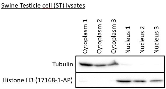
FH Robert (Verified Customer) (07-18-2019) | The Histon H3 antibody gives a signal in the three nuclear samples, but not in the cytoplasmic samples.
 |